

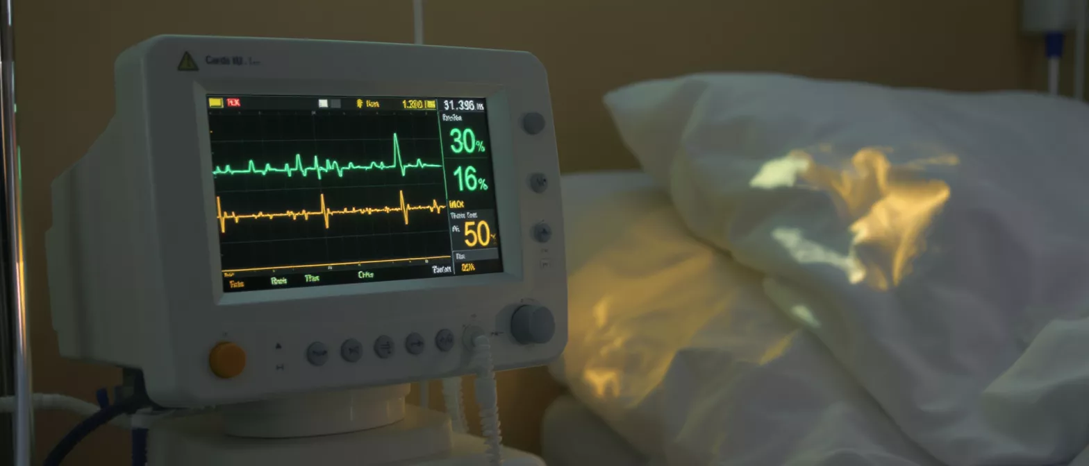
Haemodynamic Monitoring
Learn how haemodynamic monitoring tracks blood flow and heart function after surgery, including arterial lines, PA catheters, and ICU techniques.
What is haemodynamic monitoring in cardiac surgery?
Haemodynamic monitoring is the continuous assessment of blood flow, blood pressure, and fluid status within a patient's cardiovascular system. For a patient recovering from heart surgery, it provides the critical care team with a real-time picture of how effectively the heart is pumping blood to the body's vital organs. This vigilance allows clinicians to detect subtle changes before they become serious complications.
What do haemodynamic parameters tell the surgical team? These measurements reveal the precise balance between oxygen delivery and oxygen demand. By tracking the pressure inside the heart chambers and the volume of blood ejected with each heartbeat, doctors can ensure that the recovering heart muscle is neither overworked nor starved of essential fluids.
When you or a loved one is in the intensive care unit after cardiac surgery, you will notice several monitors displaying waveforms and numbers. These displays are the visual language of advanced cardiovascular assessment tools, translating internal physiological data into actionable information that guides medication adjustments, fluid management, and overall care planning.
Why is cardiovascular monitoring critical after surgery?
Following heart surgery, the cardiovascular system is in a highly vulnerable state. The heart has just undergone significant physical stress, and its ability to maintain stable blood pressure and optimal cardiac output can fluctuate. Cardiovascular monitoring provides an early warning system, allowing the medical team to intervene immediately if the heart's performance begins to decline.
How does perioperative haemodynamic monitoring protect the patient? It protects the patient by ensuring that the heart's workload remains balanced during the crucial healing period. If the heart pumps too hard, it risks damaging the surgical repairs; if it pumps too little, the kidneys and brain may not receive adequate oxygen. Monitoring ensures the clinical team maintains this delicate physiological balance.
Continuous observation is the cornerstone of postoperative cardiac care. It removes the guesswork from recovery management, providing objective data that dictates when to administer intravenous fluids, when to introduce blood pressure medications, and ultimately, when a patient is stable enough to leave the intensive care unit.
How invasive haemodynamic monitoring techniques work
Invasive haemodynamic monitoring involves placing specialised catheters or lines directly into the patient's blood vessels or heart chambers. These lines are connected to external pressure transducers, which convert the physical pressure of flowing blood into electrical signals displayed on the bedside monitor as continuous waveforms.
What is the purpose of invasive blood flow monitoring? The primary purpose is to obtain highly accurate, beat-to-bebeat blood pressure readings and direct cardiac pressure measurements. Unlike standard arm cuffs, invasive lines provide immediate feedback on rapid changes in cardiovascular status, which is essential when administering fast-acting intensive care medications.
While the idea of invasive lines can be frightening for families, they are standard, routine tools in cardiac surgery recovery. They are inserted under strict sterile conditions and remain in place only for as long as strictly necessary, usually being removed within the first day or two as the patient stabilises.
Understanding haemodynamic parameters and blood flow
Haemodynamic parameters are the specific numerical values derived from the monitoring equipment. Key metrics include heart rate, blood pressure, cardiac output (the total volume of blood pumped per minute), and stroke volume (the amount pumped per beat). Together, these figures illustrate the overall efficiency of the cardiovascular system.
How do clinicians interpret these haemodynamic parameters? Clinicians look at the relationships between the numbers rather than viewing them in isolation. A low blood pressure reading, for example, might be interpreted differently depending on whether the cardiac output is also low or if the patient's fluid status indicates dehydration versus a heart struggling to pump effectively.
- Cardiac Output: Total blood volume pumped per minute, indicating overall heart function.
- Systemic Vascular Resistance: The resistance blood vessels offer to blood flow, guiding blood pressure therapy.
- Stroke Volume: Blood ejected per heartbeat, reflecting heart muscle strength and fluid status.
What is an arterial line used for in the ICU?
An arterial line, often called an art-line, is a thin catheter usually placed into the wrist artery. It is one of the most common forms of ICU haemodynamic monitoring. This line provides a continuous, highly accurate reading of the patient's blood pressure with every single heartbeat.
Why choose arterial line monitoring over a standard cuff? Standard blood pressure cuffs only measure pressure every few minutes, which is insufficient immediately following heart surgery. An arterial line captures beat-to-beat variations, allowing the clinical team to instantly see how the heart and blood vessels are responding to medications or changes in fluid levels.
Additionally, the arterial line allows nurses to draw frequent blood samples without using a needle. This significantly improves patient comfort during the intensive care stay, as multiple arterial blood gas tests are often required to monitor oxygenation and ventilation postoperatively.
Central venous pressure and right heart function
Central venous pressure (CVP) measures the blood pressure in the vena cava, the large vein that returns blood to the right side of the heart. A specialised central venous catheter is usually inserted into a vein in the neck or chest to measure this pressure, which reflects the patient's overall fluid volume.
How does central venous pressure guide fluid management? CVP helps doctors determine if the heart is receiving enough blood to pump effectively. If the CVP is low, the patient may be dehydrated and require intravenous fluids. If the CVP is high, it might indicate that the right side of the heart is struggling to handle the incoming blood volume.
By carefully tracking central venous pressure alongside other parameters, the surgical team can fine-tune the exact amount of intravenous fluid a patient receives. This prevents fluid overload, a condition that can strain a recovering heart and potentially lead to fluid accumulation in the lungs.
How a pulmonary artery catheter guides recovery
A pulmonary artery catheter, commonly known as a Swan-Ganz catheter, is a sophisticated device advanced through the right side of the heart into the pulmonary artery. It provides direct measurements of pressures within the heart chambers and calculates cardiac output, offering a comprehensive picture of cardiovascular performance.
When is a pulmonary artery catheter necessary? This level of detailed cardiac output monitoring is typically reserved for patients with complex cardiac conditions, severe heart failure, or those undergoing high-risk surgical procedures. It is a powerful diagnostic tool used when standard monitoring does not provide enough data to safely manage a critically ill patient.
By directly measuring pressures inside the heart, the pulmonary artery catheter acts as a precise intracardiac pressure monitor. It allows the intensive care team to differentiate between left-sided and right-sided heart dysfunction, ensuring that targeted therapies are applied exactly where they are needed most.
Non-invasive and advanced cardiac output monitoring
Not all haemodynamic assessment requires invasive lines. Non-invasive monitoring techniques use external sensors, ultrasound technology, or specialized cuffs to estimate cardiac output and blood flow. These methods are increasingly utilised as technology improves, offering safer alternatives with fewer risks of infection.
What are the best non-invasive haemodynamic monitoring options? Echocardiography is a primary tool, using sound waves to visually assess how well the heart muscle contracts and fills. Additionally, technologies like pulse contour analysis and oesophageal Doppler monitors provide continuous estimates of blood flow without needing to puncture blood vessels.
- Echocardiography: Uses ultrasound to provide real-time images of heart structure and pumping function.
- Pulse Contour Analysis: Derives cardiac output from an existing arterial line's waveform.
- Doppler Ultrasound: Measures blood flow velocity through the cardiovascular system non-invasively.
Recognising and treating haemodynamic instability
Haemodynamic instability occurs when the cardiovascular system fails to maintain adequate blood pressure or cardiac output. In the postoperative period, this can result from bleeding, heart muscle stunning, arrhythmias, or shifts in body fluids. Recognising this instability instantly is the primary purpose of continuous monitoring.
How is haemodynamic instability managed in the ICU? Management requires a rapid, systematic approach guided by the monitoring data. If the monitors reveal low pressures, the team may administer fluids, blood products, or prescribe intravenous medications called inotropes to strengthen the heart's contraction, or vasopressors to constrict blood vessels and raise blood pressure.
The presence of comprehensive monitors allows the clinical team to treat instability proactively rather than reactively. By observing the trends in the haemodynamic parameters, doctors can often adjust treatments before the patient experiences severe symptoms, maintaining a stable environment for the heart to heal.
Recovery and perioperative haemodynamic monitoring
Perioperative haemodynamic monitoring encompasses the entire surgical journey, from the operating room to the intensive care unit and finally to the general ward. As a patient recovers and stabilises, the intensity of monitoring is gradually stepped down, moving from invasive lines to standard blood pressure cuffs.
What happens as haemodynamic monitoring is discontinued? As the heart regains its independent strength and stable function, the arterial and central lines are safely removed. The medical team continues to observe vital signs closely, but the focus shifts from critical care interventions to rehabilitation, mobility, and preparing the patient for a safe return home.
Understanding the role of these monitors helps alleviate the anxiety that often accompanies a stay in the intensive care unit. Seeing the numbers fluctuate can be alarming for families, but recognising that these postoperative cardiac surveillance systems exist precisely to guide a safe, uneventful recovery brings vital reassurance during the healing process.






