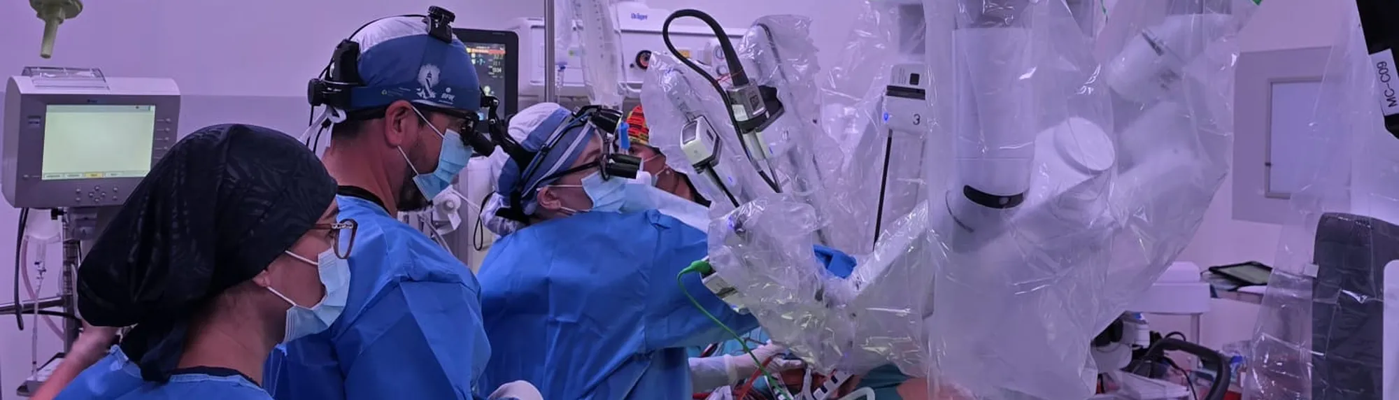
Robotic heart surgery
Robotic heart surgery in Mexico with Dr Alejandro Molina, who performed Guadalajara's first robotic heart operation.
Mitral valve repair and other cardiac procedures with the Da Vinci system — minimal invasion, maximum precision, and formal training in robotic surgery.
If you are reading this, it is likely that you or someone you love already knows that heart surgery lies ahead. That is not an easy thing to carry. There are questions, there is uncertainty, and there is almost always fear.
The first thing I want to say is this: I understand exactly what you are feeling. I have had that conversation with hundreds of patients and their families. And what I always tell them is that the fact they are looking for information, comparing options and asking questions already puts them in a very different place from those who face this alone and without guidance.
I am here to accompany you through that process.
I am Dr. Alejandro Molina, a cardiothoracic surgeon trained in Mexico, Canada and France. I performed the first robotic mitral valve repair in the whole Ángeles Health System, and the first robotic heart surgery in the history of Guadalajara. Heart surgery with the Da Vinci system is still extremely rare in Mexico; very few surgeons in the country are trained and certified to perform it.
What is robotic heart surgery?
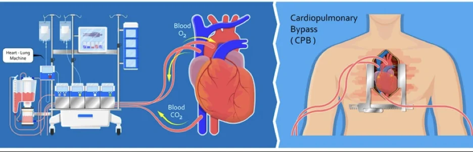
For decades, operating on the heart meant opening the sternum completely — the bone in the centre of the chest — stopping the heart and connecting the patient to a machine that did its work while we operated. It was, and remains, an extraordinarily effective operation. But it is also one of the most demanding on the human body.
Robotic surgery changes that equation.
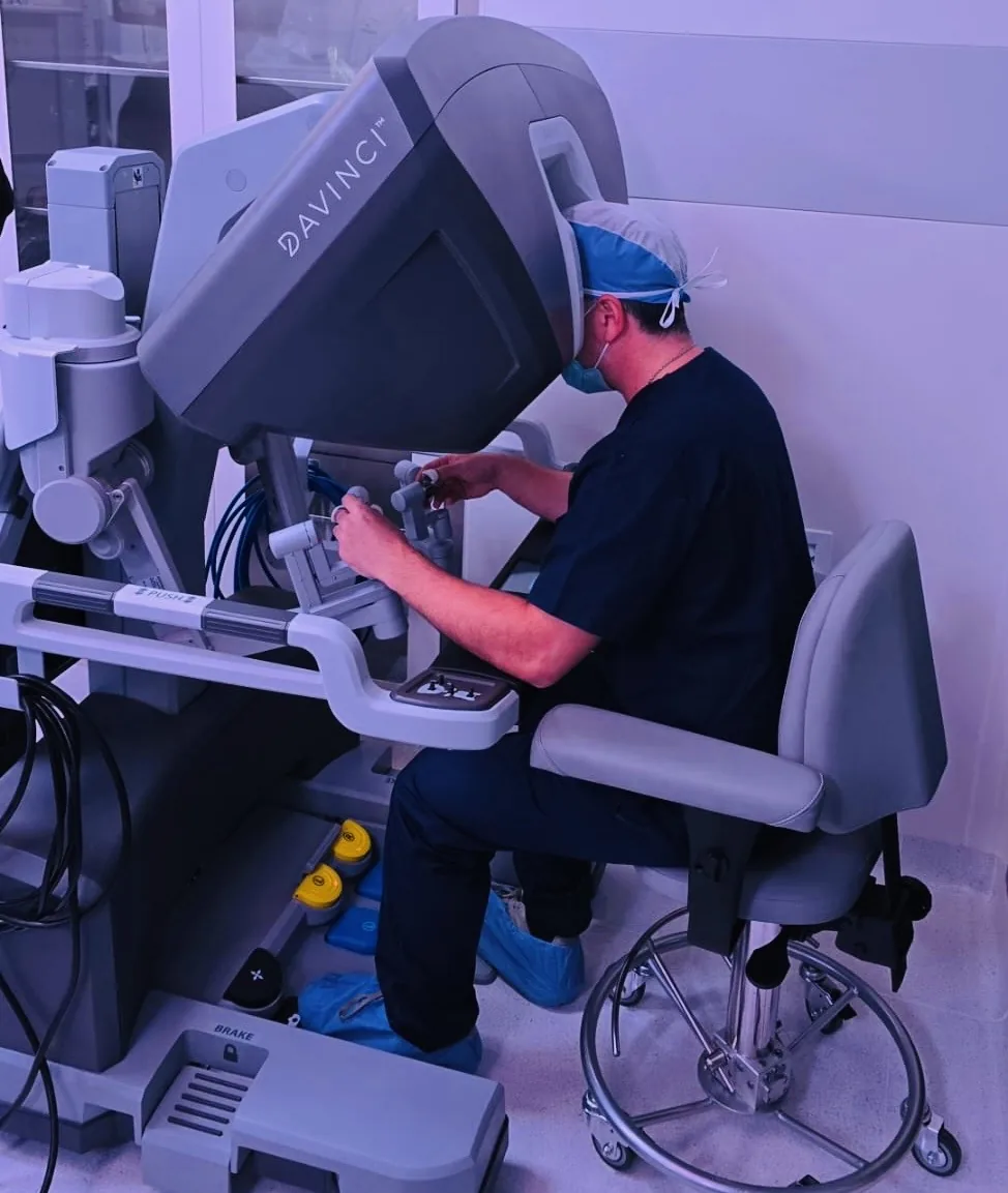
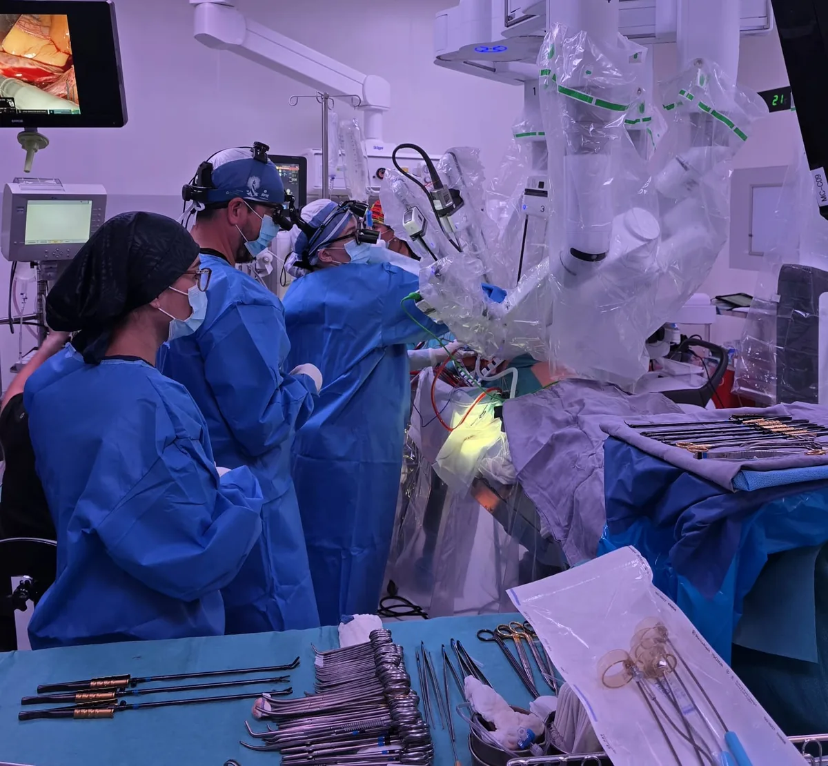
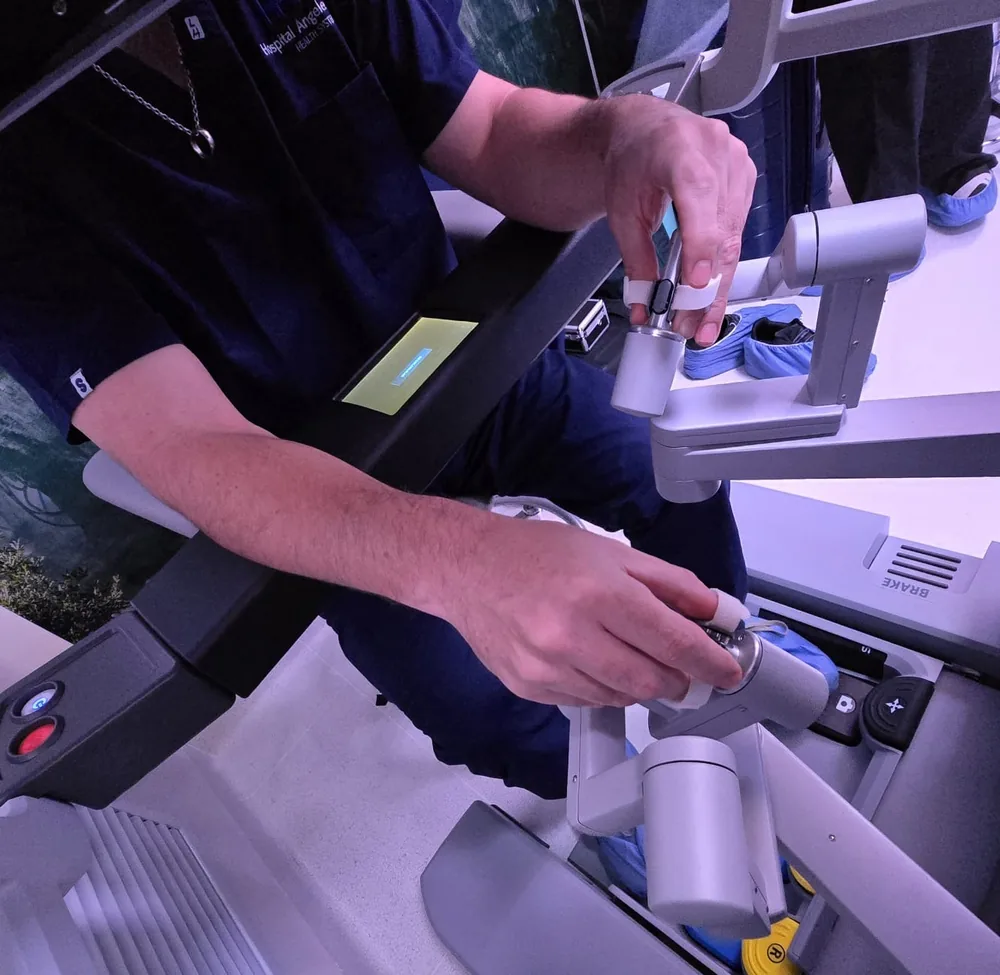
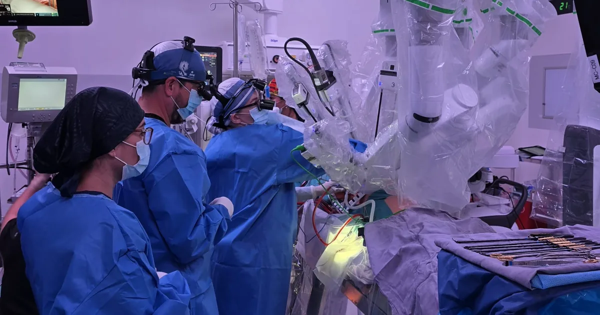
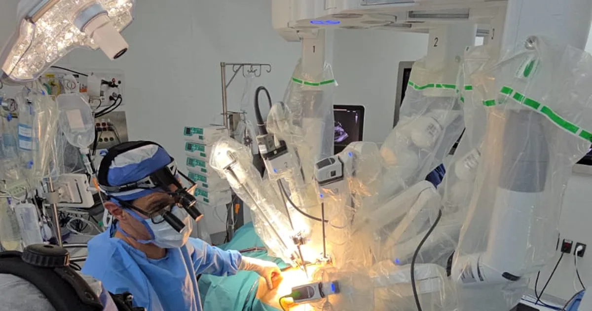
With the Da Vinci system we operate through small incisions between the ribs — without opening the sternum, without touching the bone. I work from a console with a magnified three-dimensional view of the inside of the chest. The robot's arms translate my movements with a precision that human hands, in a space that confined, could not achieve alone.
The result for the patient is concrete: less pain, less bleeding, and a recovery measured in weeks rather than months.
One clarification I always make: the robot does not operate on its own. Every movement is controlled by me. The Da Vinci system is an extension of my hands — an extraordinary tool, but guided at every moment by human judgement and clinical experience.
Which conditions can it be used for?
The most established application is repair of the mitral valve — the valve that controls the flow of blood between the two left chambers of the heart.
When this valve does not close properly, a condition called mitral regurgitation, blood flows back in the wrong direction. Over time the heart begins to work harder than it should, and that can lead to arrhythmias, heart failure or even a stroke. The good news is that when it is detected in time and treated correctly, the results are very good.
Robotic repair lets us correct this while preserving the patient's original valve — something clinical studies show is superior to replacement with a prosthesis, both in survival and in long-term cardiac function.
We also perform the following with a robotic approach
Heart valves
- Mitral valve replacement, when repair is not possible
- Tricuspid valve repair and surgery
Coronary revascularisation
- Hybrid revascularisation surgery — combining a minimally invasive coronary bypass, generally to the left anterior descending artery using the internal mammary artery, with the placement of stents in the other arteries that require them. It joins the proven durability of a bypass to the most important artery with the lower invasiveness of catheterisation for the rest.
Structural defects
- Atrial septal defect — a hole in the wall separating the atria of the heart, present from birth in some patients
Cardiac tumours
- Cardiac myxoma — the most frequent primary tumour of the heart, which can obstruct blood flow or shed fragments into the circulation
- Tumours of the pericardium, the membrane surrounding the heart
What difference does it make to your recovery?
This is by far the question I am asked most. And it makes a great deal of sense — because for most people the prospect of recovery matters as much as the operation itself.
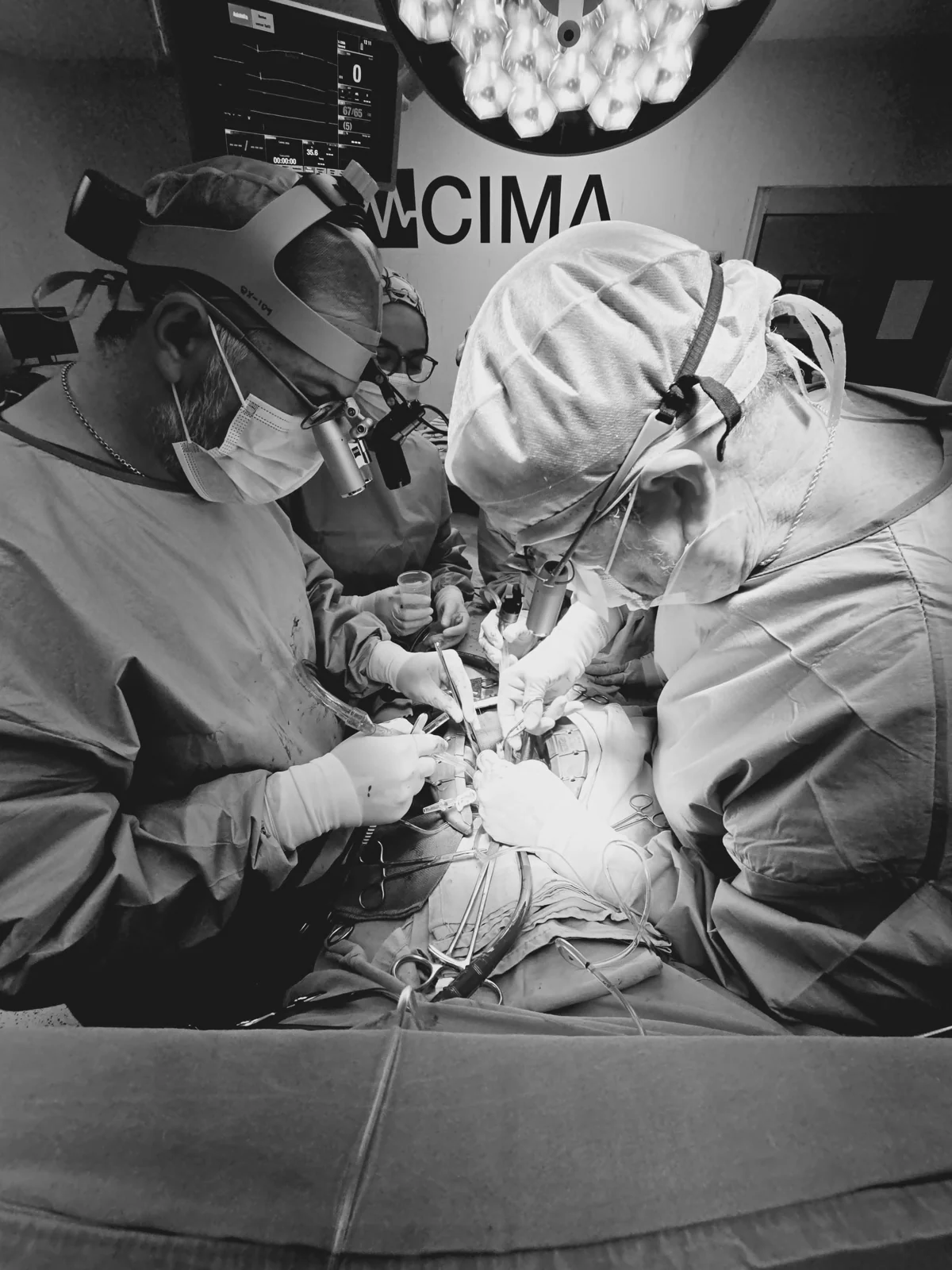
In conventional open-heart surgery the sternum is cut and needs between 6 and 12 weeks to knit. During that time you cannot drive, cannot lift anything, cannot make any physical effort. The pain in the first few days is considerable.
In robotic surgery the sternum is not touched. The incisions are small. Postoperative pain is significantly less. Most patients are home in 3 to 5 days, and within two to three weeks they are back to their normal lives.
For many people — those who work, those with children or grandchildren to look after, those who simply do not want to be out of action for months — that difference changes everything.
Real cases
Will I be a candidate for robotic surgery?
Not every patient is a candidate, and I want to be direct with you about that. Careful patient selection is as important as the surgical technique. There is no operation that is right for everyone.
What we assess before making any decision:
- The anatomy of your valve and your chest. Some variations can make robotic access more complex, or not the most suitable choice.
- The type and degree of your cardiac condition. The function of your left ventricle and the presence of other heart disease.
- Your surgical history. Previous chest surgery can change the approach.
- Your general health. Lung function, body weight and other medical conditions all form part of the full assessment.
We always make this decision as a team — cardiologist, anaesthetist and surgeon — and never without having reviewed your case in detail. There are no quick answers here, and there should not be.
What studies do I need to bring to the consultation?
A very practical question almost every patient asks me: "Doctor, what should I arrive with?"
The assessment follows a natural sequence, from the simplest to the most specific. If you already have any of these studies — even from a few months ago — bring them. They are the starting point of our conversation.
- Transthoracic echocardiogram. The first step. An ultrasound of the heart performed from the surface of the chest, with no discomfort at all. It shows the anatomy of the valves, how the chambers of the heart are working and how far the disease has advanced. Most patients already have one when they come to us.
- Transoesophageal echocardiogram. When we need to see the valve in far greater detail — particularly to plan a repair — we perform this study. A very fine probe is passed through the mouth into the oesophagus, which sits directly behind the heart. The image we obtain is markedly sharper. It is done as an outpatient, under light sedation, and the patient feels no discomfort.
- CT angiography of the chest, abdomen and pelvis, from the neck to the groin. This is the key study for planning the robotic approach. It is similar to the protocol we use for TAVI. It gives us a precise three-dimensional image of the chest, the aorta and the vessels we will use for access. With it we confirm whether the patient's anatomy is compatible with robotic surgery.
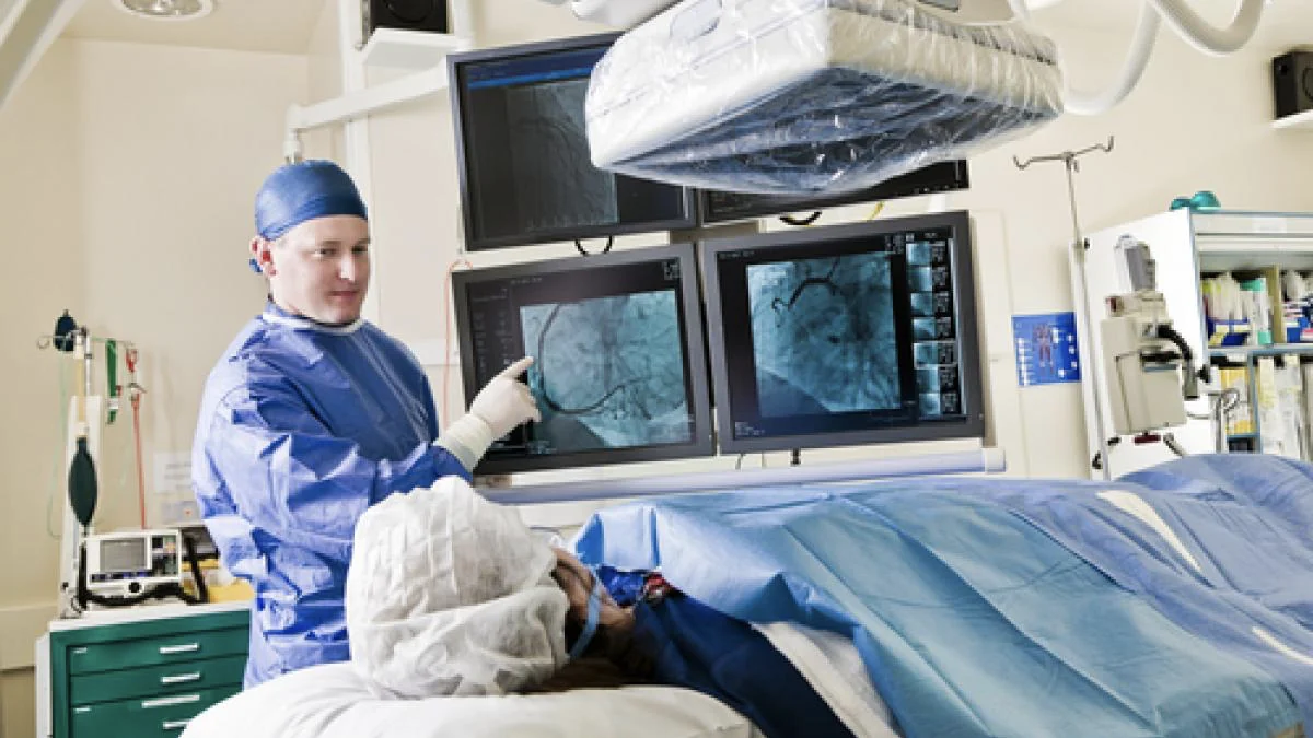
- Cardiac catheterisation, in selected cases only. It is not always necessary. We indicate it when coronary artery disease is suspected, or when we need information about pressures inside the heart that the other studies could not clarify.
- Routine laboratory tests. Full blood count, blood chemistry, clotting tests, kidney and liver function. These are the basic studies that give us the general health context before any intervention.
Don't worry if you don't have them all. We start with whatever you have.
An honest word
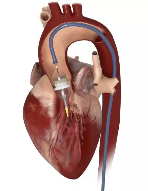
I want to be clear with you: robotic surgery is not the answer for everyone. There are cases in which conventional surgery remains the safest option. There are also cases in which a catheter procedure — such as TAVI or MitraClip — is more appropriate than any operation. Our guide to robotic, minimally invasive or open-chest surgery explains how we choose.
I spent years of training learning all of those options — conventional open surgery, minimally invasive, robotic and transcatheter procedures — precisely for that reason. Not to have a longer CV, but to be able to sit in front of each patient and recommend what genuinely suits them, rather than what I prefer to do.
When a doctor masters a single technique, that technique tends to become the answer for all of their patients. I do not want to be in that position. I want to have every tool available so that the decision is always in favour of the person in front of me.
My job is to listen to your case, review your studies, and give you an honest recommendation. I will always give it clearly, without evasion, and with whatever time you need to understand it and ask all your questions.
By the time we reach the operating theatre we have already planned every step together. The technology is powerful — but it is clinical judgement, and the trust we build with you before that day, that really matters.
Where do we perform this procedure?
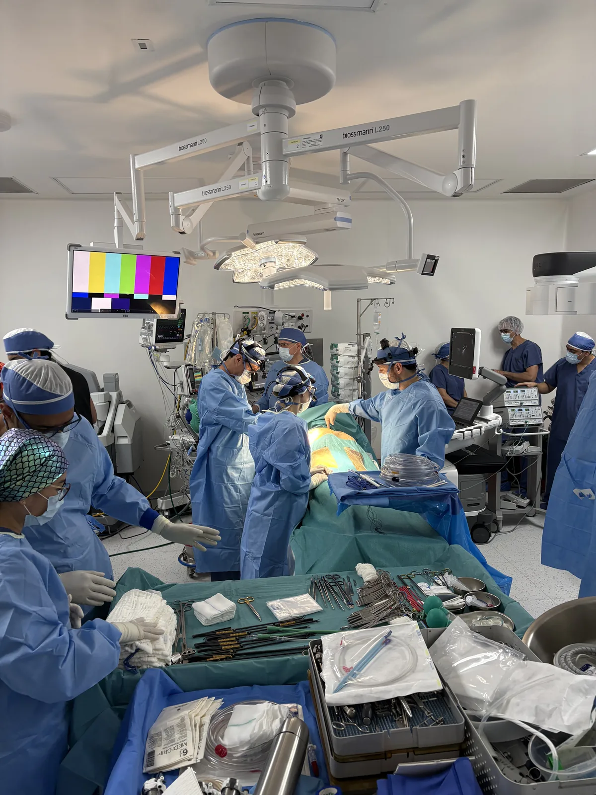
Robotic heart surgery is currently available with our team in three cities — Guadalajara, Querétaro and Hermosillo — at hospitals equipped for it, and the programme continues to grow.
If you live in another city, that is not an obstacle. A video consultation is the first step — we review your studies together, assess your case, and if appropriate we coordinate everything needed for an in-person assessment.
The next step
If you have read this far, it is because you are taking an important decision seriously. That already says a great deal about you.
The next step does not have to be a large one. It is simply a conversation — a consultation where we review your studies, I explain clearly what is happening in your heart, and we explore together the best path for your particular case.
You do not have to arrive with all the answers. Come with your questions.
For cardiologists: if you have a patient who might be a candidate for robotic heart surgery, see how to refer a patient.
About the author
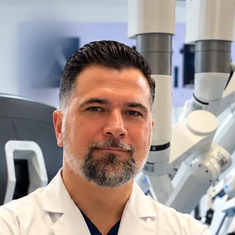
Dr. Alejandro Molina Romo is a cardiovascular surgeon practising in Guadalajara, Querétaro and Hermosillo. He trained in cardiothoracic surgery at the Universidad de Guadalajara, completed advanced training in cardiac surgery, minimally invasive surgery, robotic surgery and structural interventions at the University of Montreal, and a fellowship in structural cardiology and TAVI at CHU Bordeaux, France. He performed the first robotic mitral valve repair in the Ángeles Health System and the first robotic heart surgery in Guadalajara.
Medical degree: Universidad Autónoma de Guadalajara, Cédula Profesional 5757260 · Cardiothoracic surgery: Universidad de Guadalajara, Cédula de Especialidad 10768871 · Current certification, Consejo Nacional de Cirugía del Tórax, no. 447
This content is educational and does not constitute personal medical advice. Every cardiac case is unique and requires individual assessment by a specialist medical team. Always consult the doctor treating you before making decisions about your treatment, particularly if you take anticoagulants, antiplatelet drugs or other cardiac medicines.
Book a video consultation
Come with your questions. We review your studies together and give you an honest recommendation for your case.