Robotic Mitral Valve Repair at 77: The Case That Started With the Aorta
How a Heart Team planned the first robotic mitral valve repair in the Ángeles Health System: a 77-year-old patient, a hidden aortic problem, no residual leak.
Robotic surgery is usually described by what you can see: small incisions, no opened chest, a faster recovery. This case is about what you cannot see — the planning that makes it safe. Before we touched his heart, we had to deal with a problem in his aorta that nobody knew was there.
The case at a glance
- Patient: 77 years old, very active, no known heart disease
- Diagnosis: severe mitral regurgitation from a degenerative valve with ruptured chords
- Hidden finding: an ulcer in the wall of the abdominal aorta
- Plan: treat the aorta first through a catheter, then repair the valve robotically
- Result: no residual leak in the repaired valve
A very active patient with new symptoms
The patient is a 77-year-old man: independent, a professional, and in the habit of walking for an hour every day. He had no known chronic illness, apart from a recent diagnosis of an enlarged prostate.
Towards the end of 2025 he began to notice that he could do less than before. Exertion left him short of breath, and he had occasional palpitations. His cardiologist heard a very clear murmur over the mitral valve — the kind of finding that calls for an echocardiogram straight away.
What the studies showed
The mitral valve sits between the two left chambers of the heart and should close completely with every beat. In his case it no longer did. The echocardiogram showed severe mitral regurgitation: with each heartbeat, a large amount of blood was flowing backwards.
The cause was degenerative valve disease. The valve tissue had become redundant and floppy, and some of the chords that hold the leaflets in place — like the strings of a parachute — had ruptured. The rear leaflet, in particular, was prolapsing back into the left atrium, which had already enlarged under the strain. The good news was that the heart muscle was still pumping normally. That is exactly the moment to operate: before the heart starts to suffer lasting damage.
Valves like this one are usually repairable. And when a mitral valve can be repaired well, repair is preferable to replacement: the patient keeps their own valve, avoids the long-term issues of a prosthesis, and long-term results are better.
The finding that changed the plan
Before any robotic or minimally invasive operation, we study the aorta and the arteries of the abdomen and legs with a CT scan. There is a practical reason for this. In these operations, the heart-lung machine is usually connected through the blood vessels of the groin, so that during the operation blood is pumped backwards up through the aorta. Those vessels need to be able to take it.
His coronary arteries were clean. But the CT showed something nobody had suspected: an ulcer in the wall of the abdominal aorta, with a small tear in its inner layer. It was causing him no symptoms and no complications. But with blood flowing backwards through that segment during surgery, it was a weak point we were not willing to leave unprotected.
This is where a Heart Team earns its name. Rather than abandon the less invasive approach, or simply hope for the best, we treated the aorta first: a covered stent was placed inside the damaged segment through a catheter, sealing it from the inside. With the aorta protected, the robotic operation could go ahead safely.
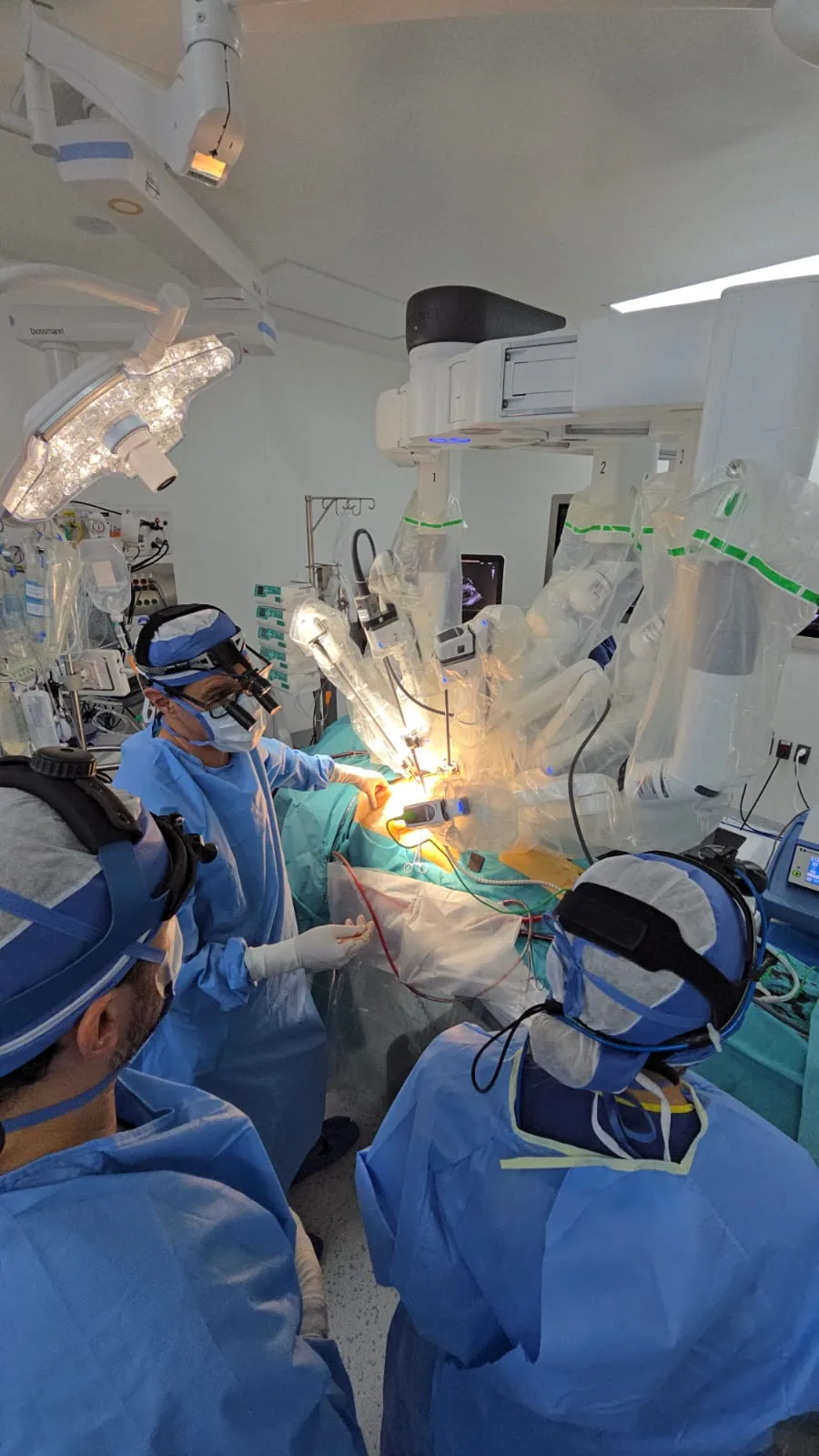
The technology is impressive. But what makes robotic surgery safe is the decisions made before anyone sits at the console.
The operation
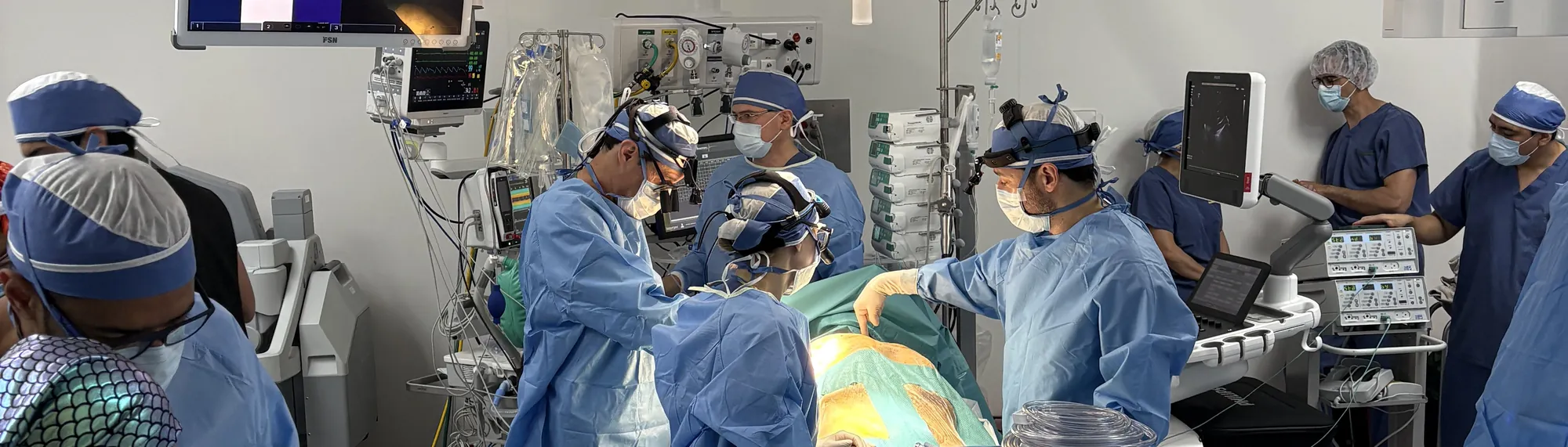
In January 2026, at Hospital Ángeles Centro Sur in Querétaro, we repaired his mitral valve with the Da Vinci Xi surgical system. It was the first robotic mitral valve repair in the Ángeles Health System.
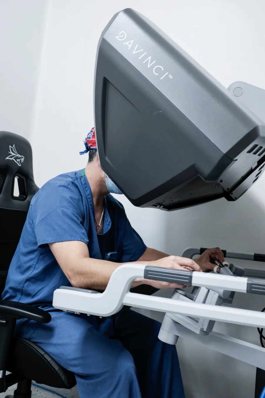
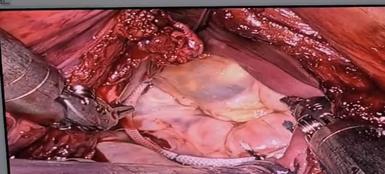
The operation was planned and rehearsed in detail beforehand. Through small incisions between the ribs, and without opening the sternum, we removed the damaged segment of the rear leaflet, brought the healthy tissue back together, and placed a ring to reshape and support the valve. A replacement with a biological valve was prepared as a back-up plan in case the repair did not work. It was not needed.
Recovery
His breathing tube was removed in the operating room, before he left it. He spent two days in intensive care, following a protocol of enhanced recovery after surgery. From the point of view of his heart, the recovery was uneventful.
There was one complication, and it had nothing to do with his heart: his enlarged prostate caused an episode of acute urinary retention. The urology team placed a catheter, and he went home eight days after surgery with the catheter in place until his prostate operation a few weeks later.
The echocardiogram after surgery showed no residual leak in the repaired valve, and his recovery has been very good.
We share this detail because it is real. Recovery is not always a straight line, even when the heart operation goes perfectly — and being open about that is part of being trustworthy.
What this case shows
- Age alone does not decide. At 77, an active patient in good general health can be an excellent candidate for robotic surgery. What matters is the heart, the blood vessels and the overall condition — not the number.
- Planning comes first. A careful CT found a problem that could have caused a serious complication. Treating it first is what made the robotic approach safe.
- Repair is the goal. When the valve can be repaired well, the patient keeps their own valve for life.
- It takes a team. Cardiology, endovascular treatment, anaesthesia, the surgical team and urology all played a part in this one patient’s recovery.
Read more about robotic heart surgery, about how to choose between robotic, minimally invasive and open-chest surgery, about the options for replacing a heart valve, or about the first robotic heart surgery in Guadalajara.
The next step
If you have been told you have mitral regurgitation, or a murmur that needs investigating, bring your studies to a consultation — in person in Querétaro, Guadalajara or Hermosillo, or by video from anywhere. We will tell you honestly whether your valve can be repaired, and which approach makes sense for you.
For cardiologists: if you have a patient with mitral disease who might be a candidate for robotic or minimally invasive repair, see how to refer a patient, or send me the studies directly.

About the author
Dr. Alejandro Molina Romo is a cardiovascular surgeon practising in Guadalajara, Querétaro and Hermosillo. He trained in cardiothoracic surgery at the Universidad de Guadalajara, completed advanced training in cardiac surgery, minimally invasive surgery, robotic surgery and structural interventions at the University of Montreal, and a fellowship in structural cardiology and TAVI at CHU Bordeaux, France. He performed the first robotic mitral valve repair in the Ángeles Health System and the first robotic heart surgery in Guadalajara.
Medical degree: Universidad Autónoma de Guadalajara, Cédula Profesional 5757260 · Cardiothoracic surgery: Universidad de Guadalajara, Cédula de Especialidad 10768871 · Current certification, Consejo Nacional de Cirugía del Tórax, no. 447
This content is educational and does not constitute personal medical advice. It describes one patient’s case, shared anonymously and with his consent; every patient’s course is different. Every cardiac case requires individual assessment by a specialist medical team. Always consult the doctor treating you before making decisions about your treatment.
Book a video consultation
Come with your questions. We review your studies together and give you an honest recommendation for your case.