3D Endoscopic Camera in Minimally Invasive Heart Surgery
3D endoscopic camera heart surgery repairs valves through tiny incisions with faster recovery.
If you or a family member has been told you need heart surgery, one of the first questions is usually the same: how big will the incision be? For decades, the answer was a full sternotomy — opening the breastbone down the middle. Today, a 3d endoscopic camera heart surgery makes it possible to repair or replace heart valves through incisions of just a few centimetres, while the surgical team watches a high-definition three-dimensional image on a screen. This guide explains what this technology is, which procedures use it, what recovery actually looks like, and how to decide whether it is the right route for you. We are a multidisciplinary cardiac team led by Dr Alejandro Molina, with consulting rooms in Guadalajara, Querétaro and Hermosillo, and we perform conventional, minimally invasive, robotic and transcatheter procedures — always recommending the less invasive route only where the evidence supports it for that specific patient.
What Is a 3D Endoscopic Camera?
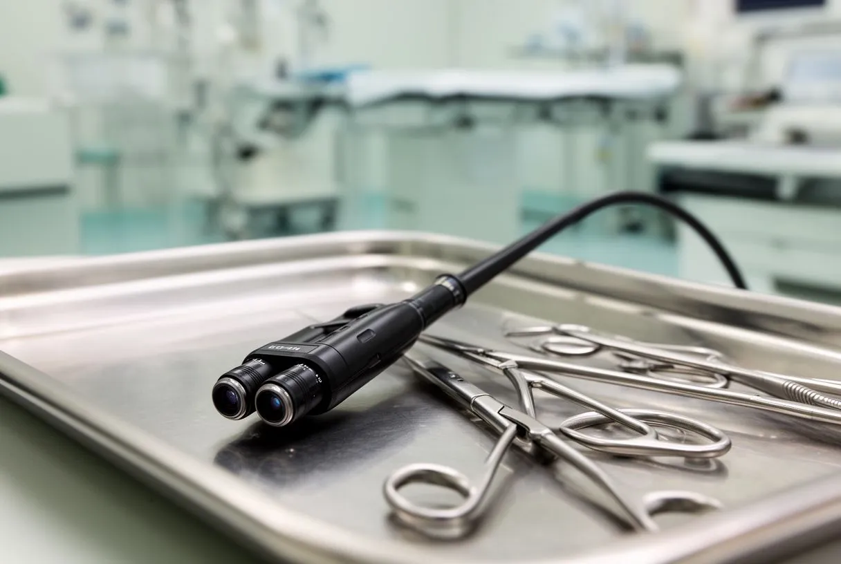
An endoscopic camera is a tiny video camera mounted at the tip of a thin, rigid tube called an endoscope. The surgeon inserts this tube through a small incision and uses the live image it transmits to see inside the chest without opening it fully. In a 3D system, the camera captures two slightly different images at once — one for each eye — and the surgical display merges them so the team perceives true depth, much as you do with your own vision.
Why depth matters inside the chest. Standard two-dimensional endoscopy flattens the image, so judging how far a structure sits from the camera takes experience and careful instrument movement. A three-dimensional view restores natural depth perception. When a surgeon is working millimetres from a coronary artery or sewing a delicate valve leaflet, that extra dimension changes what the hands can confidently do.
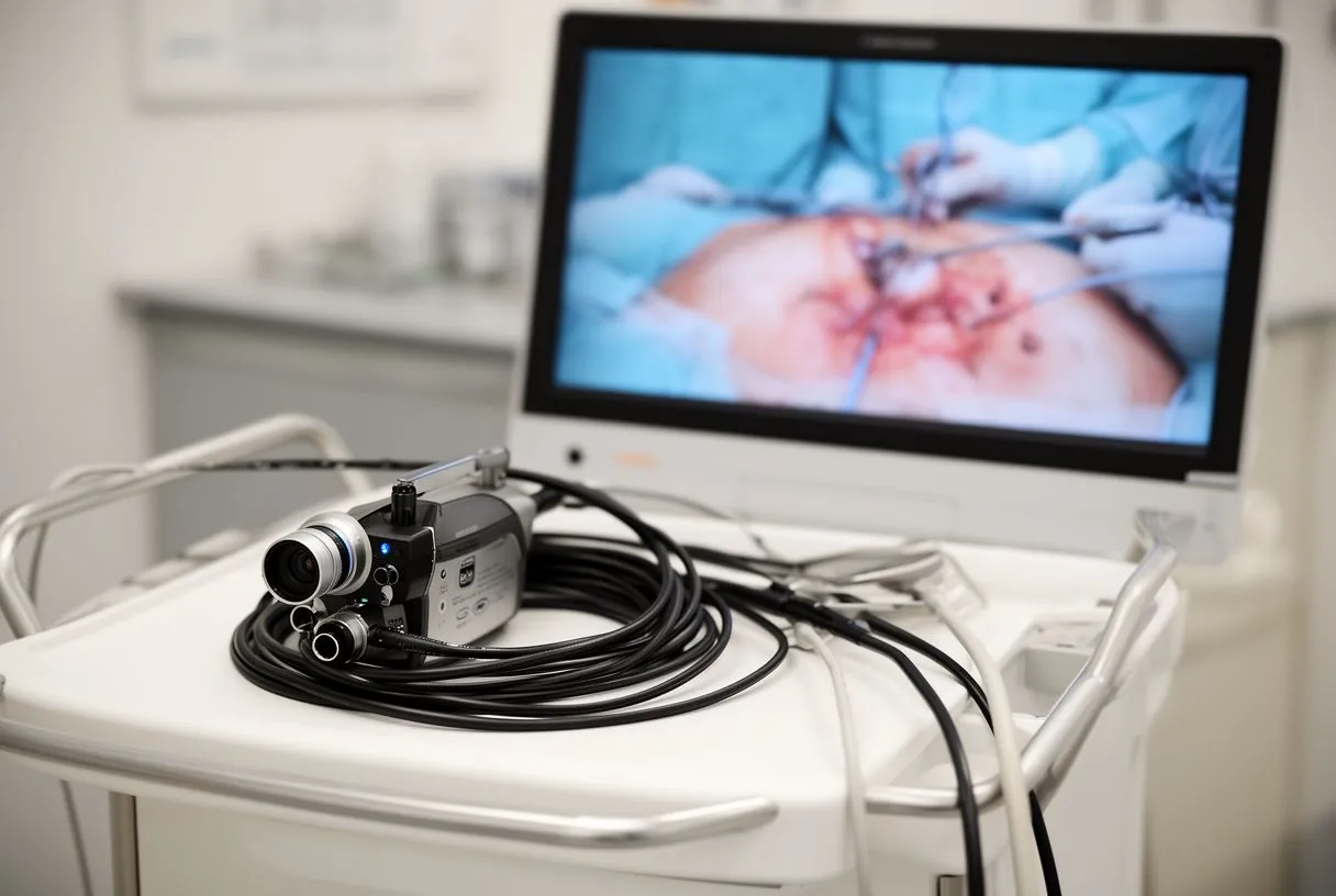
What the system actually includes. A full setup is more than the camera itself: it includes the scope, a light source, a high-definition display screen, and image processing that keeps colour and detail accurate. Modern systems operate in full HD or better, which matters because the surgeon is operating from the screen rather than looking directly at the tissue.
How it compares to the naked eye. In open surgery, the surgeon looks directly at the heart. With endoscopy, the image can actually be magnified beyond what the eye could see, so small structures appear larger on screen. The trade-off is that the surgeon loses direct touch and must rely on instrument feedback and visual cues — a skill that requires specific training.
How Does a 3D Endoscopic Camera Heart Surgery Work?
A 3d endoscopic camera heart surgery follows a carefully planned sequence, and understanding it helps take away much of the fear of the unknown. Before the operation, the team reviews your imaging — echocardiogram, CT scan, catheterization — to confirm the anatomy is suitable for a small-incision approach. Every case is reviewed by the multidisciplinary team against international practice guidelines before a recommendation is given, and the patient receives a clear proposal with the alternatives, with the final decision always theirs.
On the day of surgery. You are placed under general anaesthesia. Rather than splitting the breastbone, the surgeon makes one small incision — often on the right side of the chest for mitral valve work, or a mini-incision at the top of the sternum for aortic valve access. The endoscope is inserted through one of these openings, and long, thin instruments enter through the others.
During the operation. The heart is typically stopped and a heart-lung machine takes over circulation, just as in open surgery. The difference is purely in access: the surgeon works through the small ports while watching the 3D image. The camera can be angled to look around corners and beneath structures, which is sometimes easier than with direct vision.
After the operation. Once the repair or replacement is complete, the heart is restarted, the ports are closed, and you go to the intensive care unit. Because the breastbone is intact or only partially divided, the chest is mechanically far more stable than after a sternotomy — one of the main reasons recovery tends to be faster.
What Is an Endoscopic Camera Used for in Cardiac Care?
Endoscopic visualization is now used across most areas of cardiac surgery where a small-access approach is appropriate. The technology is not a treatment in itself — it is the surgeon's eyes — and it is chosen when the procedure and the patient's anatomy allow it.
Valve surgery. Mitral valve repair is the classic indication. The valve sits on the left side of the heart, and the right chest offers a direct route to it with excellent visibility through the endoscope. Aortic valve replacement can also be done through a mini-sternotomy or right-sided approach in selected patients.
Coronary and other procedures. In selected cases, bypass grafts on the front of the heart can be performed minimally invasively, and endoscopic techniques are also used for harvesting veins or arteries for grafts — taking the graft vessel through small leg or wrist incisions instead of one long one. Some teams also use endoscopic guidance for ablation procedures aimed at atrial fibrillation, an irregular heart rhythm, performed alongside other valve operations.
Diagnostic and support roles. Endoscopes also help the team inspect the inside of the chest before closing, confirm that bleeding has stopped, and position chest drains — small steps that reduce complications. In our consultations we take the time to explain what the studies actually show, in plain words, and which of these techniques applies to your particular case.
3D Endoscopic Camera Benefits for Heart Patients
The benefits of a 3d endoscopic camera minimally invasive heart surgery fall into two groups: those you feel in the first weeks, and those that matter over the long term. Both are worth understanding before you decide.
Short-term advantages. With a small incision and no fully divided breastbone, patients generally experience less post-operative pain, a lower chance of wound infection at the incision site, less blood loss, and a shorter hospital stay. Breastbone complications — the slow healing problems that can follow a sternotomy — are largely avoided. For many patients, being able to drive, return to work and sleep comfortably weeks earlier is the difference that matters most.
Cosmetic and psychological factors. A small incision on the side of the chest heals to a few centimetres, often hidden under the arm line.
Longer-term considerations. The technology also aids the surgeon: magnified 3D vision can support very precise repair of valve tissue, and precise mitral repair has durable long-term results when performed by experienced teams. The honest framing is this: minimally invasive access offers real advantages, but the quality of the repair and the experience of the team matter more than the size of the incision. Recent clinical experience with minimally invasive approaches, including detailed early-period outcomes after such operations, is documented in the literature, such as this study of the early period after minimally invasive cardiac surgery Reanimatology.
- Less pain: no divided breastbone means far less chest discomfort in the first weeks.
- Lower wound risk: small incisions have lower infection rates than a full sternotomy wound.
- Shorter stay: many patients leave hospital sooner and regain independence faster.
- Precision for the surgeon: magnified 3D vision supports delicate valve repair work.
Which Heart Procedures Use a 3D Endoscopic Camera?
Not every heart operation suits a small-incision approach, and part of a good consultation is explaining exactly why a particular route is or is not recommended for you. Here are the main procedures where a 3d endoscopic camera mitral valve repair or other endoscopic technique is commonly considered.
Mitral valve repair and replacement. This is where 3D endoscopic surgery is most established. The mitral valve, which sits between the two left chambers of the heart, is reached through a small right-chest incision. Endoscopic magnification is genuinely useful for the fine sewing that valve repair requires, and repair — when feasible — is preferred over replacement because it preserves your own valve tissue.
Aortic valve replacement. A 3d endoscopic camera aortic valve replacement, or a mini-access version of it, reaches the aortic valve — the valve between the left pumping chamber and the main artery to the body — through a partial upper sternotomy or a right-sided incision. Published centre experience shows this approach can be performed safely in appropriate patients, as described in this report on minimally invasive aortic valve replacement Cirugía y Cirujanos. For patients who are not candidates for any surgical approach, transcatheter aortic valve replacement — TAVR — offers a catheter-based alternative, and our team performs both, choosing based on evidence for each patient.
Other applications. Endoscopic techniques also support atrial septal defect closure in selected cases, surgical ablation for atrial fibrillation combined with valve surgery, and minimally invasive harvest of graft vessels for coronary bypass. The common thread is that each option is weighed against the conventional route in your specific case — our multidisciplinary team reviews every case together before recommending a less invasive route, precisely because smaller access is not automatically better for every patient.
3D Camera Versus Robotic Heart Surgery: What Is the Difference?
Patients often ask about robotic vs endoscopic heart surgery, because both are "small incision" approaches and the marketing around them can be confusing. Understanding the distinction helps you ask the right questions.
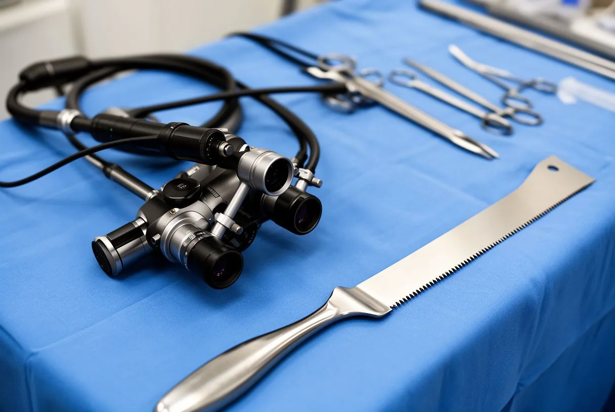
Who holds the instruments. In endoscopic surgery, the surgeon holds the long instruments directly, watching the 3D screen. In robotic surgery, the surgeon sits at a console away from the table and controls mechanical arms that hold the instruments, translating hand movements into smaller, tremor-free motions inside the chest. The robotic system also provides its own 3D vision — in fact, a 3D endoscopic camera is the eye of the robot as well.
What each is good at. Robotic systems offer enhanced wrist-like instrument movement and are particularly suited to very intricate mitral valve repair. Direct endoscopic surgery avoids the cost and setup of the robot, and many procedures — including aortic valve work — are well served by the direct endoscopic approach. Neither is universally superior; the right choice depends on the procedure, the anatomy, and the team's experience.
What this means for you. Ask any team you consult which approach they use for your specific diagnosis and why, and how many of those procedures they have performed. Dr Molina performed the first robotic mitral valve repair in the Ángeles Health System and the first robotic heart surgery in Guadalajara, and our team performs both robotic and direct endoscopic procedures — which means the recommendation you receive is about your heart, not about the equipment we happen to own.
Are 3D Endoscopic Camera Procedures Safe?
This is the most important question, and it deserves an honest answer: minimally invasive cardiac surgery is safe and well established in experienced hands, but it carries the same fundamental risks as any heart operation, and it adds a few of its own.
Shared risks with open surgery. Any operation on a stopped heart with a heart-lung machine carries risks of bleeding, stroke, infection, kidney stress, and rhythm problems such as atrial fibrillation. These risks depend heavily on your age, other conditions, and the urgency of surgery. Modern risk tools such as EuroSCORE II — a scoring system surgeons use to estimate operative risk — help quantify this before any decision is made.
Risks specific to the minimally invasive approach. Working through small ports means less direct access if sudden bleeding occurs, and converting to an open sternotomy must sometimes be done — experienced teams plan for this possibility from the first incision. There is also a learning curve: published evidence consistently shows that outcomes depend on surgeon and centre experience with the specific technique.
How the risk is actually managed. Careful patient selection is the biggest safety factor. Patients with severely diseased lung tissue on the operative side, severe chest deformity, or extensive calcified arteries may be better served by a conventional approach. We encourage second opinions and treat seeking one as a normal part of deciding about heart surgery — a team confident in its recommendation will always welcome it. General information on surgical risks and how to prepare is available from patient-oriented resources such as Healthline.
What Is the Recovery Time After Endoscopic Heart Surgery?
Endoscopic heart surgery recovery time is one of the main reasons patients choose this route — and also one of the areas where expectations need to be realistic. Recovery is faster, but it is still open-heart surgery.
The first days. You will wake in the intensive care unit, often sitting up in a chair within the first day. Chest tubes — small drains that remove air and fluid from around the heart — are usually removed within the first day or two. Most patients move to a regular ward room quickly and are walking the corridor within days.
Weeks one to six. The hospital stay after a minimally invasive valve operation is often shorter than after a sternotomy. At home, the first weeks are about gentle walking, gradually increasing, and avoiding lifting anything heavy. Because the breastbone is intact, the classic six-week sternal restriction is usually shorter or lighter — but your team will give you specific instructions, and they differ from those after open surgery.
Return to normal life. Many patients return to desk work within a few weeks and to full activity over one to three months, depending on the procedure and their overall health. Driving is usually permitted once you are off strong pain medication and can turn the wheel without discomfort. Cardiac rehabilitation — a supervised exercise and education programme — is strongly recommended regardless of the surgical approach, and it is one of the best predictors of a good long-term outcome.
What to watch for. Contact your team if you develop fever, increasing redness or discharge at the incision, new or worsening shortness of breath, or chest pain that is different from your usual post-operative soreness. After surgery, questions about what is normal during the recovery milestones are always worth asking.
What Does 3D Endoscopic Camera Heart Surgery Cost in Mexico?
Cost is a real and reasonable concern, especially for international patients, and it is worth understanding how a 3d endoscopic camera cost Mexico comparison actually works — because the camera itself is one component of a much larger picture.
What you are actually paying for. The total cost of heart surgery includes the surgeon and team fees, hospital and intensive care time, anaesthesia, the perfusionist who runs the heart-lung machine, implants such as valve prostheses, disposable technology including the endoscopic system, and your recovery care. The camera system itself is hospital infrastructure, not a line item on your bill — hospitals invest in it, and the cost is distributed across all procedures.
Why minimally invasive can change the total. Although the technology costs more per case in equipment, shorter intensive care stays and shorter hospital admissions can offset part of that. The exact balance varies by hospital, so it is fair to ask for a detailed written estimate broken down by component.
Insurance and international patients. Many Mexican insurance policies cover minimally invasive cardiac surgery when it is medically indicated, and international patients should check whether their policy covers planned surgery abroad. At the first contact we offer a free video consultation: we get to know the patient, talk through the condition, and say which studies are still needed so no unnecessary ones are ordered — and a face-to-face visit follows only when the patient decides to go ahead.
- Ask for itemised estimates: surgeon, hospital, implant, and recovery costs should each be listed.
- Check insurance coverage: confirm in writing whether minimally invasive and robotic procedures are covered.
- Plan travel time: international patients should budget for pre-operative tests, the procedure itself, and a recovery period before flying home.
How Do You Choose a Minimally Invasive Heart Surgeon?
If you are searching for the best minimally invasive heart surgeon Guadalajara has to offer — or anywhere else — it helps to know what "best" actually means in this field, because the answer is more specific than a name or a website.
Volume and specific experience. In cardiac surgery, outcomes are closely linked to the experience a team has with the specific procedure you need. Ask directly: how many mitral repairs through small incisions does this team perform per year? What are their results? A surgeon who performs a high volume of conventional open surgery but only occasional minimally invasive cases is a different proposition from a dedicated minimally invasive team.
Training and verification. Formal training in minimally invasive and robotic cardiac surgery is a real credential worth checking. Dr Molina trained in cardiothoracic surgery at the Universidad de Guadalajara, in cardiac, minimally invasive, robotic and structural surgery at the University of Montreal, and in structural cardiology and TAVR at CHU Bordeaux; our team also trained at Jena, Bad Berka and Buffalo.
How the team decides with you. The most important quality is not the equipment but the process. A good team reviews every case against international guidelines, presents the conventional and less invasive options side by side, explains the trade-offs honestly, and tells you plainly when a conventional approach is actually the safer choice. Beware of any team that offers only one route — the small-incision route is a tool, not a philosophy.
Questions to bring to a consultation. What approach do you recommend for my diagnosis, and why? What are the alternatives? What is your team's experience with this specific procedure? What does recovery look like for someone with my health profile? What happens if the minimally invasive approach has to be converted to open surgery? A good team will answer all of these without hesitation.
Your next step. If you or a family member has been diagnosed with valve disease, coronary disease, an aortic condition, or a structural heart problem, the technology discussed in this article may well be part of your treatment — but only a proper evaluation can tell you which route is lowest risk for your situation. A 3d endoscopic camera heart surgery is not automatically the answer, and neither is a sternotomy; the answer is the procedure, done by an experienced team, that best fits your heart and your life. We encourage you to discuss your case with a qualified cardiac team — you can start with a free video consultation with our team in Guadalajara, Querétaro or Hermosillo, where we will explain what your studies show, answer every question, and lay out your options clearly before any decision is made. The final decision is always yours.