

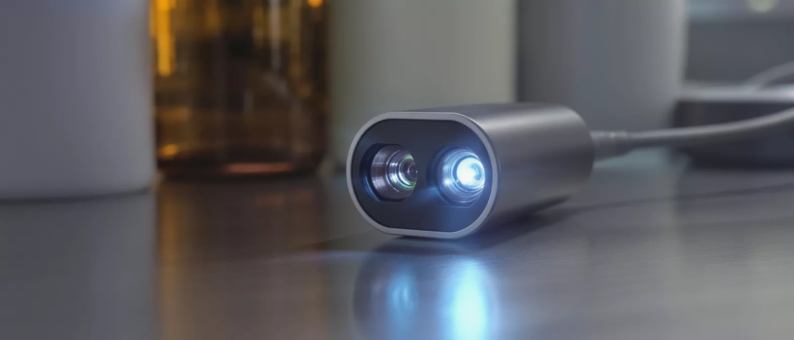
3D Endoscopic Camera
Learn how a 3d endoscopic camera enhances depth perception and precision in minimally invasive heart surgery.
What is a 3D Endoscopic Camera?
A 3d endoscopic camera is a specialized medical device used during minimally invasive procedures to provide surgeons with a high-definition, three-dimensional view of the internal body. When facing cardiac surgery, it helps to understand that this technology acts as the surgeon's eyes inside the chest cavity without requiring a large incision. The camera transmits live, stereoscopic images to a monitor in the operating room.
How does the camera fit into heart surgery? In specific minimally invasive cardiac procedures, the surgeon makes small keyhole incisions between the ribs. The endoscopic camera is inserted through one of these small ports, illuminating and capturing the surgical field so the surgeon can operate with precise control. This avoids the need to open the sternum completely.
This technology represents a significant advancement in medical visualization. By offering a true-to-life perspective of the surgical field, the surgical team can navigate complex anatomical structures with enhanced confidence. For the patient, this means a highly precise approach designed to minimize trauma to surrounding tissues.
How 3D Endoscopic Camera Systems Work
3d endoscopic camera systems operate on the principle of stereoscopy, mimicking human binocular vision. The camera head contains two separate optical channels, effectively acting as two eyes. These dual lenses capture the surgical field from slightly different angles simultaneously.
How are the images processed for the surgeon? The two separate video feeds are sent to a specialized processing unit that synchronizes them into a single cohesive 3D image. The surgeon views this high-resolution output on a 3D medical monitor while wearing specialized glasses, which creates a natural perception of depth.
Because the brain receives two distinct perspectives, it naturally merges them into a multi-dimensional picture. This allows the operating team to gauge the exact distance and spatial relationships between delicate tissues. The system is designed to replicate the natural hand-eye coordination the surgeon relies upon.
Why Depth Perception Matters in Surgery
Why is endoscopic camera depth perception important? Depth perception allows a surgeon to accurately judge the distance between surgical instruments and delicate internal structures. In traditional 2D surgery, where the image is completely flat, the surgeon must rely heavily on touch and experience to navigate complex three-dimensional spaces, which can be mentally taxing during long procedures.
Restoring natural depth perception is crucial when operating near the heart and major blood vessels. A 3d endoscopic camera provides the spatial awareness needed to guide instruments safely around critical structures. This visual accuracy helps prevent accidental collisions and ensures precise movements.
When looking at advanced 3d laparoscopic camera systems, the enhanced depth perception directly translates to better surgical control. The surgeon can identify tissue layers more easily and manipulate them with the exact force required. This reduces the risk of unintended trauma to the heart muscle.
3D Endoscopic Camera Benefits for Patients
The primary advantage of this technology for patients is the combination of surgical precision and minimal access. Because the surgeon can see the surgical site clearly in three dimensions, they can perform intricate maneuvers through tiny incisions. This results in less physical trauma compared to traditional open-heart approaches.
How does the 3D camera impact recovery? When a surgeon uses minimally invasive surgical visualization equipment, patients typically experience less bleeding during the operation and less pain afterward. The smaller incisions heal faster, which can lead to a shorter hospital stay and a quicker return to everyday activities.
Furthermore, the high-definition clarity of modern endoscopic camera technology helps the surgical team work more efficiently. A procedure completed with enhanced visual accuracy often means less time under anesthesia. This comprehensive approach supports a smoother postoperative healing journey.
Minimally Invasive 3D Camera Applications
In the context of cardiac care, a minimally invasive 3d camera is typically utilized for specific, targeted procedures. These can include minimally invasive mitral valve repairs, the removal of cardiac tumors, or the closure of atrial septal defects. The camera allows the surgeon to access the heart without a full sternotomy.
Can this technology be used for all heart surgeries? While highly effective, 3d endoscopy equipment is not suitable for every cardiac case. Complex or emergency surgeries may still require a traditional open approach to provide the surgeon with direct physical access to the heart. The decision is based on patient anatomy.
The operating surgeon evaluates each patient to determine the safest and most effective surgical route. When appropriate, the use of endoscopic visualization systems offers a highly effective, modern alternative to open surgery, prioritizing patient safety above all else.
Endoscopic Visualization Systems Explained
What components make up an endoscopic visualization system? A complete setup goes beyond the camera itself. It includes a specialized light source that illuminates the dark internal cavity, a high-definition monitor capable of displaying 3D images, and the digital processing tower that manages the video signals.
These integrated components work together seamlessly to provide a continuous, clear view of the heart. The camera features a flexible or rigid scope that projects light into the chest while simultaneously capturing the returning light to build the image. The entire chain must be perfectly calibrated.
For the surgical team, these systems turn a small physical incision into a wide, well-lit panoramic view. Understanding how this equipment works helps patients and families appreciate the technological sophistication involved in modern, minimally invasive surgical camera 3d technology.
3D Surgical Visualization vs Traditional 2D Imaging
The transition from 2D to 3d surgical visualization represents a major leap in operative safety. Traditional 2D cameras show a flat image, which can make it difficult to determine the exact depth of a surgical incision or the precise position of a needle. Surgeons must mentally reconstruct the 3D space.
How does 3D imaging improve surgical accuracy? With 3D imaging, the surgeon no longer has to guess the spatial relationships between tissues. The realistic depth perception speeds up complex surgical steps, such as suturing heart tissue or tying knots, because the visual feedback is intuitive and natural.
Patients who undergo procedures utilizing high-definition 3d endoscopic imaging devices benefit from this increased efficiency. The direct visual feedback allows the surgeon to operate with greater confidence, reducing operative time and minimizing the window of vulnerability for the patient.
Are 3D Endoscopic Cameras Safe for Cardiac Procedures?
Are 3D endoscopic cameras safe for heart surgery? Yes, these cameras are designed with strict medical safety standards. They undergo rigorous testing to ensure they do not interfere with other operating room equipment, such as pacemakers or electrosurgical units, making them a reliable choice for cardiac procedures.
The use of advanced endoscopic cameras has been studied extensively in surgical literature. The data indicates that when used by properly trained surgical teams for appropriate candidates, the visual enhancements provide a safe and highly effective method for navigating the complexities of the human heart.
Ultimately, the safety of the procedure relies on the combination of this advanced technology and the specialized expertise of the cardiovascular surgical team. The camera serves as a vital tool that enhances, rather than replaces, the skilled hands of the surgeon.
What to Expect from Advanced Endoscopic Cameras
When preparing for a minimally invasive cardiac procedure, it is natural for patients and their families to have questions about the technology involved. You can expect the surgical team to use state-of-the-art 3d endoscopic imaging to guide the operation safely and precisely. The equipment is handled by highly trained specialists.
How do surgeons prepare using 3D cameras? Before the operation begins, the surgical team carefully calibrates the 3d endoscopy equipment to ensure the white balance, focus, and depth alignment are perfectly set. This preparation helps ensure that the images displayed on the monitor are a true, accurate representation of the heart.
Knowing that your surgical team has access to high-definition 3d laparoscopic camera technology can provide significant peace of mind. It means your surgeon has the best possible visual tools to perform your procedure, ensuring that every movement is guided by clear, accurate depth perception.
Conclusion on 3D Endoscopic Imaging
The integration of a 3d endoscopic camera into cardiac surgery represents a significant step forward in patient care. By providing surgeons with true depth perception and high-definition, three-dimensional views, these systems allow for precise, minimally invasive procedures that protect the patient from unnecessary trauma.
For patients facing heart surgery, understanding this technology can alleviate much of the anxiety associated with the operating room. The use of modern surgical endoscopic cameras means smaller incisions, reduced physical stress, and a pathway to a smoother, more comfortable recovery.
Ultimately, the combination of skilled surgical expertise and advanced endoscopic camera technology ensures the highest standard of care. If you have further questions about how these tools will be used in your specific procedure, it is always best to discuss them directly with your cardiac surgeon.
- Enhanced Precision: Provides true depth perception for navigating delicate cardiac structures safely.
- Minimally Invasive: Allows surgery through small incisions, reducing recovery time and physical trauma.
- Improved Safety: High-definition 3D visualization minimizes the risk of accidental tissue damage.
- Camera Head: Contains dual optical lenses to capture stereoscopic images.
- Light Source: Illuminates the internal chest cavity for clear visualization.
- Processing Unit: Synchronizes the dual video feeds for the 3D monitor display.






