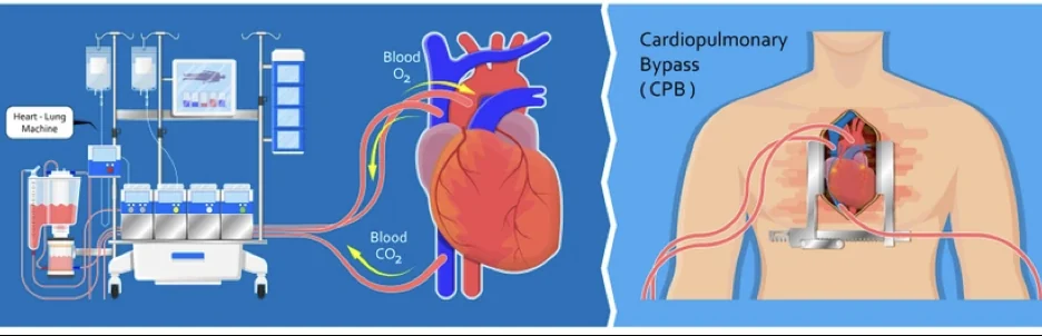
Common cardiac surgical procedures
Learn about cardiac procedures like bypass and valve surgery.
A complete guide to the operations we perform most often: coronary bypass, MitraClip, aortic aneurysms, TAVR, mitral and aortic valve disease, and cardiac tumours.
At Cirugía de Corazón we created this practical guide for patients, families and anyone interested in understanding the details of heart operations, so we can go through the process step by step, along with the risks, the benefits and the alternatives.
Introduction
One of the moments in your life, or in the life of someone you love, that can most significantly change or alter everything around you is the day you find out you may need heart surgery. Although most cardiac procedures can now be performed with a relatively low risk of complications, we should not diminish how significant this situation is. We understand how you feel. That is why we created this guide to the most common procedures we perform in heart surgery, so that you can be well informed when the time comes to make decisions or to talk about heart surgery with your doctor or your family.
Of course this guide does not replace a consultation with your specialist, nor a solid doctor–patient relationship. But we think it is a sensible first step towards your immersion in the world of heart surgery. Remember that this information serves as a reference only, that every patient is different and must be assessed as such, and that new questions may well come up by the time you finish reading. Whenever you wish, you can request a first video consultation to meet your specialist and clear up any doubts.
My name is Dr Alejandro Molina and I am a cardiovascular surgeon specialising in minimally invasive procedures. My intention is that this guide serves as a reference for you and your family, helps in making decisions, and makes the conversation easier at such a delicate point in your life. It is worth noting that this is not an academic article aimed at the medical community, but a way in for you to the world of heart surgery, in simple, clear language.
In this guide:
- Coronary bypass surgery
- MitraClip
- Aortic aneurysms
- TAVR
- Mitral valve disease
- Aortic valve disease
- Cardiac tumours
- We are here for you
Myocardial revascularisation, or coronary "bypass" surgery
1. What does revascularisation or bypass surgery refer to?
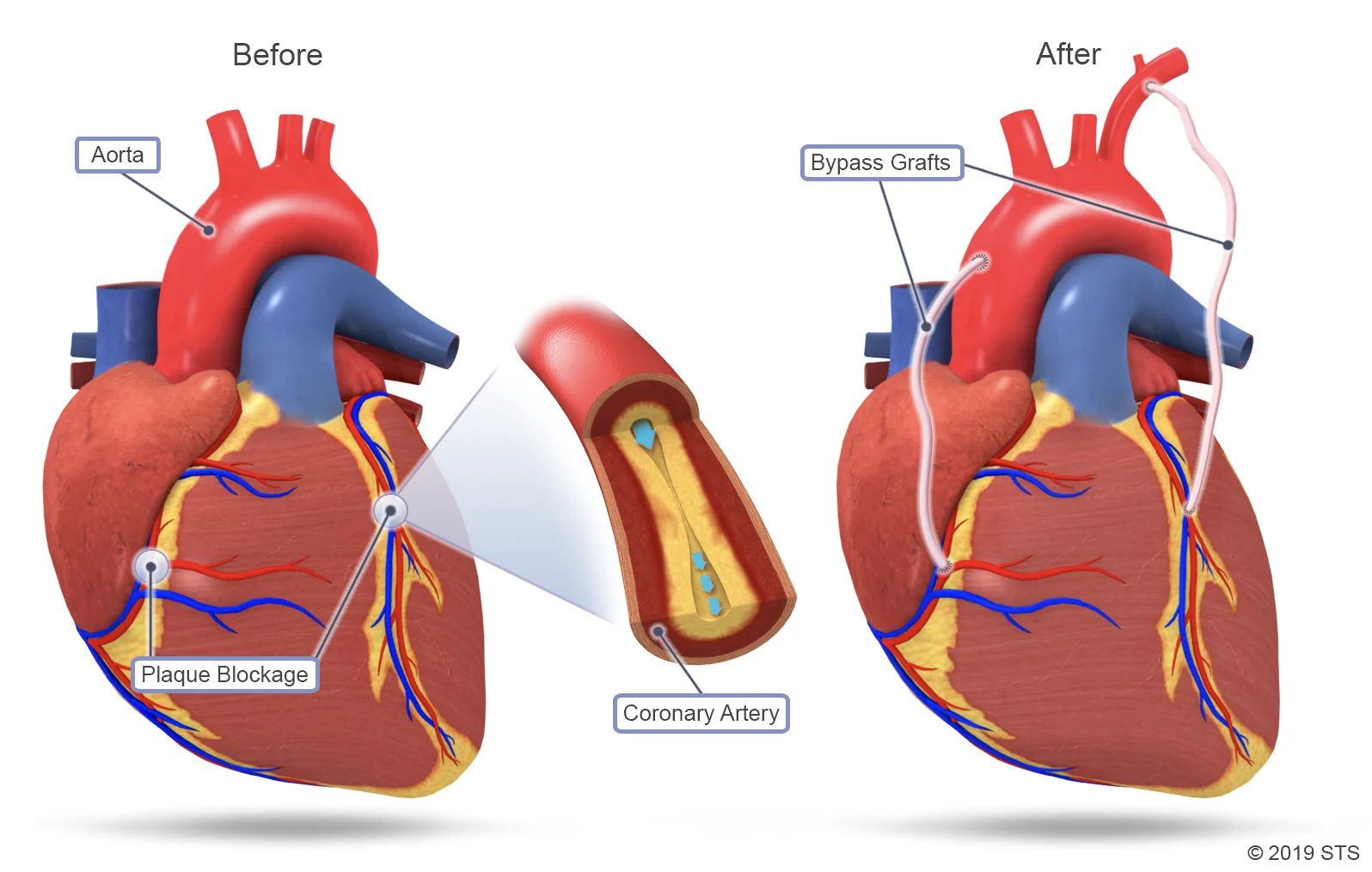
This is the surgery we perform when there is a blockage in the coronary arteries — the arteries that cause acute heart attacks when they close completely.
The coronary arteries are responsible for carrying blood and oxygen to the heart muscle so that it can contract and do its job properly. When there are varying degrees of blockage in these arteries, there can be signs of angina (chest pain) through to an acute myocardial infarction.
Revascularisation or bypass surgery is aimed precisely at restoring the flow that has been compromised or lost completely. In practical terms this surgery allows us to restore a continuous flow of blood and oxygen to the heart so that it can go on working properly.
This is the most common operation, and the one most performed in cardiac surgery centres around the world even today.
2. How is this kind of surgery performed?
In the great majority of cases this procedure is performed through an incision in the sternum, what we commonly know as open-heart surgery. In selected cases minimally invasive or hybrid procedures can be used.
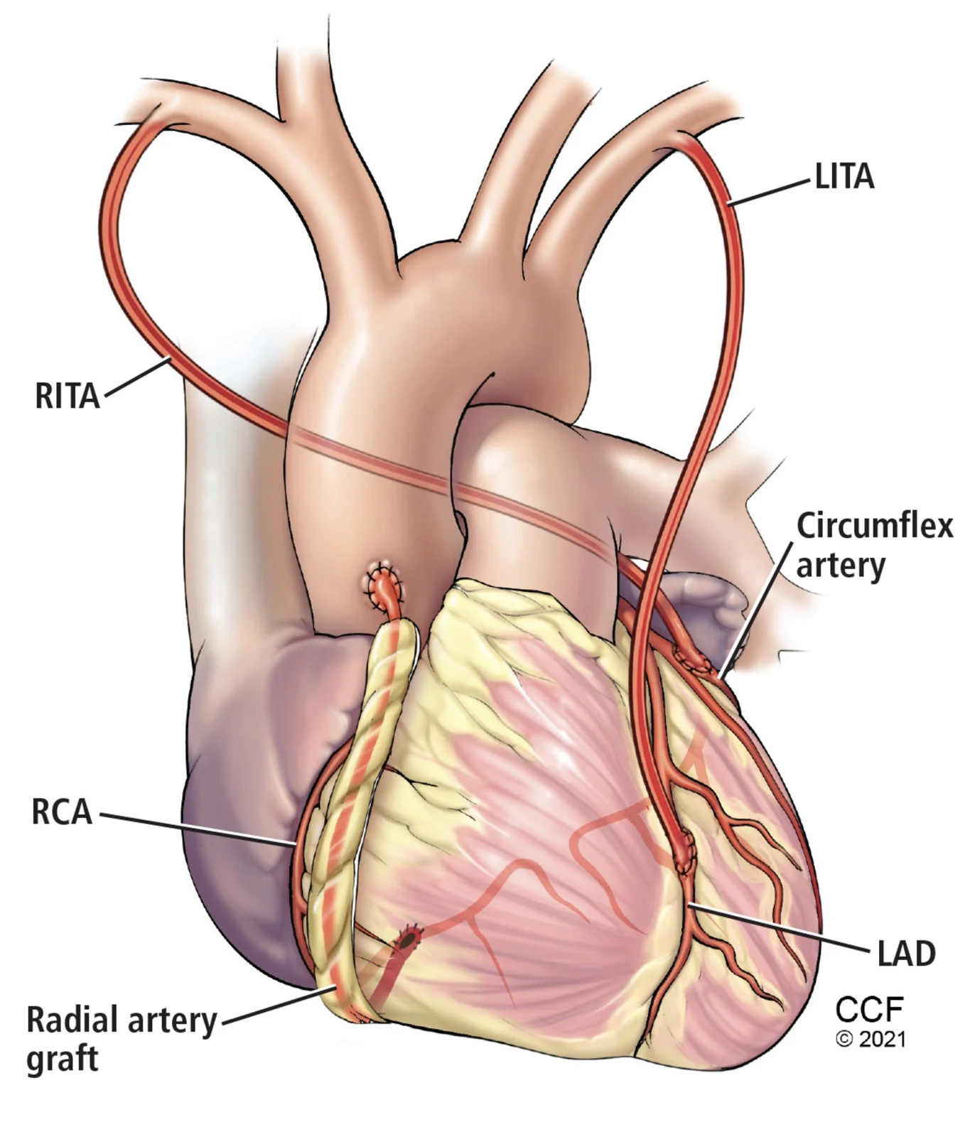
To create the coronary grafts or bypasses we use various arteries and veins, depending on how many the patient needs. The artery most frequently used is the left internal mammary artery, which is almost always used to revascularise the most important artery of the heart, the left anterior descending. Besides the mammary artery, we commonly use the radial artery in the left arm, the right-sided mammary artery, and very frequently the saphenous veins from the legs. This depends on each patient's anatomy.
3. How long does the procedure take?
The procedure in theatre usually takes between four and six hours, although this depends on a number of anatomical and haemodynamic factors during the intervention.
Once it is finished you will go to intensive care for monitoring and immediate management after the revascularisation, where a team of experts will take charge around the clock. Time in intensive care generally ranges between 36 and 48 hours, but it depends on how each person progresses. Total time in hospital averages 4–7 days, though again we should not generalise. We have to be careful and treat each person individually at every stage of this procedure.
4. How do I know whether I am a candidate for coronary bypass surgery?
As with any heart surgery, the first thing we have to assess is whether there really is an indication for heart surgery. To do that we carry out imaging studies that let us establish the individual anatomical and functional characteristics of the heart's arteries.
The most common studies are:
- Stress test. Carried out by a cardiologist, this test lets us assess both the symptoms brought on by exercise or exertion and the electrical behaviour of the heart during it. From this we can form a reasonable clinical suspicion about the state of your coronary arteries.
- Cardiac catheterisation. This study lets us see the anatomy and the sites of blockage in the coronary arteries. It is essentially a map of the arteries, from which we can determine their state and judge whether they require any intervention if blockages are present. The procedure usually takes one to three hours and may require admission in some cases.
- Transthoracic echocardiogram. Carried out by a cardiologist specialising in imaging, usually taking between 30 minutes and an hour, with no admission required. It lets us assess the heart valves and determine whether there is an indication to replace or repair any of them during the same bypass operation. Function is also assessed.
- Coronary CT angiography. Sometimes performed instead of catheterisation, and on other occasions to confirm or complement what we have seen in other studies. It lets us take various measurements and assess the coronary anatomy, and helps us plan the operation. It does not normally require admission.
- Other studies: electrocardiogram, blood tests, viability or ischaemia testing of the heart, lung function tests, and other studies that may be indicated.
Once we have all the indicated imaging studies, a group of several specialists will determine whether you are a candidate for myocardial revascularisation. The final decision about the procedure will always be yours and your loved ones'. It is taken once a level of mutual trust has been established in the doctor–patient relationship.
5. What are the benefits of coronary bypass surgery?
This operation aims to restore the flow of blood and oxygen to the coronary arteries. Once that is achieved, a long-term benefit is seen both in symptoms and in quality of life. The cardiology literature has also demonstrated a protective effect against further heart attacks, by offering alternative routes or collateral circulation to the coronary arteries.
6. What are the risks associated with this intervention?
Although CABG is a common and effective procedure, it also carries certain risks and complications, which can include:
- Lung complications. Surgery can increase the risk of pneumonia, particularly in people with pre-existing lung disease.
- Bleeding. CABG involves cutting the sternum, which can cause excessive bleeding. Sometimes a blood transfusion is needed to make up the loss.
- Infection. As with any surgery, there is a risk of infection at the incision and in the surgical wound.
- Clotting problems. Surgery can increase the risk of blood clots, which can cause complications such as a stroke or a heart attack.
- Kidney failure. CABG can affect kidney function, particularly in people with pre-existing kidney disease.
- Neurological complications. Rarely, surgery can cause brain injury, which may show as problems with memory, concentration, motor skills or speech.
- Anaesthetic risks. The general anaesthetic used during surgery can have complications, particularly in older people or those with chronic conditions.
- Allergic reactions. Some people may have an allergic reaction to the medicines used during surgery.
It is important to point out that these risks are relatively rare and that, in general, CABG is a safe and effective procedure for treating coronary disease. Even so, it is important that you discuss all the risks and benefits with your doctor before deciding about surgery.
MitraClip
1. What is a MitraClip?
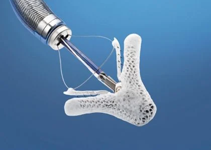
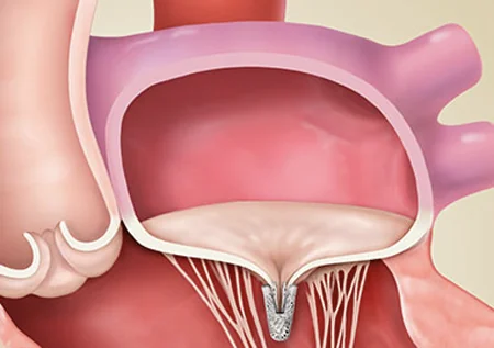
A MitraClip is a new device that lets us repair or improve the function of a mitral valve that is diseased or leaking. Remember that the heart valves work by opening and closing their doors, letting blood pass in one direction only — blood should not flow backwards. Where the valve is diseased, flow can be seen going backwards, against the normal direction. When that happens we say the valve is regurgitant. In cases where we consider the degree of regurgitation severe, we generally think about some kind of intervention to repair the problem. The basic principle of the MitraClip is precisely to bring the leaflets, the "doors" of this valve, closer together to restore normal function.
2. How is this procedure performed?
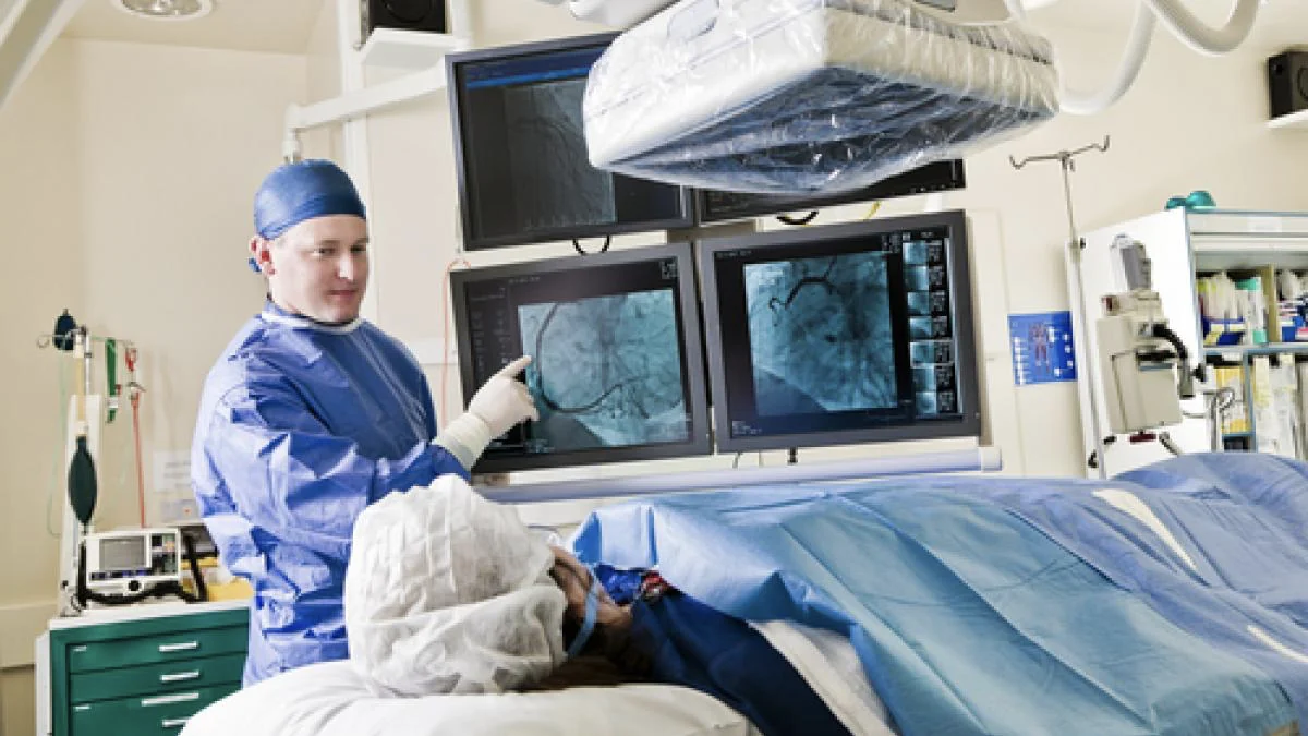
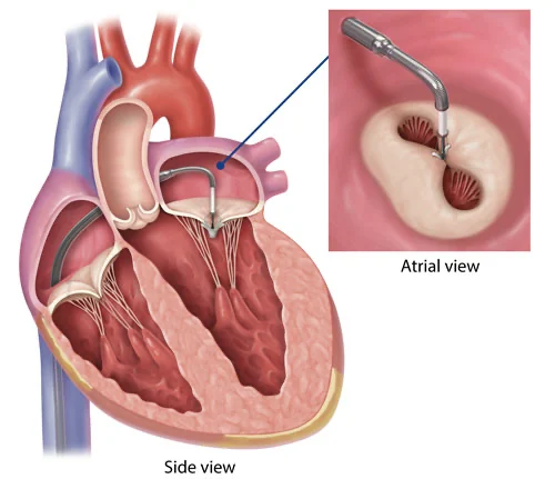
The procedure is carried out in a catheterisation laboratory or a hybrid theatre. It requires hospital admission and, although it is not surgery as we traditionally understand it, it is still considered a surgical procedure. It is performed through a needle puncture in the groin. This procedure does require general anaesthesia and a transoesophageal echocardiogram.
Through a special catheter we advance the device from the groin, through the vena cava, until we reach the heart and can access the valve. Accessing the valve, assessing it and achieving proper placement requires a large team of specialists, each with very specific roles, working together. Once the correct position has been confirmed, we release the MitraClip in its final position. Various imaging studies are performed to confirm that the result is optimal. It is common for more than one MitraClip to be needed to achieve the expected result. At the end of the procedure the catheters and special devices are removed.
3. How long does it take?
The procedure in the catheterisation laboratory usually takes between two and three hours, although this depends on a number of anatomical and haemodynamic factors during the intervention.
Once it is finished you will go to intensive care for monitoring and immediate management after the MitraClip, where a team of experts will take charge around the clock. Time in intensive care generally ranges between 24 and 36 hours, but it depends on how each person progresses. Total time in hospital will depend on each person's characteristics and other conditions.
4. How do I know whether I am a candidate for a MitraClip?
As with any heart surgery, the first thing we have to assess is whether there really is an indication for heart surgery. To do that we carry out imaging studies that let us establish your individual anatomy.
- Transthoracic and then transoesophageal echocardiogram — a study through the mouth under light sedation that lets us see the function and anatomy of the heart and of each valve. It is carried out by a cardiologist specialising in imaging, usually takes between 30 minutes and an hour, and does not require admission.
- Cardiac catheterisation. Because of the age at which a MitraClip is normally indicated, a cardiac catheterisation will be indicated. It is essentially a map of the arteries, from which we can determine the state of the coronary arteries and judge whether they require any intervention if blockages are present. The procedure usually takes one to three hours and may require admission in some cases.
- Other studies: electrocardiogram, blood tests, in some cases a stress test, lung function tests, and other studies that may be indicated.
Once we have all the indicated imaging studies, a group of several specialists will determine whether you are a candidate for a MitraClip. The final decision about the procedure will always be yours and your loved ones', taken once a level of mutual trust has been established in the doctor–patient relationship.
Special considerations for MitraClip
Because this is a relatively new device, the indications for placing it are currently limited to patients who have severe mitral regurgitation and who are considered too frail or too high a risk for conventional surgery. Even in those cases this therapy is not for everyone, which makes a multidisciplinary approach vital in deciding and in selecting the patients considered possible candidates.
There are individual cases in which the benefits and risks of a MitraClip against conventional surgery need to be discussed.
5. What are the benefits of MitraClip therapy?
The MitraClip was designed to be minimally invasive, and to offer optimal treatment to patients who were considered too frail to withstand surgery. In other words, it serves a group of patients to whom surgical treatment could not previously be offered. By avoiding traditional or "open-heart" surgery, we avoid any risk associated with that intervention. Recovery time tends to be shorter, and the risk of complications during the procedure is generally very low.
As experience has grown, the indications have also widened to younger and less frail patients.
This should always be discussed at length with your surgeon, covering the benefits and risks of each type of intervention.
Trust is fundamental in making these decisions.
Aortic aneurysms
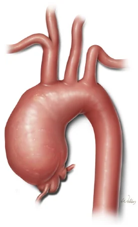
An aortic aneurysm is a bulge that appears in the wall of the main blood vessel, the aorta, which carries blood from the heart to the body. Aortic aneurysms can occur anywhere along the aorta and can be tube-shaped (fusiform) or round (saccular).
What different types of aneurysm are there?
- Abdominal aortic aneurysm. This occurs along the part of the aorta that passes through the abdomen.
- Thoracic aortic aneurysm. This occurs along the part of the aorta that passes through the chest cavity.
Some people can have both types of aortic aneurysm.
Having an aortic aneurysm increases the risk of a tear in the inner layer of the aortic wall, known as an aortic dissection.
What symptoms does an aortic aneurysm cause?
It is important to know that they are often symptomless, and can stay silent and undiagnosed for years. In cases where symptoms do appear, we may see difficulty breathing, a hoarse voice, or pain in the chest or neck.
What complications can arise from an aortic aneurysm?
The most severe and catastrophic are dissection of the aorta — a sudden separation of the layers of the aorta — and complete rupture of the aneurysm, which carries a mortality of roughly 90% if it is not treated immediately.
Problems can also arise with the aortic valve, angina (chest pain), or on occasion compression of nearby structures.
Is there treatment for this condition?
Yes. In many cases the treatment is surgical, involving heart surgery, and it can sometimes be done through small incisions or minimally invasively. On other occasions management is entirely endovascular, that is to say without opening the patient, working through a catheter or percutaneously. In some cases management may be with medication and regular monitoring alone.
What matters most is timely diagnosis and multidisciplinary assessment by a dedicated team. Every person is different and management must be personal and suited to each individual's characteristics.
Some of the treatment possibilities for aortic aneurysms are set out below:
- Surveillance. For small aneurysms, under 5 cm, that cause no symptoms, careful and regular monitoring may be recommended to detect any change in size.
- Open surgery. Open surgery is a common option for aortic aneurysms measuring 5.5 cm or more. During the operation an incision is made in the wall of the abdomen or the chest to reach the aorta and repair or replace the affected section.
- Aortic endograft. Also known as an aortic stent, this refers to a minimally invasive technique in which an artificial prosthesis is inserted inside the aneurysm to reinforce the wall of the aorta and prevent rupture. This option is suited to aneurysms of the descending thoracic aorta, and its use is becoming increasingly common in other parts of the aorta.
- Stress reduction. Stress reduction techniques such as yoga or meditation can be useful in controlling blood pressure and reducing the risk of rupture.
- Medication. Medicines such as beta blockers can lower blood pressure and reduce the risk of the aneurysm rupturing. Anticoagulants can also help prevent blood clots forming in the aneurysm.
It is important to stress that the choice of treatment will depend on the size, location and shape of the aneurysm, as well as the patient's general health. Specialists such as cardiologists or vascular surgeons can help determine the best course of action for each patient.
TAVR
What is TAVR?
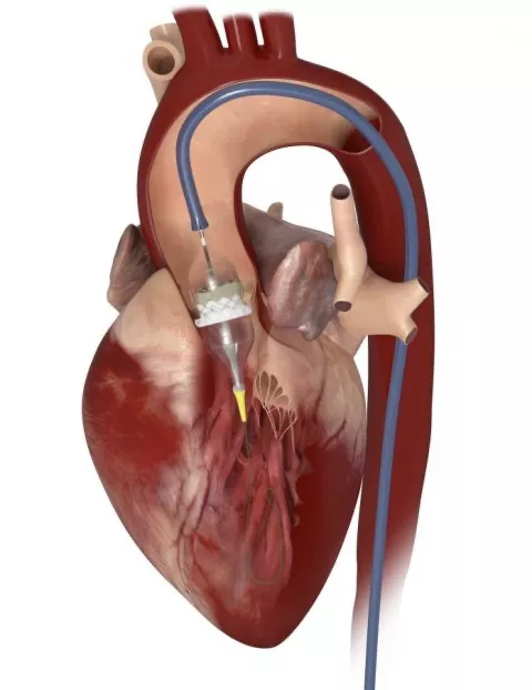
It refers to the implantation or replacement of the aortic valve percutaneously, or by catheter — TAVR stands for Transcatheter Aortic Valve Replacement. In practical terms, it is the replacement of a diseased heart valve without needing to open the chest or perform traditional open-heart surgery.
We invite you to read our complete guide to TAVR, where you can learn everything about this procedure.
Of course this information does not replace a face-to-face consultation with your doctor, but it can be an excellent starting point for understanding the procedure better, answering the most common questions, and making well-informed decisions.
Mitral valve disease
Mitral valve disease is a problem with the valve situated between the chambers of the left side of the heart, the left atrium and the left ventricle.
Mitral valve disease includes the following:
- Mitral regurgitation. The leaflets of the mitral valve do not close properly, which causes blood to flow backwards, so that part of the blood in the left ventricle returns towards the lungs instead of moving on into the aorta and carrying oxygenated blood to the rest of the body.
- Mitral stenosis. The leaflets of the mitral valve thicken or stiffen, and may fuse together. This narrows the valve opening, reducing blood flow from the left atrium to the left ventricle. The atrium has to work much harder to push blood in the right direction, causing it to dilate and blood to back up in the lungs.
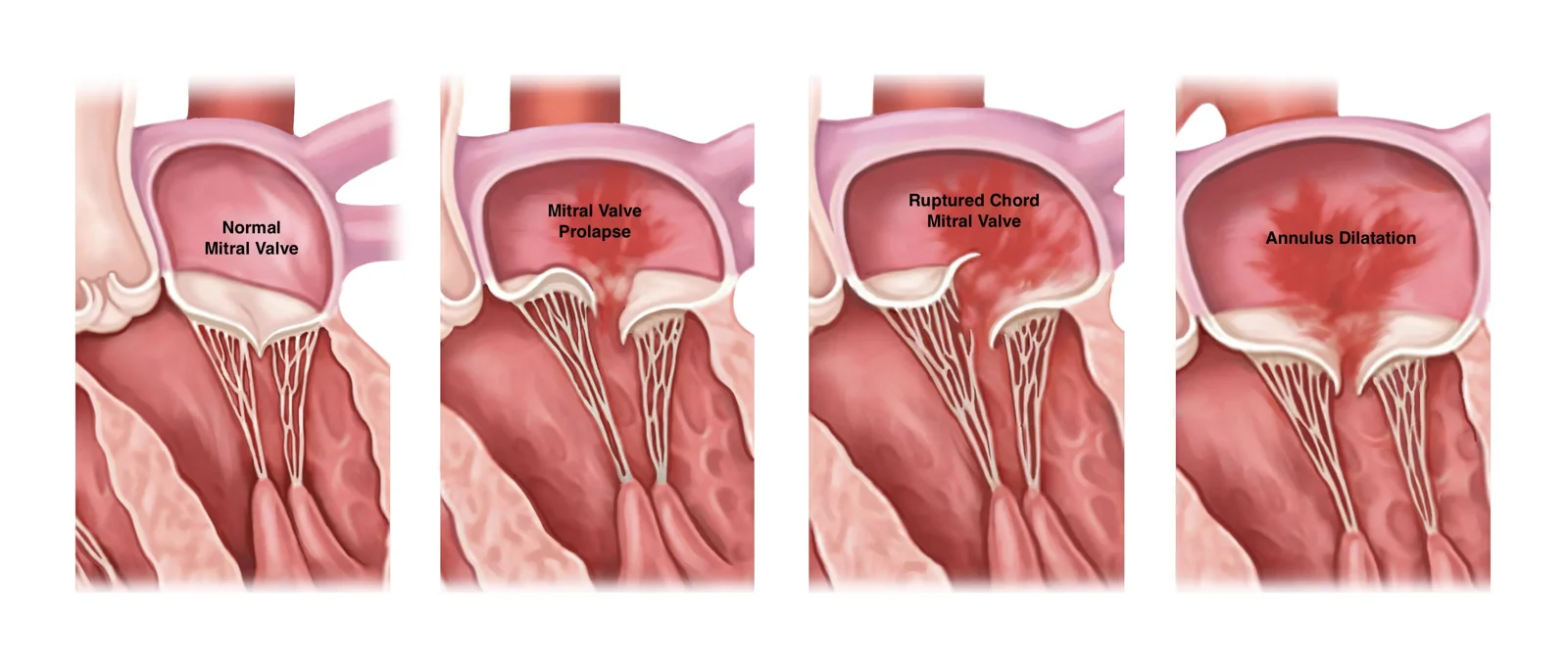
Treatment of mitral valve disease depends on the severity of the condition and how it progresses. Sometimes surgery is recommended to repair or replace the mitral valve.
Symptoms
Some people with mitral valve disease may have no symptoms for many years, or may never have any.
The signs and symptoms of mitral valve disease can include:
- Fatigue
- An irregular heart sound (a heart murmur)
- Irregular heartbeat
- Shortness of breath
- Chest pain
- Palpitations
- Cough
Causes
To understand the causes of mitral valve disease, it helps to know how the heart works.
The mitral valve is one of the four heart valves that keep blood flowing in the right direction. Each valve has leaflets that open and close once during every heartbeat. If a valve does not open or close properly, blood flow from the heart to the body can be reduced.
In mitral stenosis the valve opening narrows. The heart then has to work harder to push blood through the reduced opening. If the opening narrows too far, blood flow from the left atrium to the left ventricle can fall.
Mitral valve disease has many causes. Some forms can be present at birth (a congenital heart defect).
It can also develop later in life (acquired). Mitral stenosis, for example, is usually caused by rheumatic fever. That fever is a complication of a streptococcal infection which can affect the heart. When that happens it is called rheumatic mitral valve disease.
Other causes of acquired mitral valve disease include:
- Other cardiac conditions
- Calcification
- Tumours
- Clots
- Perforation of the valve
- Infection
- Age-related change
- Autoimmune disease, for example lupus
Risk factors
Several things can increase the risk of mitral valve disease, including:
- Older age
- Certain infections that affect the heart
- Heart attack and some kinds of heart disease
- Use of certain medicines
- A heart condition present at birth (a congenital heart defect)
- Radiation to the chest
Complications
Mitral valve disease can cause many complications. Severe mitral regurgitation, for example, makes the heart work harder, which leads to enlargement of the left ventricle and weakening of the heart muscle.
Other complications of mitral valve disease can include:
- An irregular and often rapid heart rate (atrial fibrillation)
- High blood pressure in the blood vessels of the lungs (pulmonary hypertension)
- Blood clots
- Congestive heart failure
- Stroke
What are the treatment options for this condition?
Medical management of mitral valve disease can include a range of options, depending on the type and severity of the disease. Some of the common ones include:
- Medication. Medicines may be prescribed to reduce the symptoms of mitral valve disease. They can include diuretics to reduce fluid retention, beta blockers to lower heart rate and blood pressure, and anticoagulants to prevent blood clots forming.
- Lifestyle changes. Changing the diet, increasing physical activity and stopping alcohol and tobacco are important measures that can improve symptoms and general heart health.
- Monitoring. Regular follow-up of people with mitral valve disease is important in order to assess any change in severity. Follow-up examinations can include echocardiograms, lung function tests and electrocardiograms.
- Surgery. In severe cases surgery may be necessary to repair or replace the mitral valve. Surgery is considered a treatment option when symptoms are severe or when there is a high risk of complications.
The specific treatment will depend on the cause and severity of the mitral valve disease, as well as your general health. It is important to work with your doctor to develop a suitable treatment plan and to make sure you have regular follow-up to monitor progression.
What kinds of surgical treatment are there?
- Mitral valve repair. Mitral valve repair is surgery in which the surgeon tries to repair the existing mitral valve rather than replace it. During the repair, damaged tissue can be removed and the parts of the mitral valve that are damaged or not working properly can be repaired. Where it is feasible, this technique is considered the ideal one, since it preserves your own valve and requires no replacement with an artificial prosthesis. It is important to stress that repairs are always accompanied by an annuloplasty, a procedure to reinforce the ring of the mitral valve and so prevent it dilating in future. That procedure uses a type of prosthetic ring but does not require removing the native valve; it is only a reinforcement.
- Mitral valve replacement. Mitral valve replacement is surgery in which the mitral valve is removed and replaced with a mechanical or a biological valve. Mechanical valves are durable but require lifelong anticoagulants to prevent blood clots, while biological valves are less durable but do not require lifelong anticoagulation. The decision about which to choose will depend on your age and your risk factors, and is a conversation to have with your surgeon weighing the risks and benefits of each.
- Mitral commissurotomy. This is surgery in which an incision is made in the mitral valve to separate leaflets that have fused, allowing the valve to open and close properly. It is used mainly to treat mitral stenosis, but it is very important to say that this procedure is usually palliative, used when surgery needs to be delayed or when the patient needs it urgently. It tends to be done only as a temporary measure while planning more definitive and durable treatment.
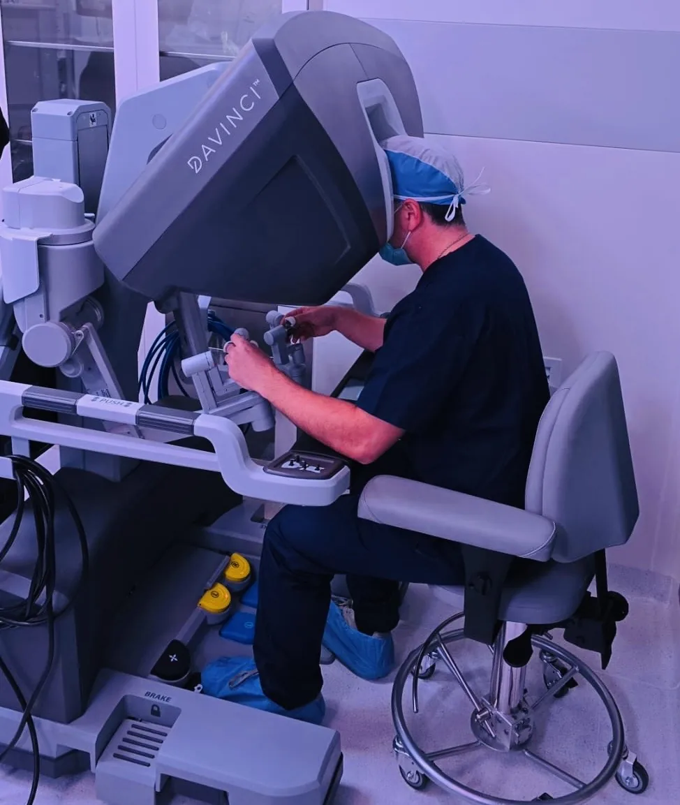
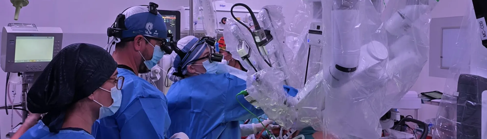
We have discussed minimally invasive cardiac surgery for carrying out these procedures, as well as the endovascular or percutaneous procedures for treating these conditions. If you wish, request a first free video consultation to clear up any questions.
Aortic valve disease
The conditions that most commonly affect the aortic valve are aortic stenosis and aortic regurgitation.
Aortic stenosis is a narrowing of the opening of the aortic valve, which makes it harder for blood to flow from the left ventricle into the aorta. The most common causes of aortic stenosis are ageing, calcification and degenerative disease of the valve.
Aortic regurgitation is a condition in which the aortic valve does not close properly, allowing blood to flow backwards from the aorta into the left ventricle. The most common causes are dilatation of the aorta, rheumatic heart disease and bacterial endocarditis.
Both aortic stenosis and aortic regurgitation can be serious and require medical treatment or even surgery if they become symptomatic or if there is evidence of damage to the heart. Regular follow-up with a cardiologist is important in order to detect and treat any problem with the aortic valve.
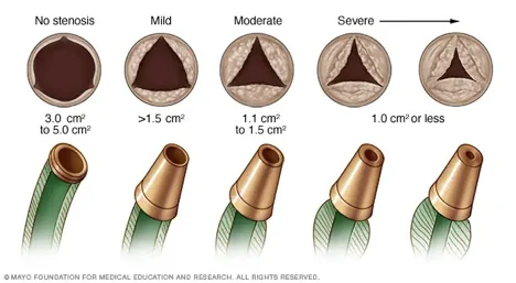
What are the symptoms of aortic valve disease?
The symptoms of aortic valve disease can vary depending on whether the condition is aortic stenosis or aortic regurgitation, and on how severe it is. Some of the common symptoms include:
- Shortness of breath. Difficulty breathing or breathlessness is a common symptom of aortic valve disease. It can occur during physical activity or even at rest.
- Chest pain or angina. Chest pain or pressure in the chest is also a common symptom. The pain may feel like tightness, burning or discomfort in the chest.
- Dizziness or fainting. Aortic valve disease can cause dizziness or fainting because of reduced blood flow to the brain.
- Palpitations. A fast or irregular heartbeat can be a symptom of aortic valve disease.
- Fatigue. Fatigue or general weakness can also be a symptom.
- Swelling in the feet or abdomen. In advanced cases, aortic valve disease can cause fluid retention in the body, which can lead to swelling in the feet or abdomen.
What is the treatment for aortic valve conditions?
Treatment for aortic valve disease depends on how severe the disease is and on the symptoms you have. In general, the aim of treatment is to relieve symptoms, improve quality of life and prevent complications.
- Medical treatment. If the aortic valve disease is mild and causes no symptoms, your doctor may choose observation and regular follow-up to monitor how it progresses. Medicines may also be prescribed to control blood pressure and reduce the heart's workload.
- Aortic valve replacement surgery. If the aortic valve disease is severe and causes symptoms, aortic valve replacement surgery may be necessary. During the operation the damaged aortic valve is replaced with a mechanical or a biological valve. The choice of prosthesis will depend on your age, your risk factors and your history. Your doctor can of course make a recommendation, but the final decision will always be yours and your loved ones'.
- TAVR. A less invasive procedure called transcatheter aortic valve replacement may also be considered, used in patients who are not candidates for traditional aortic valve replacement surgery, or in patients of a certain age for whom a less invasive procedure is chosen. During the procedure a new aortic valve is inserted through a catheter and placed at the site of the damaged valve. See our TAVR guide, devoted entirely to these procedures.
- Treating the underlying cause. If the underlying cause of the aortic valve disease is an infection or rheumatic heart disease, treating that cause may be necessary in order to treat the valve disease.
- Aortic valve repair. Sometimes, if the condition of your valve is favourable, your surgeon may recommend repairing it. Where feasible, this option may be the best one, since your own valve is preserved without needing to insert an artificial prosthesis. Note that not all centres and not all surgeons have the training to perform an aortic valve repair, and it is also important to stress that not every valve can be repaired. It depends on several factors. The option chosen should always be the one that offers the best probability of success for each individual patient.
How is surgery for these conditions performed?
These procedures may require what we call open-heart surgery, or a minimally invasive procedure. We invite you to visit the page where the details of minimally invasive surgery are discussed in depth. You will be able to learn the characteristics of these interventions and their pros and cons, and by the end you will be well informed to make a better decision. After that, if you prefer, you can book a first free video consultation.
What are the risks associated with these operations?
Aortic valve replacement surgery is a complex procedure that carries some risks. Although advances in surgical technique and technology have improved its safety, some of the risks associated with the procedure include:
- Bleeding. Aortic valve replacement surgery can cause significant blood loss, which may require transfusion during or after the operation. It may sometimes require a surgical intervention to check the bleeding; this occurs in roughly 4% of cases.
- Infection. The surgery can increase the risk of infection at the incision site or in the body generally.
- Heart rhythm problems. Surgery can increase the risk of arrhythmias. Most are transient, but in some cases they can be permanent. The most common arrhythmia after surgery is atrial fibrillation. There is also a risk of blocks in the heart's electrical conduction that may require a temporary or permanent pacemaker.
- Breathing problems. Aortic valve replacement surgery can affect the patient's ability to breathe and cause small areas of lung collapse known as atelectasis. It may require respiratory support after surgery, as well as pulmonary rehabilitation to avoid longer-term complications.
- Blood clots. After surgery the patient may be at greater risk of developing blood clots in the legs or lungs. Anticoagulation is often chosen to reduce that risk.
- Allergic reaction. The patient may have an allergic reaction to the materials used during surgery, such as heparin solution, or to a medicine.
Cardiac tumours
1. What are the most common cardiac tumours?
Cardiac tumours are quite rare, and can be either benign or malignant. The most common types of benign cardiac tumour include:
- Atrial myxoma. This is the most common type of benign cardiac tumour. It usually arises in the left atrium of the heart, and can cause symptoms such as fatigue, dizziness, palpitations and difficulty breathing.
- Cardiac rhabdomyoma. This is the most common type of benign cardiac tumour in babies and young children. It often causes no symptoms and can disappear on its own as the child grows.
- Cardiac lipoma. This type of benign cardiac tumour is uncommon and generally causes no symptoms.
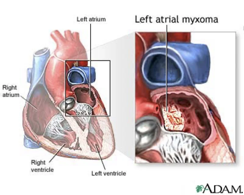
The most common malignant cardiac tumours, on the other hand, are:
- Cardiac sarcoma. This type of malignant cardiac tumour is uncommon but very aggressive. It arises in the soft tissues of the heart and can spread rapidly to other organs.
- Metastatic carcinoma. This type of tumour arises elsewhere in the body but spreads to the heart through the blood. It can cause symptoms such as chest pain, difficulty breathing and fatigue.
It is important to bear in mind that cardiac tumours are very rare, and that most of the time the symptoms a person experiences are due to another cause. Even so, the symptoms you feel should never be dismissed.
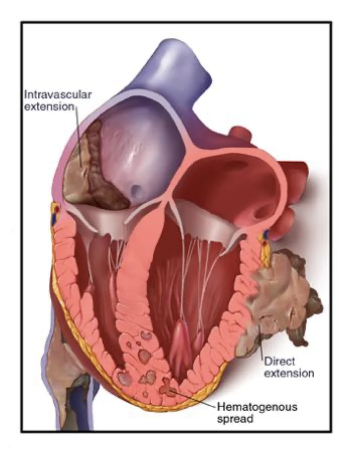
2. How are cardiac tumours treated?
Treatment of cardiac tumours depends on the type of tumour and on whether it is benign or malignant. In general, benign tumours can be treated with surgery, while malignant tumours may require a combination of surgery, radiotherapy and chemotherapy.
For benign tumours such as atrial myxoma, surgery is often the best treatment option. Most of the time the tumour can be removed completely and the patient can expect a full recovery.
For malignant tumours, treatment will depend on the stage of the cancer and whether it has spread to other parts of the body. In some cases it may be necessary to remove the tumour surgically, followed by radiotherapy and/or chemotherapy to destroy any remaining cancer cells.
In cases where the tumour cannot be removed completely, treatment will focus on controlling its growth and reducing the associated symptoms. This can include targeted therapies, radiotherapy and/or chemotherapy.
It is important to remember that treating cardiac tumours is a complex process and requires an individualised approach based on the patient's needs and health. Patients should work closely with their medical team to develop a treatment plan that is effective and safe for them.
3. Can cardiac tumours be removed minimally invasively?
Yes. In some cases cardiac tumours can be resected minimally invasively, which can have several advantages for you or your relative compared with traditional open surgery.
Minimally invasive surgery uses special surgical techniques and smaller instruments to operate through small incisions rather than a large incision in the chest. This can result in a faster recovery, less pain, less scarring and a shorter hospital stay.
In minimally invasive resection of cardiac tumours we make small incisions in the chest to reach the heart and remove the tumour. A video instrument is used to see inside the heart and guide the surgery, and special surgical tools may be used to remove the tumour.
However, not all cardiac tumours can be removed minimally invasively, and the decision to use this technique will depend on the size and location of the tumour, as well as the surgeon's experience and skill.
In general, minimally invasive resection of cardiac tumours can be a viable option for some patients, and whether it is suitable for your case should be discussed with your medical team.
4. What is the outlook?
The outlook for cardiac tumours depends on the type of tumour, whether it is benign or malignant, and the stage of the disease at the time of diagnosis.
In general, benign cardiac tumours have a favourable outlook after treatment, and most patients recover completely. Even so, some types of benign tumour, such as atrial myxoma, can have a high rate of recurrence after surgery.
Malignant cardiac tumours, on the other hand, can carry a more guarded outlook. Survival depends largely on the type of cancer, the stage at diagnosis and the effectiveness of treatment.
In general, malignant cardiac tumours can carry a more serious outlook than other types of cancer because of the tumour's location in the heart and the difficulty of treating it. Even so, advances in early detection and treatment have improved survival and quality of life for patients with malignant cardiac tumours.
It is important to bear in mind that every case is unique, and a patient's outlook will depend on many individual factors. You will need to work closely with your medical team to develop a personalised treatment plan and to monitor your progress and outlook over time.
We are here for you
At Cirugía de Corazón we created this guide to begin orienting you in this unfamiliar world, which without doubt represents an enormous challenge in your life and in the lives of those you love. We understand the moment you are going through. We are here for you.
As we said earlier, this guide does not replace a face-to-face consultation with your doctor, nor a solid doctor–patient relationship. But we know that access to the right information should be a priority, and that it can help you have a far more productive conversation with your family and with your medical team.
When you feel ready, if you decide to look for more information, you can request a video consultation with one of our specialists, or even a face-to-face appointment at the practice, to go on exploring treatment options, talk through alternatives, and work out a management plan together, individually.
Welcome to Cirugía de Corazón, where the main focus is you. You will find humane and respectful treatment, surrounded by a team of experts and a world-class service.

About the author
Dr Alejandro Molina is a cardiovascular surgeon practising in Guadalajara, Querétaro and Hermosillo. He trained in cardiothoracic surgery at the Universidad de Guadalajara, completed advanced training in cardiac surgery, minimally invasive surgery, robotic surgery and structural interventions at the University of Montreal, and a fellowship in structural cardiology and TAVR at CHU Bordeaux, France. He performed the first robotic mitral valve repair in the Ángeles Health System and the first robotic heart surgery in Guadalajara.
Medical degree: Universidad Autónoma de Guadalajara, Cédula Profesional 5757260 · Cardiothoracic surgery: Universidad de Guadalajara, Cédula de Especialidad 10768871 · Current certification, Consejo Nacional de Cirugía del Tórax, no. 447
Talk to a specialist
Request a free video consultation to clear up your questions and make a better-informed decision.