Cardiac tumours: understanding myxoma and other types
Cardiac tumours are mostly benign and treatable.
Most cardiac tumours are benign and treatable. What they are, how they are diagnosed, and what the options are for benign and malignant forms.
The day someone learns they have a tumour inside the heart, time seems to stop. It is more common than you think, and most cardiac tumours are benign with good treatment options.
If you are reading this, perhaps you or someone you love has recently been diagnosed with a cardiac tumour and you are looking for information.
I understand that the information available can be overwhelming and not always clear. Don't worry — you have come to the right place.
What is a cardiac tumour?
A cardiac tumour is an abnormal growth of tissue in the heart. It can be benign (non-cancerous) or malignant (cancerous).
Cardiac tumours are rare compared with other types of tumour, but when they occur they can raise significant concerns because of their vital location.
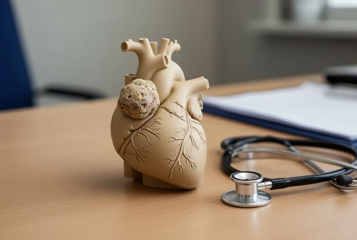
Cardiac myxoma: the most common benign tumour
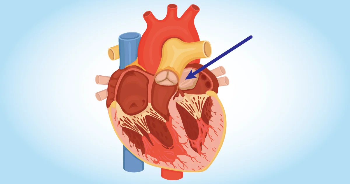
The cardiac myxoma is the most common benign tumour of the heart. Although it sounds alarming, it is important to understand that "benign" means it is not cancerous and generally does not spread to other tissues.
These tumours often develop in the left atrium, one of the upper chambers of the heart, and can interfere with normal blood flow. They are generally found attached to one of the walls of the heart known as the interatrial septum, which divides the two atria.
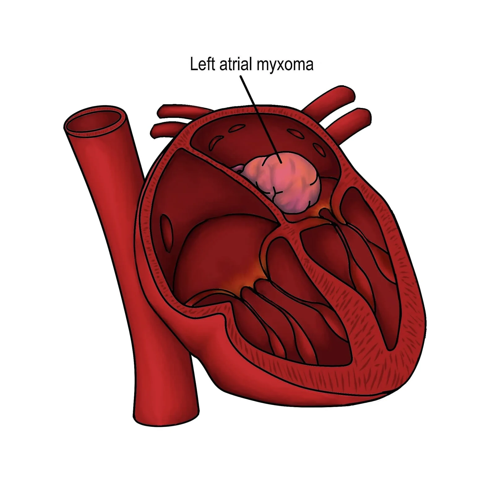
Depending on its size and growth, a myxoma can obstruct blood flow or interfere with the working of the heart valves.
In saying this tumour is benign, we do not diminish the seriousness of its presence, and we are in no way suggesting it does not require treatment. On the contrary, this represents an absolute priority because of its potential to put the patient's life at risk.
Causes and genetics
The exact cause of cardiac tumours is not always clear. In the case of myxomas, many are believed to be sporadic, which means they have no known genetic cause and are not inherited. On rare occasions, however, they can be associated with certain genetic syndromes.
Genetics plays a more prominent part in some other types of cardiac tumour, but in general more research is needed to understand the underlying causes fully.
Heart tumour symptoms: three ways a tumour shows itself
When cardiac tumours do cause symptoms, they tend to do so in three ways. Knowing them helps explain why the symptoms can be so varied, and why they are sometimes mistaken for other conditions.
- Obstruction. A tumour can partly block the flow of blood inside the heart or through a valve. This can cause breathlessness, dizziness or fainting. With a mobile myxoma, symptoms sometimes change with body position.
- Embolism. Fragments of the tumour, or clot on its surface, can break off and travel in the bloodstream. In the brain this can cause a stroke. Elsewhere it can block an artery to the legs, the kidneys or the intestine.
- General symptoms. Myxomas can release inflammatory substances. Some patients have fever, weight loss, tiredness or joint pain, which can look like an infection or a rheumatic illness.
Irregular heart rhythms and fluid around the heart are other possible signs, particularly with malignant tumours. Many heart tumour symptoms are also common in other conditions, so the echocardiogram is often the test that makes the difference.
How are cardiac tumours diagnosed?
1. Initial medical assessment
It is important to know that most of the time these tumours can remain without symptoms for months or years, with a normal life.
Symptoms can include extreme fatigue, difficulty breathing, irregular heart rhythm or chest pain. These generally occur when the tumour grows and begins to disturb the normal physiology of the heart, which can lead to an initial assessment by your doctor.
2. Imaging
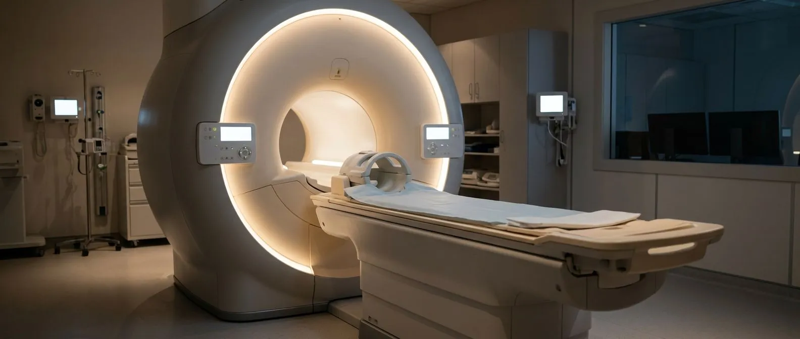
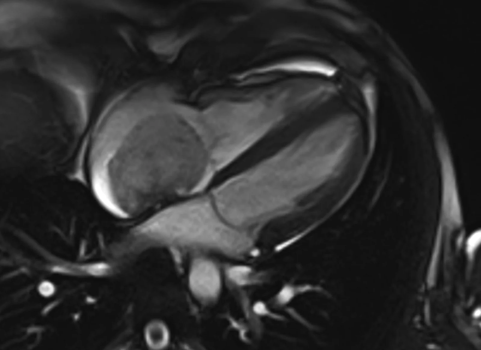
To visualise the heart and confirm the presence of a tumour, various imaging tests can be carried out: echocardiograms, cardiac MRI, CT scans and X-rays. These allow doctors to obtain clear images of the heart and its structure.
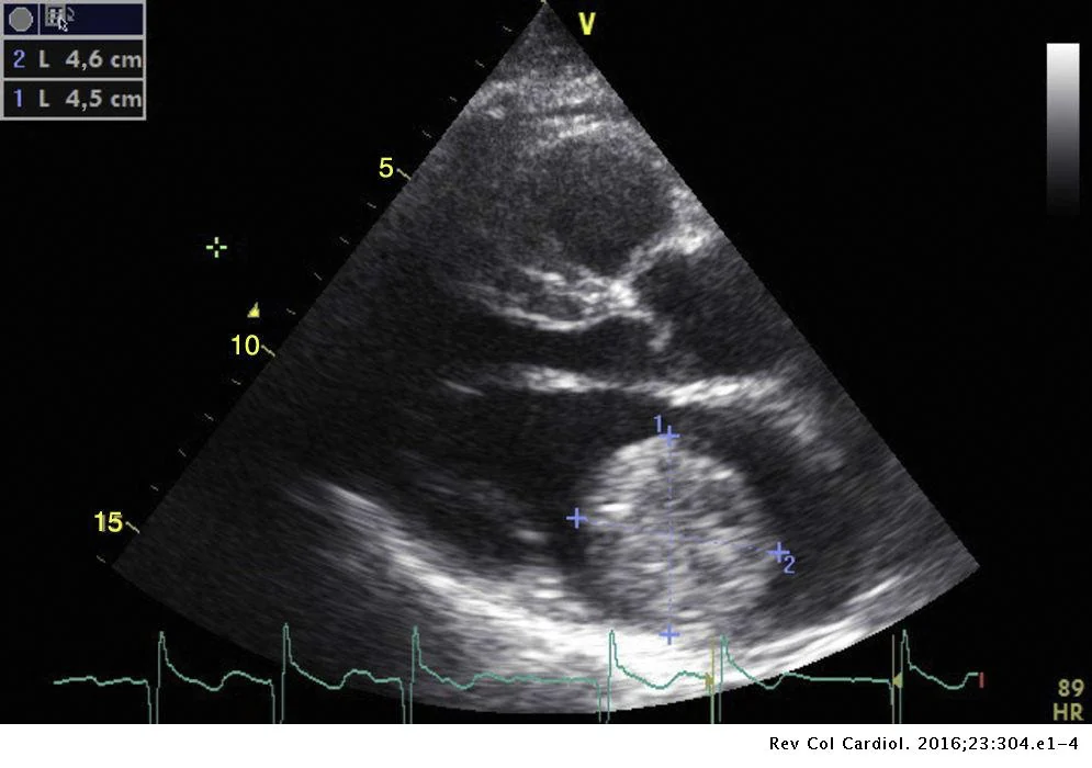
3. Echocardiogram — the most common initially
The echocardiogram is a non-invasive test using ultrasound to create moving images of the heart. This technique can identify the location, size and characteristics of the tumour, which helps in planning treatment.
4. Biopsy
In some cases a biopsy can be performed to confirm the nature of the tumour. A small sample of heart tissue is taken for analysis under the microscope. This helps determine whether the tumour is benign or malignant.
5. Multidisciplinary assessment
Once a cardiac tumour is confirmed, it is essential to consult a multidisciplinary medical team — the Heart Team — including cardiologists, cardiovascular surgeons and oncologists. This team will collaborate to develop a personalised treatment plan.
6. Genetic testing, if needed
Where there is suspicion of genetic predisposition, genetic tests can be carried out to detect mutations that may be related to the development of the tumour.
7. Staging, in malignant cases
If the tumour is malignant, staging can be carried out to determine whether the cancer has spread to other parts of the body. This helps guide treatment and estimate the outlook.
Early diagnosis and appropriate treatment are crucial to a better result.
Treating benign cardiac tumours, such as myxoma
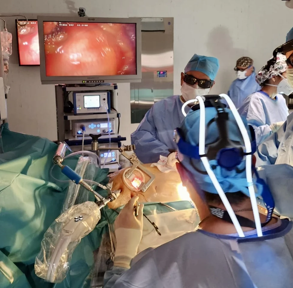
- Surgery. In most cases, surgical removal is the main treatment for benign cardiac tumours. Cardiovascular surgeons will work to remove the tumour safely and preserve normal cardiac function. Surgery can be performed through an incision in the chest or through minimally invasive techniques, depending on the location and size of the tumour.
- Medical follow-up. After surgery, regular follow-up is carried out to monitor recovery and ensure there is no recurrence.
Treating malignant cardiac tumours
- Surgery. In some cases of malignant cardiac tumour, surgery can also be an option, especially if the cancer is localised and has not spread. Removing the tumour can help improve symptoms and delay the spread of the cancer.
- Radiotherapy. Uses high-energy radiation to destroy cancer cells. It can be used as the main therapy or after surgery to eliminate any residual cancer cells.
- Chemotherapy. Involves using medicines to destroy or slow the growth of cancer cells. Although it is not commonly used for cardiac tumours because of the heart's delicate location, in some cases it can be considered.
- Targeted therapy and immunotherapy. These are more advanced therapies focusing on specific targets in the cancer cells or on the immune system, to fight the cancer more selectively.
- Supportive treatment. For some patients with advanced malignant cardiac tumours, the focus may be on managing symptoms, controlling pain and improving quality of life.
The most common malignant cardiac tumours
Malignant cardiac tumours are extremely rare compared with benign ones, but it is still important to be informed about them.
- Cardiac sarcoma. Sarcomas are malignant tumours originating in connective tissues such as muscle, fat and blood vessels. Cardiac sarcomas are uncommon but aggressive. They can begin anywhere in the heart and tend to grow rapidly.
- Carcinoid tumour. Carcinoid tumours are a type of neuroendocrine cancer that can begin in hormone-producing tissues such as the intestine or the lung. These tumours can secrete chemicals that affect the function of the heart and cause symptoms such as irregular heart rhythm and difficulty breathing.
- Cardiac lymphoma. Lymphomas are cancers originating in the cells of the lymphatic system. Although rare in the heart, they can present as primary tumours or as part of a systemic lymphoma affecting other areas.
- Germ cell tumour. These tumours can develop from embryonic cells in the testes and ovaries, but in rare cases can also affect the heart. They can be cancerous and require a combination of surgical treatment and chemotherapy.
Because of the rarity of these tumours, collaboration between cardiologists, oncologists and other specialists is essential to determining the diagnosis and the most appropriate treatment plan.
Outlook and treatment options
Most benign cardiac tumours, myxomas included, tend to have a favourable outlook after appropriate treatment. Myxomas are often removed surgically and, in most cases, patients can expect a complete recovery.
Most myxomas can now be operated on with minimally invasive techniques.
For malignant cardiac tumours, the outlook can vary with the type and stage of the cancer. Treatment may involve surgery, radiotherapy, chemotherapy or a combination. Multidisciplinary care from a specialist medical team is essential to making informed decisions about treatment.
Other types of benign heart tumour
Myxoma is the most common benign heart tumour in adults, but it is not the only one. Others are less frequent:
- Papillary fibroelastoma. A small tumour with many fine fronds, usually attached to a heart valve. It is the most common tumour of the valves. Although small, it can shed fragments and cause a stroke, so removal is often recommended, especially when it is mobile or on the left side of the heart.
- Lipoma. A tumour made of fat cells. Many cause no symptoms and are found by chance.
- Rhabdomyoma. The most common heart tumour in children. It is often linked to a genetic condition called tuberous sclerosis, and many shrink on their own over time.
- Fibroma. A firm tumour that grows within the heart muscle, seen mostly in children. It can disturb the heart rhythm.
Each type behaves differently. Some need prompt surgery, while others can simply be watched with regular imaging. Your team will explain which applies to you.
Secondary tumours: cancer that spreads to the heart
Most cancer found in the heart did not start there. Tumours that spread to the heart from elsewhere are far more common than primary malignant cardiac tumours. Lung cancer, breast cancer, melanoma, lymphoma and leukaemia are among those that can reach the heart or the pericardium, the sac around it.
These secondary tumours often cause fluid to collect around the heart, which can lead to breathlessness. Treatment is usually led by the oncologist and aimed at the original cancer. Our role as surgeons may be to drain the fluid or, in selected cases, to help obtain a diagnosis.
Heart tumour treatment: what surgery involves
For most benign cardiac tumours, heart tumour treatment means surgery. The operation is planned with the imaging studies, so the surgeon knows the size, position and attachment of the tumour in advance.
During surgery, the heart-lung machine takes over the work of the heart and lungs. The surgeon opens the chamber containing the tumour and removes it whole, handling it gently so that no fragments break off. With a myxoma, the base where it is attached is removed too, which lowers the chance of it growing back. If a small part of the wall between the atria has to be removed, it is repaired with a patch.
After the operation, most patients spend a day or two in intensive care and around a week in hospital, although this varies. The tumour is analysed under the microscope to confirm its type. Follow-up echocardiograms check that it has not returned.
We operate on patients with cardiac tumours in Guadalajara, Querétaro and Hermosillo. Whenever the position of the tumour allows, we consider a minimally invasive approach.
Don't forget
Receiving a diagnosis of a cardiac tumour can be overwhelming, but it is important to remember that there are options. Most tumours of the heart are benign and are usually operable — although urgently or semi-urgently.
Cardiac tumours are rare, and understanding their benign or malignant nature, their causes and the treatment options available can bring some calm in the middle of uncertainty. If you or someone you love is facing this challenge, remember you are not alone.
Medical science is advancing constantly, and a structured plan needs to be established at every step we take towards treatment and recovery.
About the author
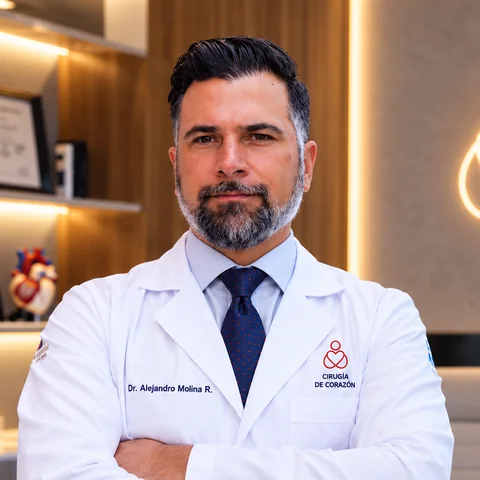
Dr Alejandro Molina is a cardiovascular surgeon practising in Guadalajara, Querétaro and Hermosillo. He trained in cardiothoracic surgery at the Universidad de Guadalajara, completed advanced training in cardiac surgery, minimally invasive surgery, robotic surgery and structural interventions at the University of Montreal, and a fellowship in structural cardiology and TAVR at CHU Bordeaux, France. He performed the first robotic mitral valve repair in the Ángeles Health System and the first robotic heart surgery in Guadalajara.
Medical degree: Universidad Autónoma de Guadalajara, Cédula Profesional 5757260 · Cardiothoracic surgery: Universidad de Guadalajara, Cédula de Especialidad 10768871 · Current certification, Consejo Nacional de Cirugía del Tórax, no. 447
Talk to a specialist
Request a free video consultation to clear up your questions and make a better-informed decision.