Cardiac Myxoma Removal: Surgery and Recovery
Cardiac myxoma removal restores heart function and prevents stroke.
When a doctor says the words "heart tumor," most patients assume the worst. Yet the most common tumor of the heart is the Cardiac Myxoma, a benign growth that is almost always curable with a single operation. A timely cardiac myxoma removal not only relieves symptoms but also removes the risk that a fragment of the tumor breaks off and causes a stroke. This guide walks you through what the tumor is, how it is found, what the operation involves, and what recovery actually looks like — in plain language, without alarm.
What Is a Benign Heart Tumor?
A benign heart tumor is a growth that does not spread to other organs, unlike a malignant cancer. A myxoma is the most common benign tumor of the heart in adults, and it usually forms inside the left atrium — the upper chamber that receives oxygen-rich blood from the lungs. It is typically a soft, gelatinous mass attached to the wall of the chamber by a small stalk, which allows it to move with the flow of blood.
Because it sits inside the heart rather than within the muscle, a myxoma can physically obstruct the mitral valve, the one-way door between the left atrium and the left ventricle. When that happens, blood flow is reduced and the valve may leak. This mechanical interference, not invasion of tissue, is what produces most of the symptoms patients notice. The National Heart, Lung, and Blood Institute describes myxomas as rare but treatable growths that generally require surgical excision once discovered (National Heart, Lung, and Blood Institute).
The good news is that myxomas are nearly always solitary and curable. Surgery for a benign heart tumor has been refined over decades, and the great majority of patients return to normal life without the tumor ever returning. Understanding exactly what the mass is doing inside your heart is the first step toward choosing treatment calmly rather than in panic.
What Are the Symptoms of Cardiac Myxoma?
Heart tumor symptoms surgery decisions depend on the pattern of complaints a patient brings to the doctor. The classic trio is constitutional symptoms — fever, fatigue, weight loss — caused by inflammatory substances the tumor releases, plus obstruction symptoms such as breathlessness, dizziness or fainting when the tumor blocks the valve, and embolic symptoms such as a small stroke or cold fingers when a fragment travels in the bloodstream.
The obstruction symptoms often have a striking feature: they can change with posture. Because the tumor swings on its stalk, some patients feel faint lying flat and better sitting up, or the reverse. Doctors sometimes hear a "tumor plop," an extra sound in the heartbeat that mimics the murmur of mitral valve disease. Patients are frequently treated for anemia, arthritis or valve disease for months before the true cause is found.
Embolism is the most dangerous presentation. A fragment of the gelatinous tumor, or a clot forming on its surface, can travel to the brain and cause a stroke, or to a limb and block circulation. In some patients, a stroke is the very first sign that anything is wrong. This is the reason a confirmed myxoma is scheduled for removal promptly rather than observed — the operation is as much stroke prevention as symptom relief.
How Is Cardiac Myxoma Diagnosed?
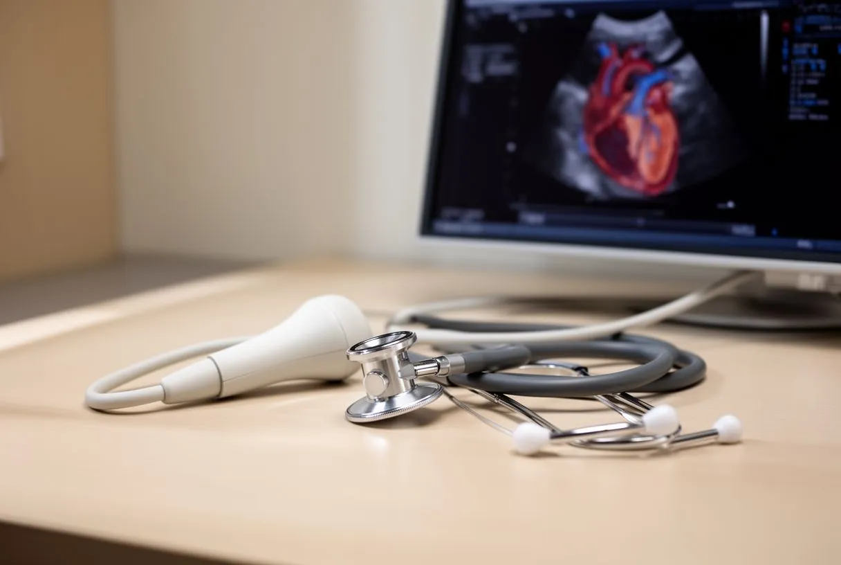
Most myxomas are discovered by echocardiogram, the ultrasound scan of the heart that shows the chambers and valves in motion. A transthoracic echocardiogram — the scan done from the chest wall — usually shows the mass clearly. When more detail is needed, a transesophageal echocardiogram, in which a small ultrasound probe is passed into the food pipe, gives sharper images of the left atrium and confirms the tumor's size, stalk and attachment point.
Additional imaging rounds out the picture. A CT scan or MRI of the heart defines the tumor's exact anatomy and rules out other masses. Blood tests may show the raised inflammatory markers that explain fevers and fatigue. Importantly, the tumor is not biopsied in the way other tumors are — sampling a myxoma inside the beating heart carries embolism risk, so diagnosis rests on imaging and the operation itself provides the final confirmation under the microscope.
In our team's experience, the diagnosis usually arrives as a surprise on an echocardiogram ordered for something else — breathlessness, a murmur or an odd fainting spell. We take the time in consultation to show patients their own images and explain in plain words what the scan shows, because an informed patient makes a calmer surgical decision.
Is Myxoma Genetic?
For the vast majority of patients, a myxoma is a sporadic event — a random growth with no family pattern and no inherited cause. Most people who have one relative with a myxoma will never see it appear in anyone else in the family. In these sporadic cases, screening of siblings and children is generally not required.
There is, however, a rare inherited condition called Carney complex, in which cardiac myxomas occur together with skin and pigmentation changes and tumors of other endocrine glands. Carney complex myxomas tend to appear at a younger age, can be multiple, can occur in either atrium or the ventricles, and have a real tendency to recur — which is why patients with this condition need lifelong imaging follow-up.
If you have had more than one myxoma, or a relative had one at a young age, mention it to your cardiac team. Recurrence after complete surgical removal is uncommon in ordinary cases, but surveillance echocardiograms are standard practice after any myxoma operation precisely so that a rare regrowth is caught while it is still small and straightforward to treat.
Cardiac Myxoma Removal vs Watchful Waiting
Unlike many conditions, this is not a decision between surgery and observation. When it comes to cardiac myxoma removal vs watchful waiting, watchful waiting is almost never recommended once the diagnosis is confirmed. The reasons are practical: the tumor can obstruct the valve suddenly, causing fainting or heart failure, and it can shed fragments that cause stroke — and neither risk can be predicted from the tumor's current size or symptoms.
Even a small, asymptomatic myxoma — one found incidentally on a scan done for another reason — carries the same dangers. Published case reports describe patients in whom a myxoma sat silently until it caused valve damage or embolism; one reported case involved a left atrial myxoma that had caused rupture of the mitral valve's supporting chordae and regurgitation, discovered alongside coronary artery disease (Annals of Cardiac Anaesthesia). Waiting converts a controlled, planned operation into an emergency one.
Every case we review is discussed by the multidisciplinary team against international practice guidelines before a recommendation is given, and the patient receives a clear proposal with the alternatives — but for a confirmed myxoma, the honest alternative is simply a matter of timing. The final decision is always the patient's, and we encourage second opinions as a normal part of deciding about heart surgery.
How Heart Tumor Removal Surgery Is Performed
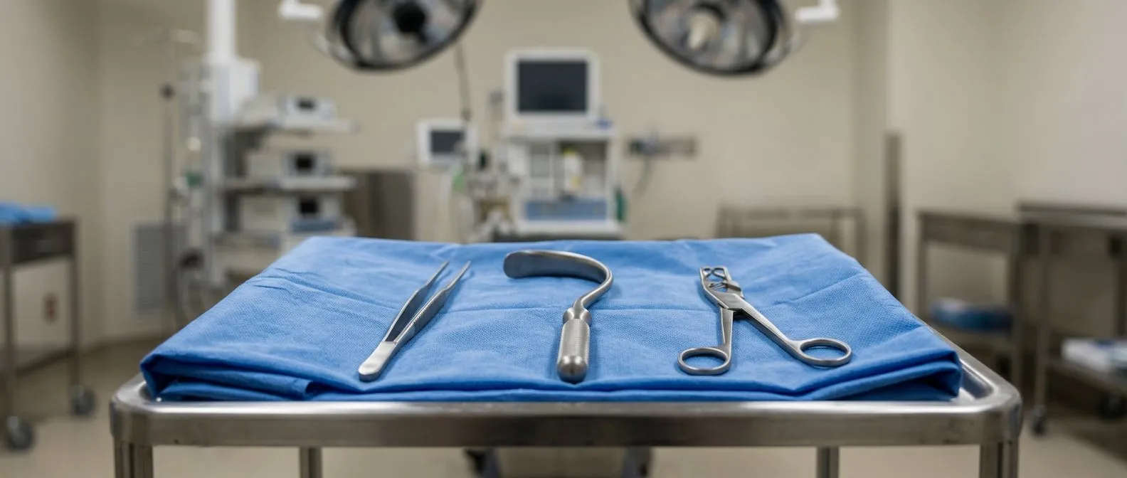
The standard open heart myxoma surgery follows the same trusted steps as other cardiac operations. The patient is put to sleep, the chest is opened through the breastbone, and a heart-lung machine takes over circulation so the heart can be temporarily stopped and made still. The surgeon opens the left atrium, removes the tumor together with its stalk and a small margin of the wall it was attached to, and closes the heart.
Taking that margin matters. Removing the base of the stalk reduces the chance of regrowth, and the surgeon inspects all four chambers for additional tumor tissue before closing. If the attachment site left a defect in the wall between the atria, that defect is patched. Some myxomas are diagnosed alongside other heart problems, and both can be addressed in the same operation; a published case report describes cardiac myxoma removal performed together with coronary artery bypass grafting in a single setting (Journal of Cardiothoracic Medicine).
The technical name for the tumor-removal operation itself is Myxoma Excision, and it is one of the shorter and more straightforward cardiac procedures when done electively. Typical operating time runs two to four hours. When the repair is complete, the heart is restarted, the heart-lung machine is withdrawn, and the patient is transferred to intensive care for close monitoring overnight.
Minimally Invasive Cardiac Myxoma Removal
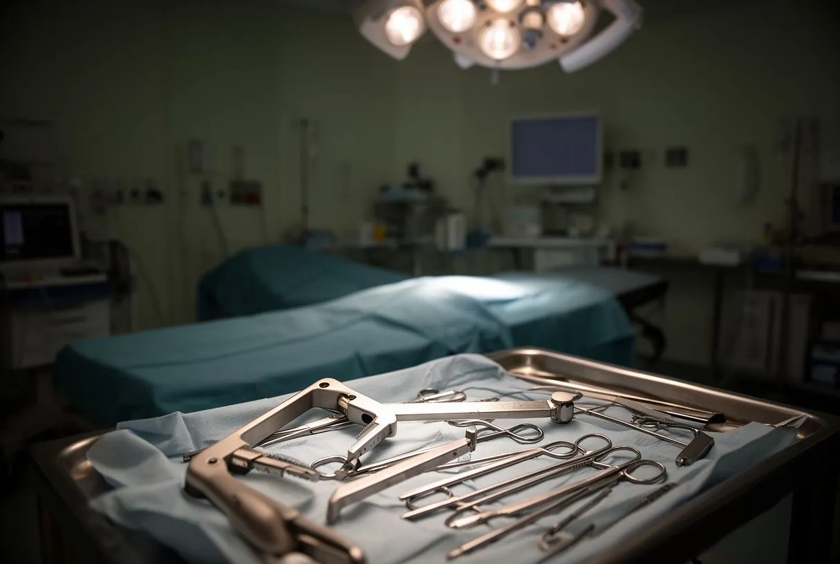
Not every myxoma requires a full breastbone incision. Minimally invasive cardiac myxoma removal is done through a small incision on the right side of the chest, typically four to six centimeters, using specialized instruments and video assistance. The heart-lung machine is still used, but the access is far smaller, which means less pain, less blood loss and a faster return to driving and daily activity.
Robotic cardiac myxoma surgery takes this approach one step further. With the robotic system, the surgeon operates instruments that translate hand movements with full precision through even smaller ports, working on a magnified three-dimensional view of the inside of the heart. For the right patient and the right tumor anatomy, robotic access allows complete removal with excellent exposure and the smallest possible footprint.
Dr Molina trained in cardiac, minimally invasive, robotic and structural surgery at the University of Montreal, and performed the first robotic mitral valve repair in the Ángeles Health System and the first robotic heart surgery in Guadalajara. Our team recommends the less invasive route only where the evidence supports it for that specific patient — tumor size, stalk position and the patient's own anatomy decide, not preference. Some myxomas genuinely do best with a conventional approach, and an honest team will say so.
What Are the Risks of Atrial Myxoma Surgery?
Atrial myxoma surgery risks are those of heart surgery in general, plus a few specific to the tumor. General risks include bleeding, infection, irregular heartbeat, stroke and the need for blood transfusion. The tumor-specific risk is embolism during the operation itself — fragments can dislodge while the surgeon manipulates the mass — which is why careful handling and complete removal of the stalk are part of standard technique.
Here is how the main risks break down in practical terms:
- Bleeding and re-exploration: some patients need a return to theatre early after surgery to control bleeding; this is monitored closely in intensive care.
- Rhythm problems: atrial fibrillation is common in the first days after any atrial surgery and usually settles with medication.
- Stroke: the risk is small in elective surgery, and the operation itself is done largely to prevent the far greater stroke risk the tumor poses if left in place.
- Recurrence: regrowth after complete removal is rare in sporadic myxomas, but follow-up echocardiograms are arranged to confirm the heart stays clear.
Before any recommendation, we review each patient's complete risk profile against international guidelines — age, other conditions, heart function and the tumor's anatomy all matter. If a heart mass removal recovery time is what worries you most, it helps to know that for an otherwise healthy patient this operation carries some of the lowest risk in cardiac surgery.
How Long to Recover From Cardiac Myxoma Surgery?
The first days after surgery are spent in hospital: one night in intensive care, then two to five days on the ward as breathing exercises, gentle walking and wound care progress. Most patients are surprised at how quickly they are on their feet — standing the day after surgery is the norm, not the exception. Drainage tubes and monitoring lines come out progressively, and pain is managed with a clear plan.
How long to recover from cardiac myxoma surgery in full depends on the approach. After conventional open heart surgery, the breastbone takes roughly six to eight weeks to knit, so driving, lifting and returning to work are paced to that timeline. After a minimally invasive or robotic removal, the small chest incision heals faster and many patients return to desk work in two to four weeks and to fuller activity sooner than that.
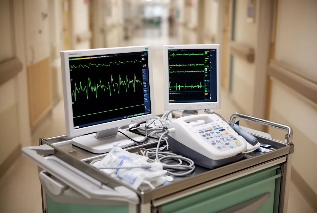
The Recovery Room — the monitored unit where you wake immediately after the operation — is where the first hours of recovery happen, with continuous observation of heart rhythm, blood pressure and breathing. Most patients walk comfortably within days, and the honest answer to "when will I feel like myself again" is usually measured in weeks, not months. A follow-up echocardiogram is arranged to confirm the heart is clear and working well.
Cost matters, and international patients routinely ask about compared to their home country.
What drives the total is the combination of the surgical approach, hospital choice and length of stay. A robotic or minimally invasive removal can shorten the hospital stay, which offsets some of the technology cost. Cardiac tumour removal at a private hospital should always be quoted as a complete package — surgeon, anesthesia, operating room, intensive care, ward days and follow-up — so there are no surprises.
Our first contact with any patient is a free video consultation: we get to know you, talk through the condition and the imaging you already have, and say exactly which studies are still needed so no unnecessary ones are ordered — and no unnecessary costs either. When you decide to go ahead, a face-to-face visit follows at our consulting rooms in Guadalajara, Querétaro or Hermosillo.
When a Heart Mass Is Not a Myxoma: Angiosarcoma and Other Malignant Tumors
Rarely, a heart mass turns out to be malignant rather than benign, so it is worth knowing the difference. Cardiac angiosarcoma symptoms include rapidly progressing breathlessness, chest pain, blood around the heart (pericardial effusion) and signs of blockage of the great vessels — and they advance far faster than myxoma symptoms. Imaging features help distinguish the two: malignant tumors tend to be bulky, invade muscle and blood vessels, and bleed internally.
What is the average life expectancy for someone with cardiac sarcoma? Honest answer: outcomes depend heavily on how far the tumor has spread at diagnosis, and primary cardiac sarcomas are difficult because they grow silently in a space where there is little room. Treatment combines surgery where feasible with chemotherapy, and prognosis is individual — which is precisely why any heart mass must be characterized carefully before a plan is made, not assumed benign or malignant from a single test.
The reassuring context for most patients reading this page is that a myxoma has a characteristic appearance on echocardiogram, a typical location and a typical stalk, and in the great majority of cases the imaging diagnosis is confident. When anything about the imaging is atypical, we investigate further before recommending surgery. If there is ever doubt, the images should be reviewed with the patient so the plan is decided together.
Choosing a Cardiac Myxoma Surgeon Guadalajara
What should you look for when choosing a cardiac myxoma surgeon Guadalajara or elsewhere? First, a team that performs the full range of approaches — conventional, minimally invasive, robotic and transcatheter — because the right approach depends on your anatomy, not on what a single surgeon happens to offer. Second, a genuinely multidisciplinary review of your case. Third, a surgeon who explains options in plain language and welcomes a second opinion rather than resisting one.
Questions worth asking at any consultation:
- Which approach fits my tumor, and why — open, minimally invasive or robotic?
- What is the expected hospital stay and recovery timeline for that approach?
- What follow-up is planned after removal, and for how long?
Dr Molina trained in cardiothoracic surgery at the Universidad de Guadalajara, and in structural cardiology and TAVR at CHU Bordeaux, with the team also trained at Jena, Bad Berka and Buffalo. That breadth matters here: a myxoma operation is individually modest, but the team around it should be able to handle any finding — valve disease, coronary disease, anything — that the operation reveals.
Life After Heart Tumor Removal Surgery
Life after atrial myxoma removal is, for most patients, simply normal life. The tumor does not return in the great majority of sporadic cases, the valve recovers its function once the obstruction is gone, and the symptoms that led to the diagnosis — the breathlessness, the fatigue, the fevers — typically resolve. Heart tumor removal success rate for myxoma is among the highest in cardiac surgery, and the operation is curative in intent and usually curative in fact.
Follow-up is straightforward. A post-operative echocardiogram confirms complete removal and valve function, and periodic surveillance continues for years as a safety net. Patients who had neurological symptoms from an embolism before surgery need coordinated neurological follow-up as well. Exercise, work and travel resume progressively as the chest heals, guided by the surgical team.
The takeaway is simple and hopeful: a myxoma is a serious finding with a genuinely good ending, provided it is removed promptly by an experienced team. If you have been told you have a heart mass, or you have an echocardiogram report with words you do not understand, the next step is not to self-diagnose — it is to sit down with a qualified cardiac team, have the images explained in plain language, and agree a plan. Our team across Guadalajara, Querétaro and Hermosillo is available for exactly that conversation, starting with a free video consultation.