Benign Cardiac Tumour: Diagnosis and Removal
Benign cardiac tumour removal: learn diagnosis steps, surgical options, and recovery timeline from Guadalajara's heart surgery specialists.
Finding out that you or someone you love has a growth on the heart is frightening, but the first thing to know is that most of these growths are not cancer. A benign cardiac tumour is a non-cancerous mass that develops in or on the heart, and benign cardiac tumour removal is a well-established operation with a long track record of success. In this guide we walk through what these tumours are, the symptoms they cause, how doctors confirm the diagnosis, what the surgery involves, and what recovery really looks like — so you can face the next steps with clear, calm information rather than fear.
What Is a Benign Cardiac Tumour?
A cardiac tumour is any abnormal mass growing inside or around the heart. When doctors say a tumour is benign, they mean it is not cancer: it does not invade surrounding tissue the way a malignancy does, and it does not spread to other organs. That distinction matters enormously, because it changes both the urgency and the outlook of treatment.
Benign does not mean harmless. Even a non-cancerous mass inside the heart can cause serious problems simply by being there. The heart is a small, hard-working pump, so a tumour can block blood flow through a valve, break off fragments that travel to the brain and cause a stroke, or interfere with the heart's electrical system and trigger rhythm disturbances. This is why a benign cardiac tumour is usually treated rather than simply watched.
Primary versus secondary tumours. A primary cardiac tumour starts in the heart itself and is rare — most masses found in the heart have actually spread there from a cancer elsewhere in the body, such as the lung or breast. In this article we focus on primary benign tumours, which are the ones patients are most often relieved to hear about, and which in adults are overwhelmingly non-cancerous. The National Heart, Lung, and Blood Institute notes that primary heart tumours are uncommon and that most are benign.
What Is the Most Common Benign Heart Tumour?
The most common benign cardiac tumour in adults is the myxoma, a soft, gelatinous growth that in about three out of four cases arises in the left atrium — the upper chamber that receives oxygen-rich blood from the lungs. Myxomas are usually attached by a small stalk to the wall that separates the two upper chambers, and that stalk lets them move with each heartbeat, sometimes swinging into the mitral valve opening.
Why location matters so much. Because a myxoma can flop back and forth with the cardiac cycle, it can intermittently block the mitral valve — the gateway between the left atrium and the main pumping chamber. This produces fainting episodes, breathlessness, and symptoms that mimic mitral valve stenosis. The mobility also raises the risk that pieces of the tumour break loose and travel in the bloodstream, which is the mechanism behind many of the neurological symptoms patients experience before diagnosis.
A rare but important exception: the germ cell tumour connection. In some patients, a mass in the chest near the heart turns out to be a Germ Cell Tumour that has spread from the mediastinum — the space between the lungs. These are a different disease entirely and are managed by oncology teams, which is why accurate tissue diagnosis matters before any plan is made. In children, benign cardiac tumours follow a different pattern, with rhabdomyomas and fibromas predominating; the pediatric literature published at Orphanet Journal of Rare Diseases describes how many childhood cardiac tumours are related to tuberous sclerosis and often regress on their own.
Benign Cardiac Tumour Symptoms to Watch For
Symptoms depend on where the mass sits, how big it is, and how mobile it is. Some benign cardiac tumours cause no symptoms at all and are discovered by accident on an echocardiogram done for another reason — an increasingly common scenario in the era of widespread imaging.
Blood-flow obstruction symptoms. When a tumour blocks a valve or a chamber, patients notice breathlessness on exertion, dizziness, fainting spells (especially with changes in position, which is a classic clue for atrial myxoma), and swelling of the legs or abdomen as blood backs up. These symptoms can come and go, which sometimes leads patients and even doctors to suspect something else first.
Embolic symptoms — the ones that demand urgency. Fragments of a friable tumour can travel to the brain, eye, kidneys, or limbs. A sudden stroke in a young person without typical stroke risk factors is one of the situations that should prompt a search for an underlying cardiac mass. Other warning signs include a transient loss of vision in one eye, unexplained kidney problems, or a cold, painful limb.
Constitutional and rhythm symptoms. Some myxomas release inflammatory substances, producing low-grade fever, unexplained weight loss, fatigue, and joint aches that can mimic an infection or rheumatic disease — patients sometimes go months being investigated for the wrong thing. Tumours near the conduction system can cause palpitations or irregular heartbeats. In our practice we always ask specifically about position-related fainting and unexplained strokes in young patients, because those two clues are what most often point us toward a cardiac mass.
How Heart Tumour Diagnosis Works
Diagnosis usually begins with the echocardiogram, the ultrasound of the heart. A transthoracic echo — the scan with the probe on the chest — picks up most masses, and a transoesophageal echo, where a slim probe is passed down the food pipe, gives much sharper pictures of the upper chambers and valves and shows the tumour's attachment point in detail.
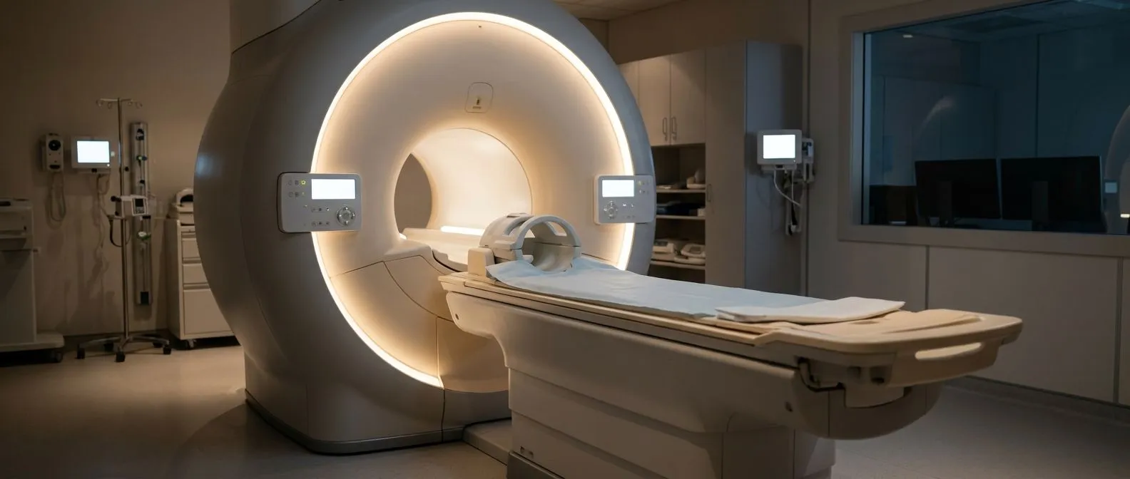
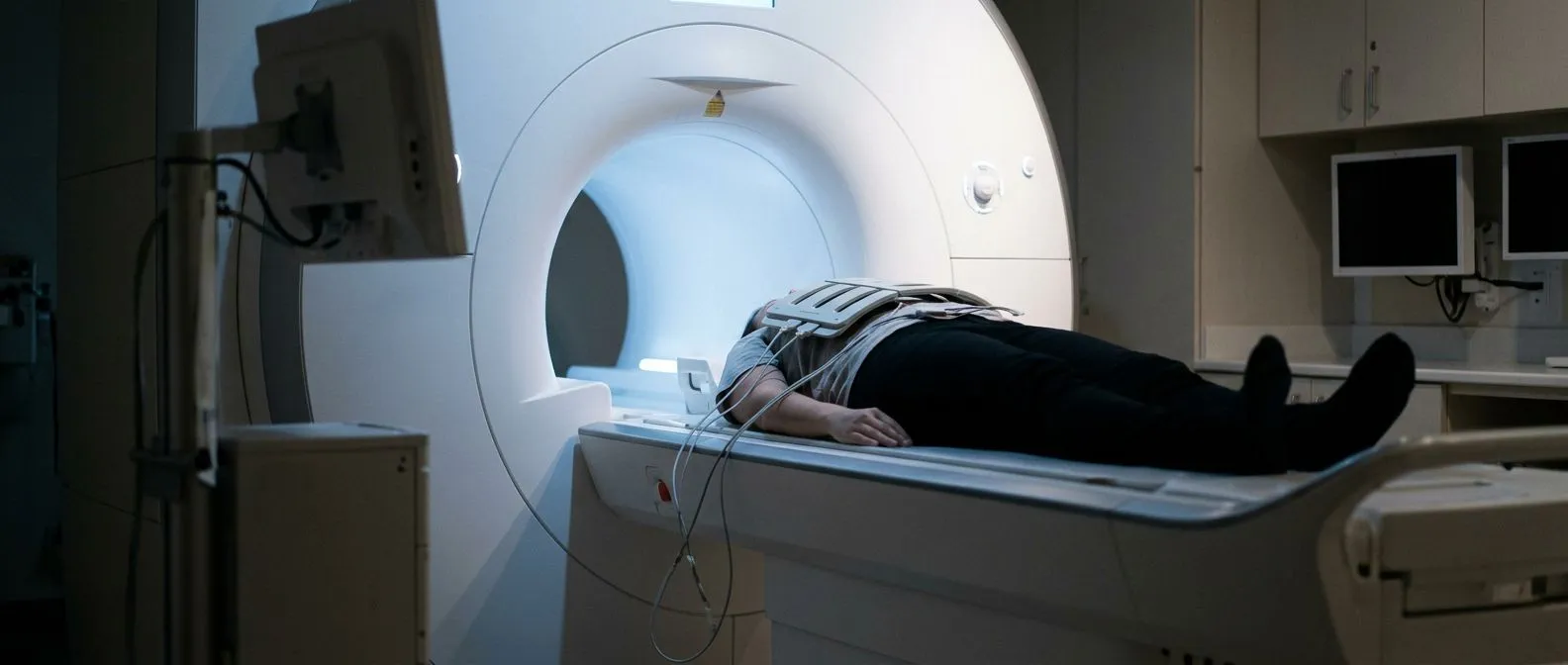
Cardiac MRI: the detail study. Magnetic resonance imaging of the heart adds information that ultrasound cannot provide: the tissue character of the mass (fat, muscle, blood, fibrosis), its exact size and extent, and its relationship to valves and surrounding structures. This detail directly shapes the surgical plan, because a tumour's composition on MRI often allows an experienced team to predict whether it is a myxoma, a fibroma, a lipoma, or something that needs a different approach.
CT and additional studies. Computed tomography may be used to rule out tumours that have spread from elsewhere in the body, to assess the coronary arteries before surgery, and to clarify calcified masses. Blood tests help in the workup too — inflammatory markers are often elevated with myxomas, and certain tumour markers become relevant when a germ cell origin is suspected. Before we recommend any study, we review which scans the patient already has and order only what is still missing, so nobody pays for imaging they do not need.
Is a Heart Tumour Biopsy Necessary?
This is one of the questions patients ask most often, and the honest answer is: usually no. For most masses that imaging shows to be typical of a benign tumour in a removable location, the standard of care is to take the whole thing out and have the pathologist examine it afterwards — the definitive biopsy happens on the removed specimen, not before.
Why pre-operative biopsy is usually avoided. Puncturing a cardiac mass carries real risks: pieces can break off and cause embolism, and a myxoma in particular is friable and mobile. If imaging characteristics are classic — a mobile, stalked, gelatinous left atrial mass in an adult — most surgical teams proceed directly to excision. Biopsy is reserved for cases where the picture is atypical or when a tumour elsewhere in the chest suggests a cancer that needs a different treatment entirely.
When tissue diagnosis does come first. If the mass looks like it may be a lymphoma, a metastasis, or a Germ Cell Tumour, a biopsy through a safe route — or sometimes a pericardial window — is the right move, because those diseases are treated with chemotherapy or radiotherapy, not surgery. This is exactly where a multidisciplinary decision protects the patient from the wrong operation.
Types of Benign Heart Tumours
Knowing which type a mass is likely to be helps you understand the treatment your team proposes. Here are the main benign tumours, each with its own behaviour and management logic:
- Myxoma: the most common in adults; gelatinous, usually left atrial, often stalked and mobile. Treated with complete surgical excision; recurrence is uncommon but follow-up imaging is standard.
- Papillary fibroelastoma: a small, frond-like growth, most often on a valve surface. It behaves like a tiny embolic risk; small asymptomatic ones may be watched, larger or symptomatic ones removed.
- Lipoma and lipomatous hypertrophy: fatty masses that are almost always found incidentally and rarely need anything beyond observation.
- Rhabdomyoma: the most common in infants and children, strongly linked to tuberous sclerosis; many shrink or disappear without surgery.
- Fibroma: a firm mass of fibrous tissue, usually in the ventricles of children; it can interfere with rhythm and often requires resection.
- Cystic tumour of the atrioventricular node: a tiny cyst at the centre of the heart that can cause sudden rhythm problems; a rare but recognised cause of sudden cardiac death.
The common thread is that type drives management. A lipoma may need nothing but an annual scan. A myxoma nearly always needs removal. Every case that reaches our team is reviewed by the whole multidisciplinary group against international practice guidelines before we propose anything, and you receive a clear recommendation with the alternatives laid out — the final decision is always yours.
When Benign Cardiac Tumour Removal Is Recommended
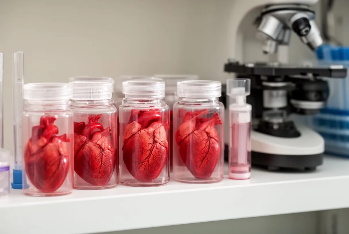
Surgery is recommended when the tumour is causing symptoms, when it poses an embolic risk — which applies to nearly all myxomas and to valve-surface fibroelastomas — or when imaging cannot exclude something more serious. A large mass, even if quiet today, is often removed because it will only grow and the risk of stroke or obstruction rises with time.
When observation is reasonable instead. Not every benign cardiac tumour needs an operation. Small asymptomatic lipomas and some fibroelastomas found incidentally can be followed with periodic echocardiograms. In children with rhabdomyomas linked to tuberous sclerosis, watchful waiting is often the correct first plan, because many of these tumours regress naturally as described in the pediatric cardiac tumour literature.
Weighing the trade-offs. Heart surgery is real surgery, so the decision balances the risk the tumour poses against the risks of the operation — which for tumour excision in experienced hands are in the same range as other routine cardiac procedures. Factors that shift the balance include the patient's age, other conditions, the tumour's size and location, and whether there has already been an embolic event. We encourage second opinions and treat seeking one as a completely normal part of deciding about heart surgery; a good decision is one you have checked from more than one angle.
What Happens During Open Heart Tumour Surgery?
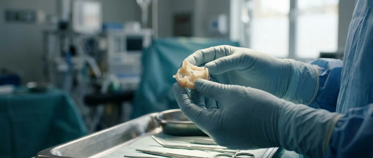
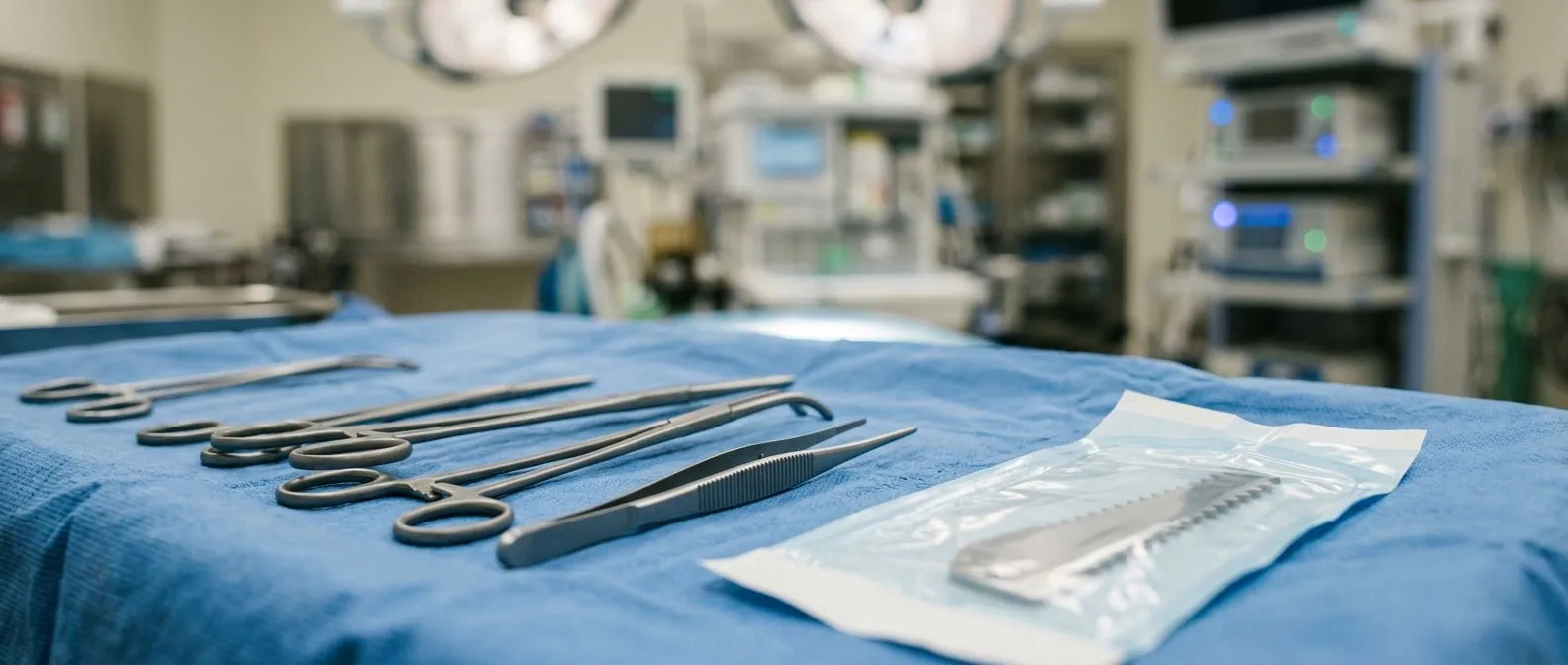
Classically, myxoma removal and other cardiac mass excision is performed as open-heart surgery under general anaesthesia. The surgeon opens the chest through the breastbone, the heart-lung machine takes over circulation, and the heart is opened through the chamber containing the tumour. The mass is removed whole, with its stalk and a small margin of the wall it was attached to, and the defect is closed — often with a small patch.
Minimally invasive alternatives. Many tumours, especially left atrial myxomas, can be removed through a small incision in the right side of the chest, or in selected cases with robotic assistance. The goals are the same — complete removal with no fragments left behind — but the incision is smaller, recovery is often faster, and the cosmetic result is better. Whether the minimally invasive route is appropriate depends on the tumour's size, position, and attachments.
What we check before choosing an approach. In consultation, our team takes the time to explain what the studies actually show in plain words — where the mass sits, what it is attached to, and why a conventional, minimally invasive, robotic, or transcatheter plan is or is not suitable — and we recommend the less invasive route only where the evidence supports it for that particular patient. Dr Molina performed the first robotic mitral valve repair in the Ángeles Health System and the first robotic heart surgery in Guadalajara, and the team trained at Montreal, Jena, Bad Berka and Buffalo.

Cardiac Tumour Removal Recovery Timeline
Most patients spend one to two nights in intensive care and leave hospital within five to seven days after an uncomplicated excision. The first week is about rest, wound care, and gradually increasing walking. By the second or third week most people are moving around the house comfortably and taking short walks outdoors.
Milestones over the first three months. Sternal precautions — no lifting more than a few kilos, no driving initially, no pushing or pulling — usually apply for six to eight weeks while the breastbone heals. Office visits track wound healing and rhythm. By around three months, most patients have returned to ordinary daily life, and many to work, depending on the job. A minimally invasive approach typically shortens the sternal precautions because there is no divided breastbone.
Follow-up imaging and recurrence. Because benign tumours — myxomas in particular — can very occasionally return, an echocardiogram is usually performed within the first year and periodically afterwards. The pathology report from the removed specimen also matters: it confirms the type of Cardiac Tumour and, in rare cases, reveals an unexpected finding that changes follow-up. Structured follow-up means recurrence surveillance is not left to chance, so ask exactly when your next scan is due.
Are Benign Heart Tumours Safe, and What Comes Next?
People often ask about the survival rate for patients with benign heart tumours. The reassuring truth is that when a benign cardiac tumour is removed completely before it has caused a major embolic event, long-term outcomes are excellent and life expectancy is generally normal — the danger is the tumour itself, not its removal. The serious risks are the ones that come from delay: stroke, valve obstruction, or dangerous rhythm problems.
What "benign" really promises you. A benign cardiac tumour is not a cancer diagnosis. It will not spread, and Cardiac Tumour Resection in experienced hands is a curative operation. The genuine uncertainties are surgical ones — the risk of any heart operation — plus the small chance of recurrence, which is why follow-up imaging exists. Understanding this framing helps many patients move from fear to planning.
Taking the next step calmly. If you have been told about a mass on your heart, or you have unexplained fainting, strokes at a young age, or persistent symptoms that imaging has never explained, the right next step is a focused review with a cardiac team rather than more internet searching. Our first contact is a free video consultation: we get to know you, talk through your condition, and tell you which studies are still needed so no unnecessary ones are ordered — and a face-to-face visit at our consulting rooms in Guadalajara, Querétaro or Hermosillo follows when you decide to go ahead.
Every case is reviewed by our multidisciplinary team against international practice guidelines before any recommendation is made, so you receive a clear proposal with the alternatives, and the final decision is always yours. A benign cardiac tumour is a serious finding, but it is also one of the most treatable problems in cardiac surgery — and benign cardiac tumour removal, done properly and followed up properly, lets most patients put the whole episode behind them. Bring your scans, bring your questions, and let a qualified team help you decide.