Heart Tumours: Types, Symptoms and Surgery
Heart tumours types range from benign myxomas to aggressive sarcomas.
Heart tumours types can be confusing to research, because the phrase covers everything from harmless fatty growths to aggressive cancers — and the difference changes everything about treatment. A heart tumour, sometimes called a cardiac tumour, is any abnormal mass growing in or on the heart. Some are discovered by chance on a routine scan. Others announce themselves with breathlessness, palpitations or a stroke. This guide walks through the main heart tumours types, the symptoms that should prompt investigation, how diagnosis works, and what surgery and recovery actually involve — written for patients and families, in plain language.
What Are Heart Tumours and How Common Are They?
Primary heart tumours are genuinely rare. A primary tumour starts in the heart itself, rather than spreading there from elsewhere. Medical literature consistently describes them as uncommon: most large hospital reviews find primary heart tumours in well under one per cent of patients examined at autopsy, and large echocardiography series detect them only rarely. Medical News Today notes that primary heart cancer is exceptionally rare precisely because heart muscle cells divide very little after childhood.
Secondary tumours are the more common story. Most masses found in the heart are metastatic — cancer that began in another organ, such as the lung, breast, kidney or a melanoma, and spread to the heart or the sac around it (the pericardium). Doctors distinguish carefully between primary heart tumours vs metastatic tumours, because the treatment plan follows the original cancer, not just the cardiac mass.
Rarity cuts both ways. The good news is that you are unlikely to have one. The challenge is that because they are rare, a heart tumour is often not the first thing considered when someone reports fatigue or breathlessness. That is why awareness of the symptoms — and of imaging that catches these masses — matters so much.
What Are the Types of Heart Tumours?
Benign heart tumours are non-cancerous growths. They do not spread to other organs, but location matters: a benign mass inside a pumping chamber can still obstruct blood flow or shed fragments that cause a stroke. The main types include myxomas (the most common in adults), papillary fibroelastomas (small, frond-like growths on valves), lipomas (fatty masses), fibromas (mostly in children), and rhabdomyomas (associated with a condition called tuberous sclerosis, also mostly in children).
Malignant heart tumours are cancerous. The most common primary malignant tumour in adults is sarcoma, which arises from connective tissue. In children, malignant primary tumours are rare but include rhabdomyosarcoma. Secondary tumours — cancers that have spread from elsewhere — outnumber all primary malignant types.
- Myxoma: benign, usually in the left atrium, most common in adults between roughly 30 and 60, sometimes run in families.
- Papillary fibroelastoma: benign, small, usually attached to a heart valve, occasionally sheds debris that causes emboli (blockages carried by the bloodstream).
- Lipoma: benign fatty mass, often found by chance, rarely needs surgery unless it causes symptoms.
- Sarcoma: malignant, most often in the right atrium, grows quickly, requires urgent specialist care.
- Metastatic tumours: cancer from another organ reaching the heart; treated mainly as part of the original cancer's care.
Knowing the type is not academic. It determines whether the plan is monitoring, surgery, chemotherapy, or a combination — which is why a precise tissue or imaging diagnosis always comes before any recommendation.
What Is a Myxoma Heart Tumour?
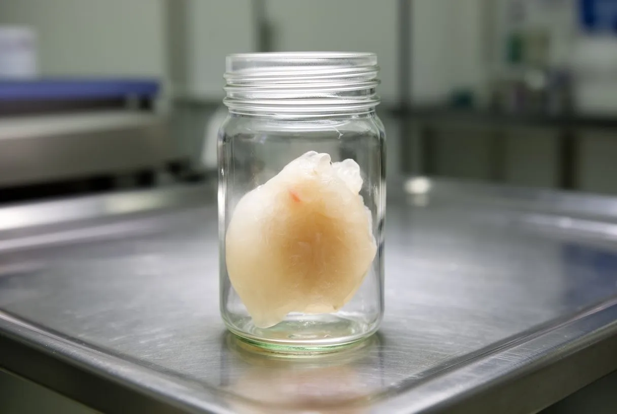
The myxoma is the classic heart tumour in adults. It is a soft, gelatinous, benign mass, most often attached by a small stalk inside the left atrium — the upper chamber that receives oxygen-rich blood from the lungs. Because it dangles on that stalk, it can move with each heartbeat, sometimes swinging toward the mitral valve and partially blocking it. This is why a myxoma heart tumour so often mimics mitral valve disease.
Symptoms follow three patterns. Some patients feel obstruction symptoms: breathlessness on exertion, fainting spells, or heart failure signs as blood flow out of the left atrium is impeded. Others present with embolic events — pieces of the tumour break off and travel to the brain, causing a stroke or transient symptoms, or to a limb. A third group has constitutional symptoms: fever, weight loss, fatigue and joint aches, caused by the tumour's inflammatory activity.
Atrial myxoma treatment is surgery, and it works well. Because myxomas can stroke or obstruct, they are removed rather than watched. The mass is excised completely, including its attachment point, and recurrence after complete removal is uncommon. Our team treats myxoma removal as a time-sensitive decision: once the diagnosis is confirmed, we bring the case before the multidisciplinary group and give the patient a clear proposal with the alternatives, and the final decision is always theirs.
What Are Cardiac Sarcoma Symptoms?
Sarcoma is cancer of the body's connective tissue. To answer the common question "what is sarcoma?": it is a family of malignant tumours arising from soft tissue — muscle, blood vessels, fat — rather than from glands or lining cells. When a sarcoma starts in the heart, it is called a cardiac sarcoma, and the most common form is angiosarcoma, which typically arises in the right atrium, the chamber that receives blood returning from the body.
Cardiac sarcoma symptoms depend on location. A tumour in the right atrium can block venous return, causing swelling of the legs, abdominal distension, and liver congestion — a picture doctors call right heart failure. Tumours near the pericardium can cause fluid to accumulate around the heart, leading to chest discomfort and breathlessness. General symptoms — weight loss, night sweats, fatigue — are common with any cancer.
Speed is the defining feature. Unlike a slow myxoma, cardiac sarcoma symptoms often progress over weeks to months. Anyone with rapidly worsening breathlessness, unexplained swelling, new arrhythmias (rhythm disturbances), or chest pain without a clear cause deserves prompt imaging. Early referral to a centre that can image, biopsy and treat quickly makes a real difference.
How Serious Is a Mass on Your Heart?
Seriousness depends on three things. First, what the mass is — benign or malignant. Second, where it sits — a small mass on a valve or near an outflow tract can be more dangerous than a larger mass in a safer spot. Third, what it is doing — obstructing blood flow, shedding emboli, disturbing rhythm, or leaking fluid into the pericardium.
A benign mass is not automatically harmless. Even benign tumours can cause strokes or sudden obstruction, so doctors rarely adopt simple watchful waiting for masses inside the pumping chambers. Superficial or asymptomatic masses — like many lipomas — may genuinely be monitored with periodic imaging instead.
Take the news seriously, not fearfully. Hearing "there is a mass in your heart" is frightening. The realistic sequence is: confirm what it is with better imaging, sometimes a biopsy, review by a multidisciplinary team, then a defined plan. In our practice, every suspected cardiac mass is reviewed against international practice guidelines before a recommendation is given, and we take the time to explain in plain words what the studies actually show — we never leave a family guessing about seriousness.
How Is a Cardiac Tumour Diagnosed?
Echocardiography is the first look. An echocardiogram uses ultrasound to show the heart beating in real time. It reveals the mass, its size, its attachment, and whether blood flow is affected. This answers the question many cancer patients ask — "why would a cancer patient need an echocardiogram?" — because metastatic disease and some chemotherapy agents can affect the heart, and an echo is the safe, repeatable way to check for masses, fluid, or function changes.
Cardiac MRI adds the detail that decides treatment. Cardiac MRI for tumour detection uses magnetic fields to characterise tissue: fat, fibrous tissue, blood clot, and tumour each look different. It shows the exact extent of the mass, which chambers are involved, and whether the pericardium is invaded — information surgery planning depends on. A CT scan may be added to check the lungs and chest.
Biopsy settles the type. When imaging cannot distinguish benign from malignant with confidence, a small tissue sample can be taken, sometimes during a catheter procedure. Interpreting a cardiac mass correctly is a skill that changes the whole treatment plan; the first contact with our team is a free video consultation where we explain which studies are still needed so no unnecessary ones are ordered.
What Are the Heart Tumour Surgery Options?
Open surgical removal is the standard for most benign tumours. Heart mass removal surgery is usually performed through a sternotomy (breastbone divided) or a less invasive approach, with the heart-lung machine taking over circulation during the operation. The surgeon removes the tumour with its attachment and repairs any affected valve. For a myxoma, this is a well-established operation with reliable long-term results.
Minimally invasive and robotic approaches can suit selected patients. Minimally invasive cardiac tumour surgery uses small incisions between the ribs, sometimes with robotic instruments. Robot-assisted removal of left atrial tumours is an established technique in experienced centres, offering less incision trauma and often faster recovery — but it is not right for every anatomy or every tumour. Our team performs conventional, minimally invasive, robotic and transcatheter procedures, and we recommend the less invasive route only where the evidence supports it for that patient — Dr Molina performed the first robotic mitral valve repair in the Ángeles Health System and the first robotic heart surgery in Guadalajara.
Malignant tumours need a wider plan. When a tumour is malignant, the discussion includes whether removal helps, whether the heart can be reconstructed, and whether chemotherapy or radiotherapy has a role. In highly selected cases of primary malignant tumours confined to one location, aggressive surgery combined with other treatment has been reported. For metastatic disease, the cardiac procedure aims at relieving symptoms — draining pericardial fluid, for example — rather than cure.
What Is Recovery Like After Heart Mass Removal Surgery?
Hospital time is measured in days, not weeks. Most patients who undergo heart tumour removal through a standard approach spend roughly five to seven days in hospital, beginning with a night in intensive care. Tubes for chest drainage and temporary pacing wires come out in the first days, and moving to a regular ward happens once the care team is satisfied the heart is stable.
Robotic and minimally invasive recovery is usually quicker. Robotic heart tumour removal recovery typically means less pain at the incision site, a shorter hospital stay, and a faster return to driving and desk work than a full sternotomy — though the internal healing timeline is similar. A robotic approach is not suitable for every patient; valve or chamber location and prior chest surgery all influence the choice.
Recovery follows recognizable milestones:
- First days: intensive care monitoring, breathing exercises, sitting up, short walks in the ward.
- Weeks 1–4: increasing walking, wound care, weaning off pain medication, driving usually around four weeks once cleared.
- Weeks 6–12: gradual return to work and normal activity, often with a structured cardiac rehabilitation programme.
- Follow-up imaging: echocardiogram within the first months to confirm the removal site is clean and valve function is preserved — important because some benign tumours can recur.
Our team walks every surgical patient through these milestones before the operation, so nobody is surprised by what day three or week six actually feels like.
Are Heart Tumours in Children Different?
Yes — the spectrum flips in childhood. The most common cardiac tumours in children are rhabdomyomas and fibromas, both benign. Rhabdomyomas are strongly associated with tuberous sclerosis, a genetic condition, and many of them shrink on their own as the child grows. Published reviews of cardiac tumours in children describe this benign predominance clearly: Orphanet Journal of Rare Diseases reviews paediatric cardiac tumours in depth and notes that malignant primary tumours in children, such as rhabdomyosarcoma, are rare.
Symptoms in children depend on age. Babies may show breathlessness, poor feeding, sweating during feeds, or arrhythmias. Older children may report palpitations, fainting, or reduced exercise tolerance. Some masses are found on prenatal ultrasound before birth. Occasionally, a rare diagnosis such as a Germ Cell Tumour can arise in or near the chest and involve the heart structures, which adds to the range of possibilities a paediatric team considers.
Management is individualized, not automatic. Many benign childhood tumours are simply monitored with serial echocardiograms. Surgery is reserved for masses that obstruct flow, cause dangerous arrhythmias, or fail to behave as expected. Families facing a paediatric cardiac mass should seek a centre with experience in both paediatric imaging and paediatric cardiac surgery.

When Should You See a Heart Specialist for a Suspected Heart Tumour?
Watch for the warning patterns. See a doctor promptly for new breathlessness on exertion, palpitations with fainting, a stroke or transient neurological event with no clear cause, unexplained fever with weight loss, or new leg and abdominal swelling. Chest pain should never be ignored, and when to see a heart specialist for chest pain is simple: urgently, especially if it occurs with exertion or comes with breathlessness.
Screening makes sense for some families. A small number of heart tumours run in families — Carney complex, for instance, produces recurring myxomas. Heart tumour screening for high risk patients means periodic echocardiography in affected families, coordinated with a genetic counsellor or specialist clinic. If a relative has had a myxoma at a young age, mention it at any cardiac appointment.
A second opinion is a normal step, not a betrayal. We encourage second opinions and treat seeking one as a normal part of deciding about heart surgery — if you are weighing a cardiac tumour resection or any major cardiac procedure, the first contact with our team is a free video consultation, and face-to-face visits at our consulting rooms in Guadalajara, Querétaro or Hermosillo follow only when you decide to go ahead. Dr Molina trained in cardiothoracic surgery at the Universidad de Guadalajara, in cardiac, minimally invasive, robotic and structural surgery at the University of Montreal, and in structural cardiology and TAVR at CHU Bordeaux, and the team also trained at Jena, Bad Berka and Buffalo.
Heart tumours are rare, but they are real, and they come in distinct types that behave very differently. Benign tumours like myxomas are usually cured by complete surgical removal; malignant tumours like sarcomas demand urgent, multidisciplinary care; and masses that spread from other cancers are treated as part of the original disease. The through-line in every case is the same: get the right imaging, get a precise diagnosis, and have the plan explained in words you understand before anything is decided. If you or a family member has been told there is a mass in the heart, do not try to self-diagnose — bring the studies to a qualified cardiac team, ask every question, and book a consultation to discuss your specific situation.