Papillary Fibroelastoma: A Benign Heart Valve Tumour
Papillary fibroelastoma treatment from Guadalajara heart valve specialists.
Being told there is a growth on your heart is frightening. The good news, in most cases, is that a papillary fibroelastoma is a benign heart valve tumour, meaning it is not cancer. It does need attention, though, because it can shed small fragments into the bloodstream. This guide explains what this tumour is, how it is found, what papillary fibroelastoma treatment involves, and what recovery really looks like, so you can face the decision with clear information rather than fear.
What Is a Papillary Fibroelastoma?
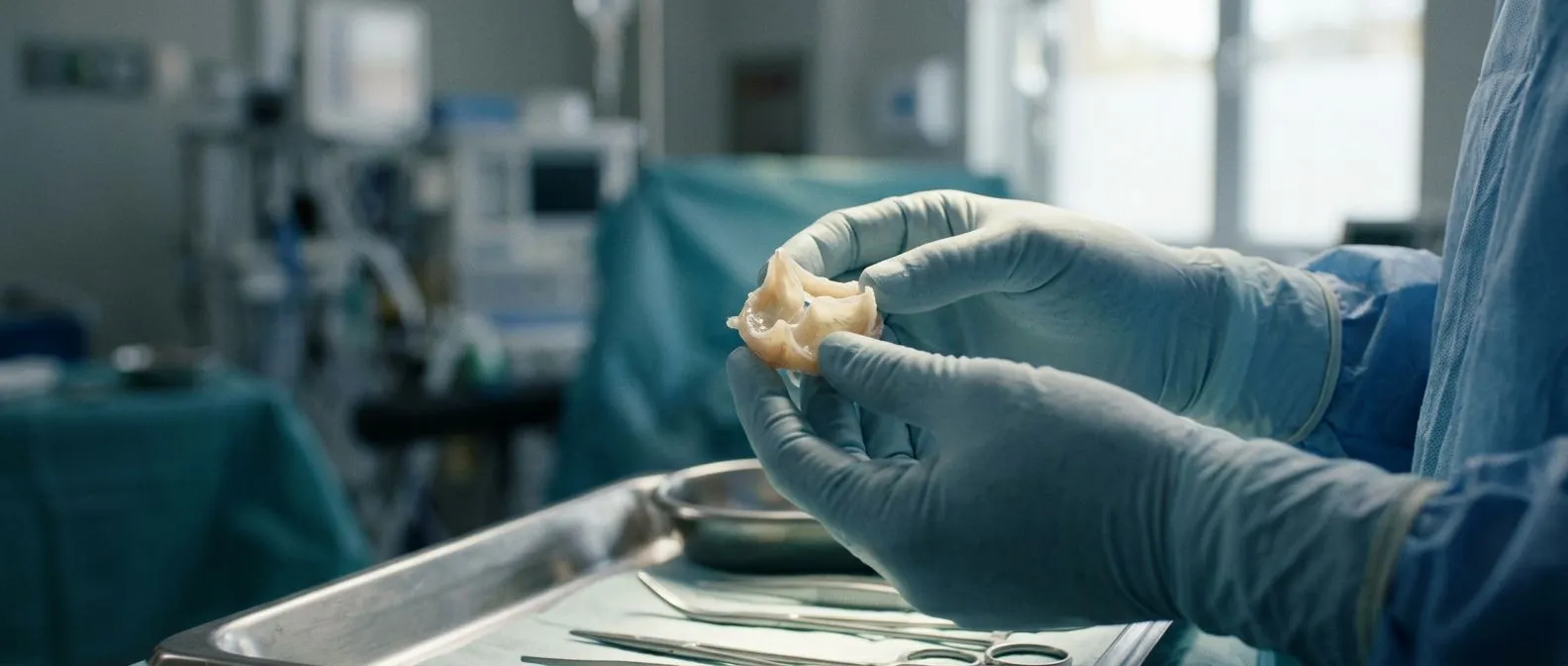
A papillary fibroelastoma is a small, benign growth that attaches to the surface of a heart valve or, less often, to the inner lining of a heart chamber. Under the microscope it looks like a sea anemone: a narrow stalk rising from the valve, crowned by fronds of loose connective tissue covered in a single layer of smooth cells. It is the second most common type of primary cardiac tumour in adults, after myxoma.
Where these tumours appear: the vast majority sit on valve tissue. The aortic valve, on the left side of the heart, and the mitral valve, which separates the left atrium from the left ventricle, are the most frequent sites. Occasionally a papillary fibroelastoma appears on a valve cusp, one of the leaflets that opens and closes with each heartbeat, or on the endocardium, the smooth lining inside the chambers.
Why it matters: even though the growth is harmless in itself, its delicate fronds can break off or trap small blood clots. Those fragments travel with the blood and can block an artery elsewhere. That is the whole reason doctors watch these tumours closely and often recommend removal, even in people who feel completely well.
How Rare Is a Papillary Fibroelastoma?
Papillary fibroelastoma is genuinely uncommon. Autopsy series suggest it may be found in a small fraction of a percent of hearts, and most of those were never discovered during life because they caused no symptoms. Diagnosis has become more frequent in recent years, simply because echocardiograms, CT scans and MRI are now routine and pick up small masses that once went unnoticed.
Rarity in numbers: among all tumours that originate in the heart, primary tumours themselves are rare, and fibroelastomas represent only a slice of those. In surgical series, they account for roughly one in ten benign cardiac tumours that are removed. For a person with no symptoms, the chance of ever having one is very small.
What this means for you: if you have been diagnosed, you are dealing with a condition most cardiologists see only occasionally, but that cardiac surgeons know well. When a valve mass is found on an echo, the first step is to confirm what it is before anyone talks about an operation.
What Are the Symptoms of a Papillary Fibroelastoma?
Most papillary fibroelastomas are silent. A large share are found by accident, during an echocardiogram performed for a murmur, chest discomfort or a routine check. The tumour itself rarely causes pain, palpitations or shortness of breath, because it usually grows slowly and does not obstruct the valve.
Warning signs to take seriously: symptoms appear when a fragment breaks loose. The most common presentations are:
- A sudden transient ischemic attack or stroke, with weakness, slurred speech or vision loss that comes on abruptly
- A new heart murmur, or a change in a murmur your doctor already knew about
- Rarely, chest pain or fainting if the tumour interferes with valve movement
Symptoms of cardiac sarcoma versus fibroelastoma: this distinction matters. A cardiac sarcoma is a malignant tumour, and its symptoms are different: it grows rapidly, invades surrounding tissue, and causes fever, weight loss and persistent fatigue along with obstruction. A fibroelastoma is a benign cardiac tumour, slow-growing, and its danger is embolism rather than invasion. The two behave nothing alike, and imaging plus pathology tell them apart.

Can Papillary Fibroelastoma Cause Stroke?
Yes, it can. This is the most significant risk of a papillary fibroelastoma, and it is the reason a diagnosis is never simply ignored. A published case of a papillary fibroelastoma of the aortic valve found in a young woman after an embolic event illustrates exactly this pattern: the tumour sat quietly on the valve until one of its fronds broke loose and travelled to the brain (Cardiovascular Ultrasound).
How embolism happens: the fronds on the tumour surface are fragile. Blood components can collect between them, forming a tiny clot, or a frond itself can detach. Either can be swept downstream with the arterial flow. If it lodges in a brain artery, the result is a stroke or a temporary attack. If it lodges in a coronary artery, the result is a heart attack.
Reducing the risk: when surgery is not immediately performed, doctors often prescribe antiplatelet or anticoagulant medication, blood thinners, to lower the chance of clot formation while the tumour is monitored. We review each case in our team meetings and discuss whether observation with medication, or removal, is the safer path for that particular person, and we always present both options.
How Serious Is a Mass on Your Heart?
Hearing the word "mass" on any heart scan is alarming, but the seriousness depends entirely on what the mass is. Heart masses fall into several very different categories, from harmless clots to malignant tumours, and a papillary fibroelastoma sits at the benign end of that spectrum.
Survival outlook: the survival rate for patients with papillary fibroelastomas is excellent once the tumour is removed. Surgical series, including a case report with a literature review of papillary fibroelastoma of the aortic valve, show that patients who undergo removal generally do well long term (Journal of Cardiothoracic Surgery). Recurrence after complete removal is rare.
What makes it serious: the danger is not the tumour growing, but the embolic risk described above. A mass that has already caused a stroke, a tumour larger than about one centimetre, or one that is mobile on its stalk, is treated more urgently. A small, asymptomatic tumour found by chance may be monitored safely in selected patients.
Getting the diagnosis right: the first step is confirming what the mass is. We take time in the consultation to explain what the studies actually show, in plain words, so the patient understands whether they are looking at a benign growth, a clot or something else before any decision is made.
How Is Papillary Fibroelastoma Diagnosed?
The diagnosis usually begins with an echocardiogram, an ultrasound scan of the heart. On the scan, a fibroelastoma appears as a small, rounded, shaggy mass attached to a valve, often with a slight shimmer as it moves with the heartbeat. Transesophageal echo, where the probe is passed down the throat, gives much sharper pictures than a scan from the chest.
Cardiac MRI and other imaging: when the echo is not definitive, cardiac MRI adds detail. A papillary fibroelastoma has characteristic features on MRI that help distinguish it from a thrombus, a myxoma or a malignant tumour. CT scanning can also help define the mass and its relationship to the valve annulus, the ring of tissue that anchors the valve.
Confirming the diagnosis: the definitive answer comes from pathology after removal, since imaging cannot be one hundred percent certain. What imaging can do is make the likelihood high enough to plan sensibly. In our team we review every imaging study together before recommending anything, because getting the diagnosis right determines whether the right plan is surgery, medication or watchful waiting.
What Are the Papillary Fibroelastoma Treatment Options?
There are two main paths after a confirmed or strongly suspected papillary fibroelastoma: surgical removal, or careful observation with medication. The choice depends on the tumour's size, mobility, location, and whether it has already caused an embolic event.
When surgery is recommended: most guidelines favour removing tumours that are symptomatic, larger than roughly one centimetre, mobile, or located on the left side of the heart, where an embolus would travel to the brain or body. Removal means the embolic risk disappears, and the valve can usually be preserved.
When observation may be reasonable: a small, immobile, asymptomatic tumour in an elderly or frail patient is sometimes watched with serial echocardiograms and blood thinners. This is a genuine option, not a compromise, and it should be discussed openly.
Minimally invasive approaches: many fibroelastomas can be removed through a small incision rather than a full sternotomy, or, in selected structural cases, via a catheter route. In our practice we perform conventional, minimally invasive, robotic and transcatheter procedures, and we recommend the less invasive route only where the evidence supports it for that patient. Dr Molina trained in robotic and structural surgery at the University of Montreal and in structural cardiology and TAVR, transcatheter aortic valve replacement, at CHU Bordeaux, and that training shapes how we weigh every option.
What Happens During Papillary Fibroelastoma Surgery?
Papillary fibroelastoma surgery is a valve-sparing procedure in most cases. The operation is performed under general anaesthesia using cardiopulmonary bypass, the heart-lung machine that temporarily takes over circulation so the surgeon can work on a still, bloodless field.
The operation itself: through a sternotomy or a smaller minimally invasive incision, the surgeon opens the relevant heart chamber or aorta, exposes the valve, and excises the tumour together with a thin layer of the tissue beneath it. The fronded head is removed in one piece where possible, so nothing is left to embolise. The underlying valve cusp is inspected and repaired if needed.
Will you need a valve replacement? Usually not. Because fibroelastomas sit on the surface rather than destroying the valve, the native valve can almost always be kept. Occasionally a Valve Degeneration case or an existing prosthetic valve is involved, and then a Valve-in-valve or repeat procedure may be considered, but for a straightforward fibroelastoma the surgeon simply shaves the tumour off and leaves your own valve working.
Before any operation: in our team every case is reviewed by the multidisciplinary group against international practice guidelines before a recommendation is given. The patient receives a clear proposal with the alternatives, and the final decision is always theirs.
What Is Recovery and Prognosis After Heart Valve Tumour Removal?
Recovery from papillary fibroelastoma removal follows the same pattern as other straightforward open-heart procedures, and for many patients it is shorter, because the valve itself is usually untouched.
The hospital stay: most patients spend one night in intensive care, then two to four more days on a regular ward. Breathing tubes come out within hours. Drainage tubes and pacing wires come out within a day or two. Walking begins the day after surgery.
The first weeks at home: expect six to eight weeks of gradual recovery. The breastbone, if divided, needs about that long to knit, so no lifting, driving or pushing during that period. Walking daily is encouraged from the start. Fatigue is normal and fades slowly.
Long-term outlook: the prognosis after surgery is very good. The tumour rarely returns, and the embolic risk is eliminated. Regular follow-up echocardiograms confirm the valve continues to work normally.
When Should You Talk to a Cardiac Team?
If you have been told there is a mass on one of your heart valves, the right next step is a conversation with a team that treats these tumours regularly, not a search for answers alone. Bring your echo report and images. Ask three questions: what do you think this is, what are my options, and what would you recommend and why.
Red flags that need urgent attention: any sudden weakness, facial droop, slurred speech or vision loss, at any time, is a medical emergency, whether or not you have a known heart mass. Call emergency services immediately.
Choosing your team and getting clarity: we encourage second opinions and treat seeking one as a normal part of deciding about heart surgery. The first contact with us is a free video consultation: we get to know the patient, talk through the condition, and say which studies are still needed so no unnecessary ones are ordered. A face-to-face visit at the consulting rooms follows when the patient decides to go ahead.
In summary, a papillary fibroelastoma is a benign heart valve tumour that usually causes no symptoms, but whose real risk is embolism, which is why it deserves a proper specialist evaluation. Treatment ranges from monitored observation to simple surgical removal, and the outlook after removal is very good. If you or a family member have been diagnosed with a papillary fibroelastoma, discuss your own situation with a qualified cardiac team before deciding anything, and consider booking a consultation so the decision is made with full information and without pressure.