Malignant Cardiac Tumour: Diagnosis and Treatment
Malignant cardiac tumour treatment from Guadalajara heart surgery specialists.
When a doctor first mentions a malignant cardiac tumour treatment plan, most families have never heard the phrase before. Cancer of the heart is genuinely rare, and it is often discovered by accident during a scan done for something else. This guide walks through what these tumours are, how they are found, what surgery and other therapies can realistically offer, and what recovery looks like afterwards. Our goal is to replace fear and guesswork with a clear picture of the decisions ahead.
Can You Have Cancer of the Heart?
Yes, you can — although the heart is one of the least likely places in the body for cancer to begin. The heart's muscle cells divide very little after childhood, and actively dividing cells are where cancers usually start. That is why primary heart tumours, meaning tumours that start in the heart itself, are uncommon compared with tumours of the lung, breast or bowel.
Why the heart resists cancer. Most heart tumours are actually "secondary" — cancer cells that travelled there from another organ, such as the lung, breast, kidney or skin. Cancer that spreads is called metastatic cancer, and the heart can be affected even though the heart muscle itself rarely becomes the origin. The National Heart, Lung, and Blood Institute describes cardiac tumours as broadly divided into benign and malignant types, with the malignant group being much less frequent but more serious.
What this means for a patient. Being told there is a mass in the heart does not automatically mean cancer. Some masses are benign myxomas or blood clots. Others prove malignant. Sorting out which one it is — safely and quickly — is exactly what a multidisciplinary cardiac team is for. In our consultation we take the time to explain what the studies actually show, in plain words, and to answer every question before any decision.
What Is a Malignant Cardiac Tumour?
A malignant cardiac tumour is a cancerous growth arising inside the heart or the pericardium, the thin sac that surrounds the heart. It can invade heart muscle, grow into the chambers, obstruct blood flow, or shed cells into the circulation. Malignant cardiac tumour treatment depends heavily on which type it is and where exactly it sits.
Primary versus secondary tumours. A primary tumour starts in the heart. A secondary, or metastatic, tumour arrives from elsewhere in the body — this type is far more common. The distinction matters because treatment priorities differ: a primary tumour may be removed surgically, while a metastatic heart tumour usually requires treating the original cancer as well.
Where tumours grow. Malignant tumours favour the right side of the heart and the pericardium. A malignant pericardial tumour can cause fluid to build up around the heart, a condition called pericardial effusion, which can squeeze the heart and needs prompt drainage. Tumours in valve areas can interfere with how valves open and close.
Related tumour types worth knowing. Rarely, a germ cell tumour — a growth arising from embryonic cells — can appear near the heart, usually in the chest between the lungs. These follow their own treatment pathways, so identifying the tumour type precisely is never optional.
What Are the Early Signs of a Heart Tumour?
Early signs are often vague, which is why these tumours are frequently missed at first. The heart keeps working, but a growing mass begins to interfere — and the symptoms depend on the mass's size and location.
- Breathlessness: shortness of breath that worsens with activity or lying flat, caused by obstruction of blood flow or fluid around the heart.
- Swelling: fluid accumulating in the legs, abdomen or around the lungs when the heart cannot fill properly.
- Palpitations or irregular rhythm: the mass irritates the heart's electrical system, producing fluttering, skipped beats or dizziness.
Symptoms that mimic other conditions. Because these signs overlap with heart failure, valve disease and pulmonary problems, patients are often treated for the wrong condition for months. Unexplained chest pain, fevers, night sweats and weight loss can also appear. If a heart mass shows up on an echocardiogram — the ultrasound scan of the heart that is usually the first test — the next step is proper characterisation, not guesswork.
Some patients feel a general fatigue that no amount of rest fixes. Others have no symptoms at all until the tumour is large. Either way, the message is the same: a mass inside the heart deserves expert evaluation, quickly but calmly.
What Is Primary Cardiac Sarcoma?
Primary cardiac sarcoma is the most common malignant tumour that starts in the heart. A sarcoma is a cancer of connective tissue — muscle, blood vessels, fat. In the heart, these tumours almost always arise from the muscle layer or the blood vessels supplying the heart, and they tend to grow quickly.
Types of cardiac sarcoma. Angiosarcoma, which arises from blood vessel cells, is the most frequent primary malignant heart tumour in adults. Others include rhabdomyosarcoma, leiomyosarcoma and undifferentiated sarcoma. Each behaves differently, and pathology — the laboratory examination of tissue — is what names it.
Why the type matters. Sarcomas are graded and subtyped because that determines whether surgery, chemotherapy, radiation, or a combination is most appropriate. Modern practice treats a cardiac sarcoma the way any soft-tissue sarcoma is treated: with a specialist team, precise imaging, and a plan agreed before the first incision. In our team every case is reviewed against international practice guidelines before a recommendation is given, and the patient receives a clear proposal with the alternatives.
The rarity of these tumours means general oncology units may see only a handful in a career. That is not a criticism — it is simply a reason to seek a centre with real experience.
What Are the Common Symptoms of Cardiac Angiosarcoma?
Cardiac angiosarcoma is the sarcoma specialists most often encounter, and it has a recognizable pattern. It usually arises in the right atrium, the upper right chamber that collects blood returning from the body, and it tends to invade the pericardium early.
The typical presentation. Patients often arrive with breathlessness, right-sided heart failure signs such as leg swelling and abdominal distension, or fluid around the heart that keeps coming back after being drained. Because the right atrium sits near the inflow of blood from the large veins, a tumour there can block venous return, producing a feeling of fullness and fatigue.
Clues that point to angiosarcoma. Recurrent pericardial effusion is the classic red flag — fluid reaccumulates because the tumour bleeds into the sac. Some patients notice haemorrhagic (bloody) pericardial fluid on drainage. Others develop symptoms of pulmonary embolism, because fragments of tumour can travel to the lungs.
None of these symptoms alone confirm angiosarcoma. But if any of them appear together — particularly recurrent bloody pericardial fluid in an adult without another explanation — imaging and biopsy follow promptly. We have seen how much anxiety the waiting period creates, and we work to shorten it: the first contact is a free video consultation where we review existing studies and say exactly which further tests are needed, so no unnecessary ones are ordered.
Cardiac Tumour Diagnosis and Treatment: Which Tests Come First?
Cardiac tumour diagnosis and treatment planning follow a sequence. Each test answers a different question: is there a mass, what does it look like, what is it made of, and can it be removed safely?
Echocardiogram first. The transthoracic echocardiogram — an ultrasound performed through the chest wall — usually discovers the mass. It shows the location, size and effect on blood flow. A transoesophageal echo, where the probe is passed down the throat, gives sharper views when needed.
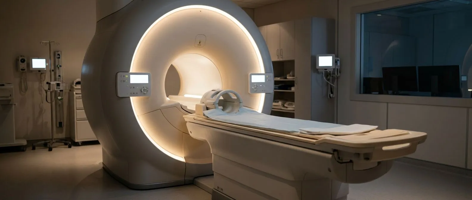
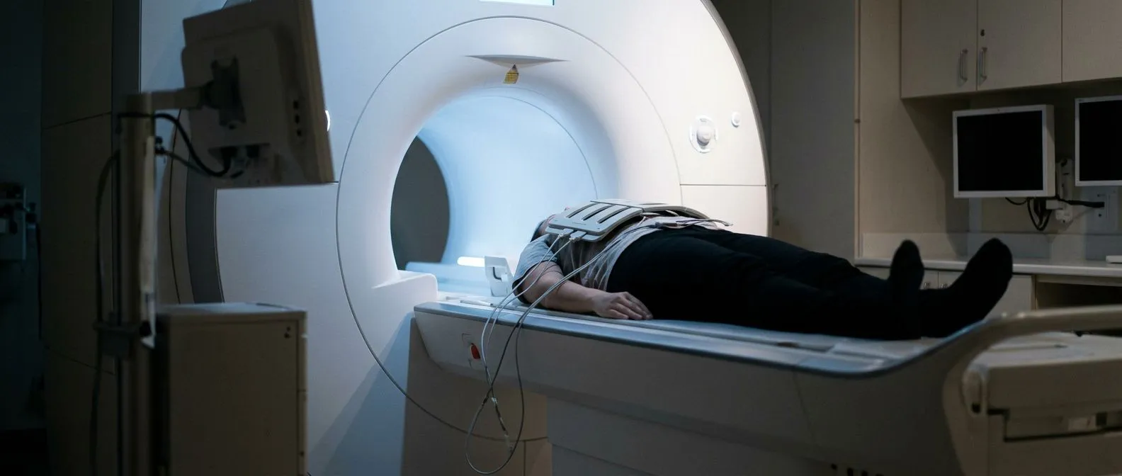
Cardiac MRI for tumour characterisation. A cardiac MRI tumour evaluation is the key next step. MRI uses magnetic fields to show tissue character in detail: fat, blood, fibrosis and active cancer look different. It also maps the tumour's exact boundaries and its relationship to valves and vessels, which surgery planning depends on. A CT scan adds information about the chest and whether other organs are involved.
Biopsy and staging. Tissue diagnosis confirms the tumour type. This may be done through a catheter-based biopsy or during surgery, depending on location and risk. Staging scans check whether the tumour has spread. For metastatic heart tumour therapy planning, knowing the original cancer matters as much as knowing the heart mass.

What the sequence looks like in practice. The steps usually run in this order:
- Echocardiogram: finds the mass and shows its effect on heart function.
- Cardiac MRI and CT: characterise the tissue, map the anatomy, and look for spread.
- Biopsy: names the tumour so the right treatment plan can be built.
A cardiac surgeon usually reviews these studies together with imaging and oncology specialists before proposing anything. The patient then receives a plain-language explanation of what the tumour is, what it is doing, and what the realistic options are.
How Does Cardiac Tumour Removal Surgery Work?
Cardiac tumour removal surgery is the mainstay of treatment for primary malignant tumours that can be resected, and it is often the only chance of cure for a localised tumour. The operation is called a Cardiac Tumour Resection, and its goal is to remove the tumour with clear margins — a rim of healthy tissue around it — while preserving as much heart function as possible.
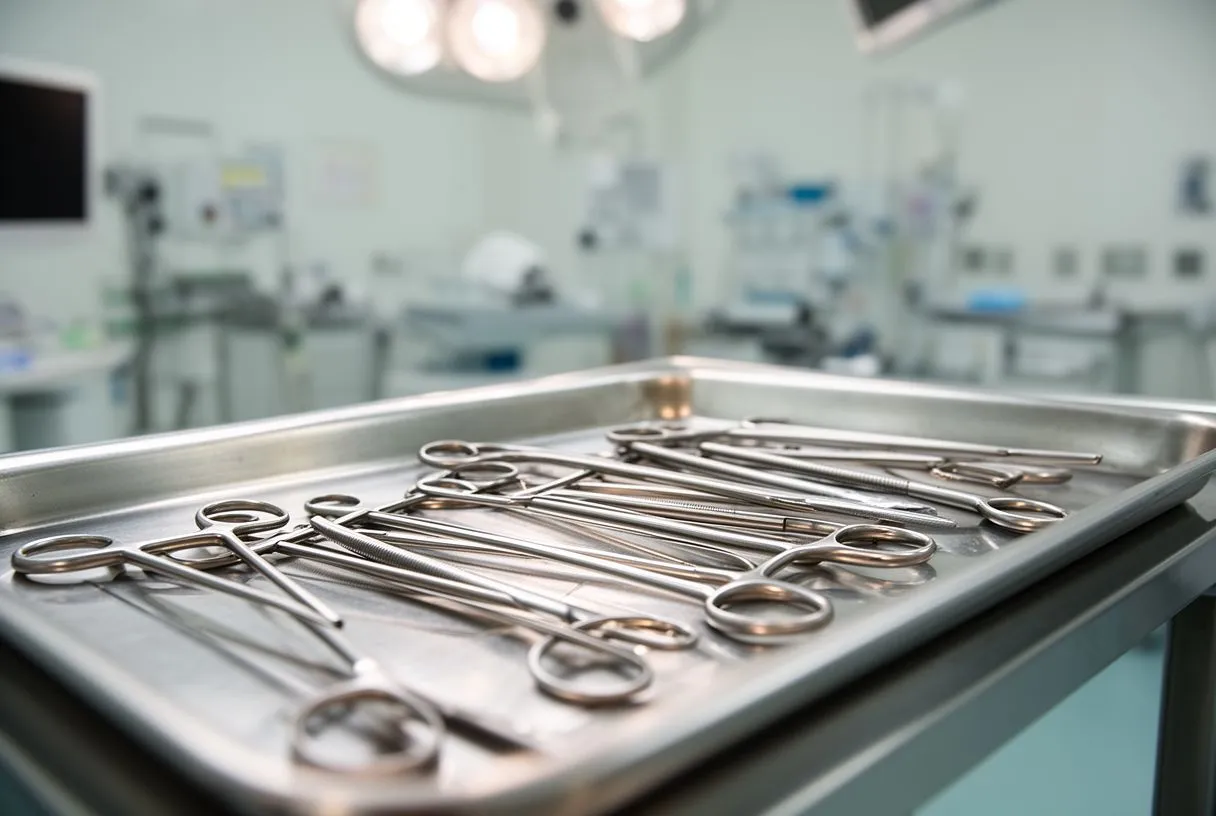
How the operation is performed. Most resections are done through a median sternotomy, an incision down the breastbone, using the heart-lung machine to circulate blood while the heart is temporarily stopped. Where anatomy allows, a minimally invasive approach through a smaller incision may be possible. In our practice we perform conventional, minimally invasive, robotic and transcatheter procedures, and we recommend the less invasive route only where the evidence supports it for that patient.
What gets rebuilt. Malignant tumours sometimes infiltrate tissue that must be removed with them. Surgeons may need to reconstruct a chamber wall with a patch, or replace or repair a valve the tumour has invaded. The operative plan is mapped on the MRI before the day of surgery.
Recovery reality. Hospital stay is typically around a week or more, longer than for simpler cardiac procedures, and full recovery takes weeks to months. Drains are removed within days, and cardiac rehabilitation — a supervised programme of gradually increasing activity — helps rebuild strength. Recovery from a cardiac tumour resection depends on whether additional treatments such as chemotherapy follow, so the surgical and oncology plans need to fit together.
Dr Molina trained in cardiothoracic surgery at the Universidad de Guadalajara and in cardiac, minimally invasive, robotic and structural surgery at the University of Montreal, and the team also trained at Jena, Bad Berka (Germany) and Buffalo (USA).
Metastatic Heart Tumour Therapy: What Are the Options?
Metastatic heart tumour therapy addresses cancer that has spread to the heart from elsewhere. Here the heart is a passenger, not the origin, and treatment follows the rules of the original cancer — lung, breast, kidney, melanoma or lymphoma.
Systemic treatment leads. Chemotherapy, targeted therapy and immunotherapy aimed at the primary cancer are the main tools. Surgery on the heart is considered when the tumour threatens immediate danger — obstructing blood flow or causing dangerous fluid — as a bridge to systemic treatment, or in select cases where the primary disease is controlled.
Managing the pericardium. When a malignant pericardial tumour or metastatic fluid build-up compromises the heart, draining the fluid brings rapid relief. A pericardial window — a small surgical opening in the sac — can prevent the fluid from reaccumulating. These are shorter procedures with a meaningful effect on quality of life.
Palliative intent is not giving up. When cure is not achievable, controlling symptoms — breathlessness, arrhythmia, fluid overload — preserves comfort and function. Guideline-based heart failure management, including the approaches set out in the 2016 ESC Guidelines for the diagnosis and treatment of acute and chronic heart failure, keeps the heart working as well as possible alongside cancer therapy.
When cancer is involved, any decision about operating on the heart should be made together with the treating oncologist, because the timing of surgery and systemic treatment can change outcomes.
Is Heart Cancer Curable?
It depends on the type and stage, and honest answers matter more than hopeful ones here. A primary malignant cardiac tumour that is found early, has not spread, and can be completely removed has a real chance of long-term control — and complete surgical removal is the single most important factor in survival for primary cardiac sarcoma.
What improves the odds. Early detection before the tumour invades surrounding structures, complete resection with clear margins, and effective follow-up treatment all shift the balance. Some patients undergo combined treatment — surgery followed by chemotherapy or radiation — to reduce the risk of the tumour returning. Cardiac tumour recurrence prevention relies on regular surveillance imaging after treatment, typically echocardiograms and periodic MRI, so that any regrowth is caught while it is still small.
What limits cure. Some tumours are diagnosed after they have spread to the lungs or other organs, or after they have grown into structures that cannot safely be removed. Even then, meaningful treatment exists: debulking surgery, systemic therapy and symptom control can extend and improve life. Primary malignant cardiac tumour treatment is individually planned because two patients with the same diagnosis can face very different situations.
Where the diagnosis is a glioma or another brain tumour rather than a cardiac one, specialist oncology guidelines such as the EANO guidelines on the diagnosis and treatment of diffuse gliomas of adulthood shape that treatment — a reminder that tumour care today is highly specialised by type, and the same is true in the heart.
Choosing a Heart Tumour Specialist in Guadalajara: What to Ask
Finding a heart tumour specialist in Guadalajara — or anywhere — comes down to three things: experience with complex cardiac surgery, genuine multidisciplinary review, and clear communication. Because these tumours are rare, the questions you ask matter as much as the answers.
Questions worth asking at any consultation. Bring these with you, in writing if it helps:
- Experience: how many cardiac tumour resections has this team performed, and what was the pathology in those cases?
- The plan: what exactly is the proposed operation, what alternatives were considered, and why was this one chosen?
- Coordination: who will manage chemotherapy or radiation, and how do the two teams communicate?
Second opinions are normal. We encourage second opinions and treat seeking one as a normal part of deciding about heart surgery — no reputable team should make you feel otherwise. A responsible surgeon will tell you plainly if a tumour is not operable and what the alternatives are.
Our approach at Cirugía de Corazón. We are a multidisciplinary team of cardiac surgeons led by Dr Alejandro Molina, with consulting rooms in Guadalajara, Querétaro and Hermosillo. Every case is reviewed by the team against international practice guidelines before a recommendation is made, and the final decision is always the patient's. The first contact is a free video consultation: we get to know the patient, talk through the condition, and identify exactly which studies are still needed — no unnecessary testing. Dr Molina also performed the first robotic mitral valve repair in the Ángeles Health System and the first robotic heart surgery in Guadalajara, and the team trained at Jena, Bad Berka (Germany) and Buffalo (USA).

Malignant cardiac tumour treatment is rare territory, but it is not uncharted. Diagnosis follows a logical sequence — echo, MRI, biopsy — and treatment combines surgery, oncology and careful follow-up. The most important steps are an accurate diagnosis and a team that explains every option honestly before anything happens. If you or a loved one has been told there is a mass in the heart, do not self-diagnose and do not wait in silence: book a consultation with a qualified cardiac team, bring every study you have, and make sure your questions are answered before you decide. Dr Molina and our team are available for a free video consultation to start that conversation.