

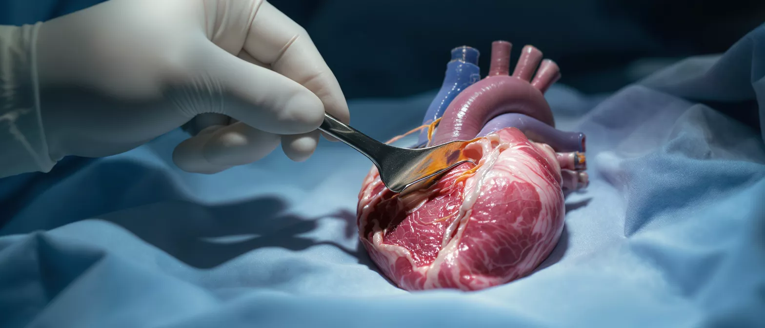
Cardiac Tumour Resection
Learn how cardiac tumour resection removes heart tumours.
What is cardiac tumour resection
Cardiac tumour resection is a highly specialised surgical procedure performed to remove an abnormal mass or growth from within the heart. When a patient hears the word "tumour," it is entirely natural to feel a wave of anxiety, but it is deeply reassuring to understand that the vast majority of primary growths found in the heart are entirely benign. The goal of this surgery is to safely excise the mass, restore normal blood flow, and protect the overall function of the heart.
This procedure demands exceptional precision and advanced surgical expertise. Because the heart is a constantly moving, highly pressurised organ, operating on it requires a carefully controlled environment. Surgeons use sophisticated techniques to temporarily stop the heart's beating, diverting the patient's blood circulation through a heart-lung machine. This allows the surgical team the clear, motionless field needed to meticulously remove the tumour without damaging the surrounding delicate cardiac tissues.
Understanding the fundamental nature of this surgery helps alleviate much of the fear patients and their families experience. By definition, a resection aims to completely cut away the problematic tissue while preserving the maximum amount of healthy heart muscle. It is a testament to modern cardiac oncology and surgical advancements that these once-daunting procedures are now performed with remarkable success rates and positive long-term outcomes.
Why heart tumour removal is necessary
Preventing blood flow obstruction: As a tumour grows inside the heart's chambers, it can physically block the flow of blood, leading to fainting, shortness of breath, or even sudden cardiac arrest. Removing the mass is critical to restoring unobstructed circulation and ensuring the heart can pump efficiently to the rest of the body.
Eliminating the risk of embolism: Fragile, gelatinous tumours, particularly myxomas, can shed small fragments into the bloodstream. These fragments can travel to the brain, lungs, or limbs, causing severe complications like a stroke. Surgical heart tumour removal completely eliminates this risk by extracting the entire mass before it can fragment.
Beyond the immediate mechanical dangers, leaving a tumour untreated allows it to continuously grow and further interfere with the heart's electrical pathways, causing dangerous arrhythmias. Whether the mass is benign or malignant, the physiological strain it places on the cardiovascular system makes surgical resection the most definitive and effective treatment available.
How cardiac tumour surgery works
The process begins with the patient under general anaesthesia. The surgical team makes an incision to access the heart, typically through the sternum in a traditional open-heart approach. Once the chest is open, the patient is connected to a cardiopulmonary bypass machine. This specialised equipment temporarily takes over the job of the heart and lungs, oxygenating the blood and pumping it through the body so the heart can be safely stopped and emptied for the procedure.
With the heart still and protected, the surgeon carefully opens the appropriate chamber to locate the tumour. The surgical team utilises high-resolution imaging and direct visualisation to ensure they have clear margins around the mass. The tumour is meticulously cut away from the heart wall or valve, taking care not to damage the underlying cardiac structures. Sometimes, a small piece of healthy tissue surrounding the base is also removed to ensure absolutely no tumour cells are left behind.
After the tumour is fully extracted, the surgeon repairs any defects in the heart wall or septum, often using a small synthetic or biological patch. The heart is then closed, the bypass machine is gradually weaned off, and the heart resumes its normal beating rhythm. The removed tissue is immediately sent to a pathology laboratory for detailed analysis to determine its exact cellular nature.
Types of heart tumours treated
Cardiac tumours are generally categorised by their origin: primary tumours, which begin within the heart itself, and secondary tumours, which have spread from elsewhere in the body. Understanding the specific type of growth heavily influences the surgical strategy and the long-term medical outlook.
Myxomas and benign masses: Myxomas are the most common primary heart tumour in adults. They typically arise in the left atrium and are attached to the wall by a stalk. Though benign, their physical presence poses significant risks of stroke and mechanical obstruction, making prompt surgical removal necessary.
Malignant cardiac masses: Malignant tumours, such as cardiac sarcomas, are rarer and more aggressive. Resecting these masses is significantly more complex, often requiring the removal of surrounding cardiac tissue to achieve clear margins. In these cases, heart tumour treatment options often combine surgery with systemic therapies to manage the broader disease effectively.
Heart tumour diagnosis and imaging
Accurately identifying a cardiac mass is the crucial first step before any surgical intervention is planned. Because the symptoms of a heart tumour can mimic valve disease or heart failure, advanced imaging is required to confirm the presence, location, size, and attachment points of the growth.
- Echocardiogram: Uses sound waves to create a moving picture of the heart, allowing surgeons to see the tumour's size and how it affects blood flow in real time.
- MRI (Magnetic Resonance Imaging): Provides highly detailed, cross-sectional images of the heart's soft tissues, helping to distinguish a benign myxoma from a malignant sarcoma.
- CT Scan: Offers precise anatomical mapping of the chest and heart structures, which is vital for planning the physical surgical approach.
Once imaging confirms the presence of a mass, the cardiac surgery team analyses the data to determine if intracardiac tumour removal is the safest and most effective course of action. This meticulous diagnostic phase ensures that the surgical plan is perfectly tailored to the patient's unique anatomy.
Cardiac tumour symptoms to watch for
The symptoms of a cardiac mass often depend entirely on its location within the heart. A growth in the left atrium might cause mitral valve obstruction, leading to shortness of breath, especially when lying flat. Conversely, a tumour in the right side of the heart might block blood returning from the body, causing swelling in the legs and abdomen.
Systemic and embolic signs: Many patients present with generalised symptoms such as unexplained fever, profound fatigue, joint pain, and unintended weight loss. These are often caused by the body's immune response to the tumour. Additionally, if tiny pieces of the tumour break off, they can cause sudden, unexplained strokes or transient ischemic attacks (TIAs).
It is important to remember that these symptoms can overlap heavily with more common cardiac conditions like heart failure or valve disease. However, when standard treatments fail to resolve these issues, or when imaging incidentally reveals a mass, it triggers a rapid referral to a cardiac surgery specialist for further evaluation.
Recovery time after myocardial tumour resection
The immediate recovery after cardiac tumour surgery begins in the intensive care unit (ICU). Here, the patient is closely monitored for the first 24 to 48 hours to ensure the heart is beating strongly and that no bleeding occurs. Once stabilized, the patient is moved to a standard cardiac ward to begin light movement and breathing exercises, which are critical for preventing post-operative complications like pneumonia.
The total cardiac tumour recovery time varies from patient to patient, but a typical hospital stay ranges from five to seven days following an open resection. Full recovery, including the healing of the breastbone and the return of normal energy levels, generally takes six to eight weeks. During this period, patients must adhere to strict lifting restrictions and slowly ease back into their daily activities under their surgeon's guidance.
Cardiac rehabilitation plays a vital role in regaining strength and confidence. Patients are often enrolled in a supervised exercise program designed to safely elevate the heart rate and rebuild cardiovascular endurance. Choosing a specialist cardiac surgeon in Mexico ensures access to excellent post-operative care and rehabilitation support throughout the healing journey.
Risks and safety of cardiac mass removal
Like all major cardiac procedures, tumour resection carries inherent surgical risks. These can include bleeding, infection at the incision site, and adverse reactions to anaesthesia. Additionally, because the heart must be temporarily stopped and manipulated, there is a small risk of damage to the heart muscle, valves, or the cardiac electrical system, which could require a pacemaker post-operatively.
Preventing tumour fragmentation: The most critical safety concern during the operation is the inadvertent shedding of tumour fragments into the bloodstream. Surgeons mitigate this risk by carefully handling the tumour, removing it intact within a protective bag whenever possible, and thoroughly irrigating the heart chambers before closing.
Despite these risks, cardiac mass removal is generally considered a highly safe and definitive procedure when performed by an experienced surgical team in a dedicated cardiac centre. The benefits of removing a tumour—preventing strokes, resolving heart failure symptoms, and eliminating the risk of further mechanical obstruction—far outweigh the surgical risks for the vast majority of patients.
Life after benign heart tumour surgery
For patients who have had a benign tumour, such as a myxoma, completely removed, the long-term prognosis is exceptionally positive. Once the mass is excised, the mechanical obstruction is immediately resolved, and the heart can return to its normal pumping function. The systemic symptoms, such as fever and fatigue, typically disappear entirely within a few weeks of the surgery.
While the risk of a benign myxoma returning at the exact site is incredibly low, there is a small possibility of developing a new primary tumour in the future. Therefore, patients will undergo periodic echocardiograms as an outpatient to monitor the heart's health. These routine check-ups are standard, preventative measures designed to offer long-term peace of mind.
Most patients return to their normal lives, work, and physical activities without any lingering restrictions once fully healed. The psychological relief of knowing the tumour is gone, combined with the restoration of physical energy, often leads to a profound improvement in overall quality of life. Many patients find it helpful to seek out a post-surgery cardiac support resource to navigate their emotional and physical recovery confidently.
Finding a cardiac tumour specialist in Guadalajara
When facing a cardiac tumour diagnosis, the expertise of the surgical team is the most critical factor in ensuring a successful outcome. Dr Alejandro Molina and the dedicated team at Cirugía de Corazón possess the highly specialised training and advanced technological resources required to perform these delicate resections with exceptional precision and care.
Choosing to undergo heart surgery is a monumental decision, but patients in Mexico and from abroad have access to world-class cardiac care right in Guadalajara. The facility is equipped with state-of-the-art imaging and surgical theatres, ensuring that every aspect of the procedure, from diagnosis to recovery, meets the highest international standards of cardiac oncology.
If you or a loved one has been diagnosed with a cardiac mass, do not delay in seeking expert evaluation. Early diagnosis and prompt surgical intervention are the keys to preventing severe complications. Contact our clinic today to schedule a comprehensive consultation and learn how our expert surgical team can safely guide you through this procedure and back to a healthy, active life.






