Cardiac Tumour Surgery: Diagnosis and Removal
Cardiac tumour surgery explained by Guadalajara specialists—diagnosis, removal options, recovery timeline, and costs in Mexico.
Hearing the word "tumour" anywhere near the heart is frightening, and most people have never heard of a cardiac tumour before the day a scan shows one. The good news is that many heart tumours are benign, meaning they are not cancer, and cardiac tumour surgery to remove them is a well-established procedure with a long track record. This guide walks you through what these tumours are, how they are found, what the operation involves, and what recovery actually looks like, so you can face the conversation with your cardiac team from a position of understanding rather than fear.
What Is a Cardiac Tumour?
A cardiac tumour is an abnormal growth of tissue inside the heart or attached to its valves and chambers. Tumours in the heart are rare compared with elsewhere in the body, partly because heart muscle cells divide very little after birth. Doctors divide them into two broad groups: primary tumours, which start in the heart itself, and secondary tumours, which are cancer that has spread from another organ.
Primary heart tumours are mostly benign. The most common primary tumour in adults is the myxoma, a soft, gelatinous growth that is not cancer. Despite being benign, it can still cause serious problems by blocking blood flow or shedding fragments into the bloodstream, which is why removal is usually recommended once it is found. Rare primary tumours can also arise from congenital origins, such as a Germ Cell Tumour, which occasionally presents in the chest near the heart.
Secondary tumours are more common overall. Cancers from the lung, breast, kidney, or lymphatic system can reach the heart through the bloodstream or by direct spread. When that happens, treatment usually focuses on the original cancer, with heart surgery playing a supporting role rather than the lead one.
Cardiac tumours in children have their own distinct behaviour and spectrum of types, and specialists note that the picture in paediatric patients differs from adults in both the tumour types involved and the urgency of treatment, as reviewed in the literature on Cardiac tumours in children.
What Are the Symptoms of a Heart Tumour?
Heart tumours are masters of disguise. They often mimic far more common conditions, which is why diagnosis is frequently delayed. The symptoms depend mainly on where the tumour sits and how big it is.
Obstruction symptoms. A tumour inside a pumping chamber, especially the left atrium, can partly block the flow of blood through the mitral valve. This can cause breathlessness on exertion, fainting spells, or a feeling of the heart racing, and it can be mistaken for valve disease or heart failure.
Embolic symptoms. Some tumours shed small fragments, or tiny clots form on their surface, and these travel to the brain, limbs, or other organs. A stroke in a young, otherwise healthy person with no obvious risk factors is one of the classic ways a hidden heart tumour first announces itself. An echocardiogram is usually requested when a younger patient has an unexplained embolic event, because it can change the whole diagnostic picture.
General and constitutional symptoms. Fever, unexplained weight loss, fatigue, joint aches, and a raised inflammatory marker on blood tests can all accompany a heart tumour, especially a myxoma. Many patients are investigated for months for infection or rheumatologic disease before the heart is imaged. Some people also notice a change in how they feel lying flat, when the tumour shifts position inside the chamber.
How Are Cardiac Tumours Diagnosed?
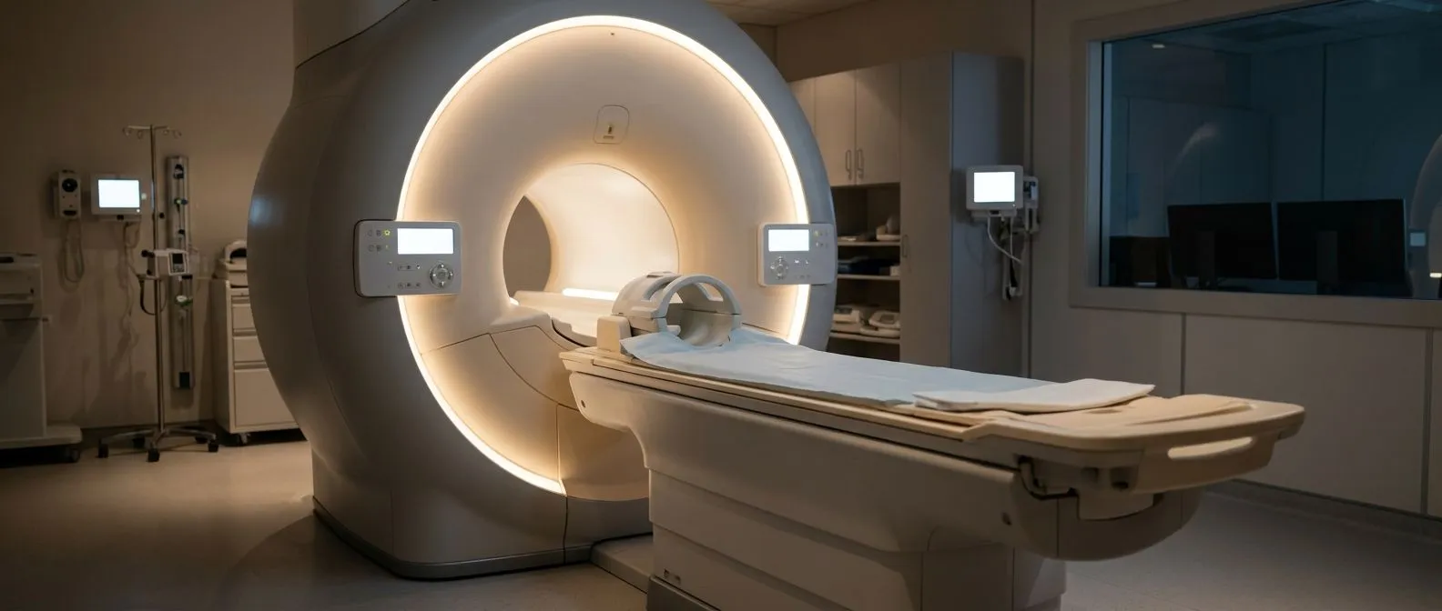
Diagnosis almost always begins with an echocardiogram, the ultrasound scan of the heart. It is painless, uses no radiation, and will show the location, size, and movement of most tumours. Two versions exist: a standard scan from the chest wall, and a transoesophageal echo (TOE), where a thin probe is passed down the food pipe to obtain far clearer pictures of the posterior chambers and valves.
Advanced imaging refines the plan. Once an echo shows a mass, cardiac MRI or CT scanning usually follows. MRI is especially useful because different tissue types look different, helping the team predict whether a mass is a benign myxoma, a blood clot, or something malignant before surgery. CT adds detail about the coronary arteries, which matters if a bypass graft may be needed during the operation.
Blood tests and biopsy have a limited role. Tumour markers can point toward certain rare diagnoses, and in selected cases a small tissue sample may be taken, but most masses in the heart are characterised by imaging and confirmed by pathology after removal. Every case we see is reviewed by the multidisciplinary team against international practice guidelines before we recommend any specific imaging pathway, so patients get the studies they need and not the ones they don't.
Types of Cardiac Tumours and Which Ones Need Surgery
Not all heart masses are the same, and the treatment decision depends heavily on the type. Here are the main categories and the usual approach to each:
- Myxoma: the most common benign adult tumour, usually in the left atrium on a small stalk; almost always removed surgically because of the risk of stroke and obstruction.
- Papillary fibroelastoma: a small, frond-like benign growth often attached to a valve; monitored if tiny, removed if it has caused symptoms or is large.
- Rhabdomyoma: the most common paediatric tumour, associated with tuberous sclerosis; many shrink on their own, so surgery is reserved for those causing obstruction or rhythm problems.
- Sarcoma: a malignant primary tumour requiring prompt tissue diagnosis and usually combined treatment, as outlined in the section on malignant disease below.
Classification matters because it sets expectations. A benign myxoma removed completely is very often cured. A malignant tumour needs a broader strategy involving cancer specialists. The literature on cardiac tumours in children shows that paediatric and adult disease follow different natural histories, which is why age-appropriate specialist review matters.
Is a Mass on a Heart Valve Dangerous?
This is one of the most common worries patients bring to a consultation, and the honest answer is: it depends on what the mass is. Not every mass on a valve is a tumour.
Valve masses can be several things. A vegetation from infective endocarditis (a bacterial infection of the valve) is far more common than a tumour, and it looks similar on ultrasound. Thrombus, a blood clot, can also sit on a valve, especially on mechanical prostheses. Distinguishing between these matters because the treatments are completely different: infection needs antibiotics and sometimes valve surgery, a clot needs anticoagulation, and a papillary fibroelastoma usually needs observation or simple excision.
Papillary fibroelastomas rarely grow and rarely embolise. These small benign tumours, most often on the aortic or mitral valve, are usually discovered by chance. Current practice is to observe small, symptom-free ones with periodic echo, while larger ones, or any that have caused a stroke or transient symptoms, are removed. The surgery itself is usually straightforward and the valve is almost always preserved.
What makes a valve mass urgent? Rapid growth, associated fever, stroke or limb symptoms, or interference with valve function all shift the timeline from watchful waiting to prompt treatment. When we review a valve mass in the consultation, we take the time to explain in plain words what the echo actually shows and which of these features, if any, are present, before any decision about surgery.
How Cardiac Tumour Removal Surgery Works
Cardiac tumour resection is performed under general anaesthesia in a hospital theatre equipped for open-heart procedures. The standard approach is a median sternotomy, an incision down the breastbone that gives the surgeon direct access to the heart.
The heart-lung machine takes over. During the operation, a pump-oxygenator circuit temporarily does the work of the heart and lungs, allowing the surgical team to stop the heart and open a chamber safely and without blood obscuring the view. For tumours of the left atrium, the surgeon enters through the left atrium, visualises the mass, and removes it together with its stalk and a small margin of the attached wall, because myxomas can regrow if any base tissue is left behind.
Valves and chambers are checked at the same time. Once the tumour is out, the team inspects all four valves and the other chambers for additional lesions, since a small minority of patients have more than one tumour. The heart is then closed, weaned off the bypass machine, and the chest is closed. Typical operating time runs from two to four hours depending on the tumour's location and whether any valve work is needed.
Pathology is the final step. Every removed specimen is examined by a pathologist, who confirms the type and confirms the margins are clear. Dr Molina has performed the first robotic mitral valve repair in the Ángeles Health System, and the same precision we bring to valve work is what we apply to tumour removal, especially when the tumour sits near the mitral valve.
Minimally Invasive and Robotic Cardiac Tumour Surgery
Open sternotomy is not the only option anymore. For suitable tumours, minimally invasive approaches avoid cutting the breastbone entirely.
Minimally invasive access uses a small side incision. A right mini-thoracotomy, an incision of a few centimetres between the ribs on the right side, gives access to the left atrium for tumour removal. Recovery is faster, pain is less, and the cosmetic result is far better. Not every tumour is suitable, however: large masses, tumours in awkward positions, or patients needing simultaneous bypass may still require the standard approach.
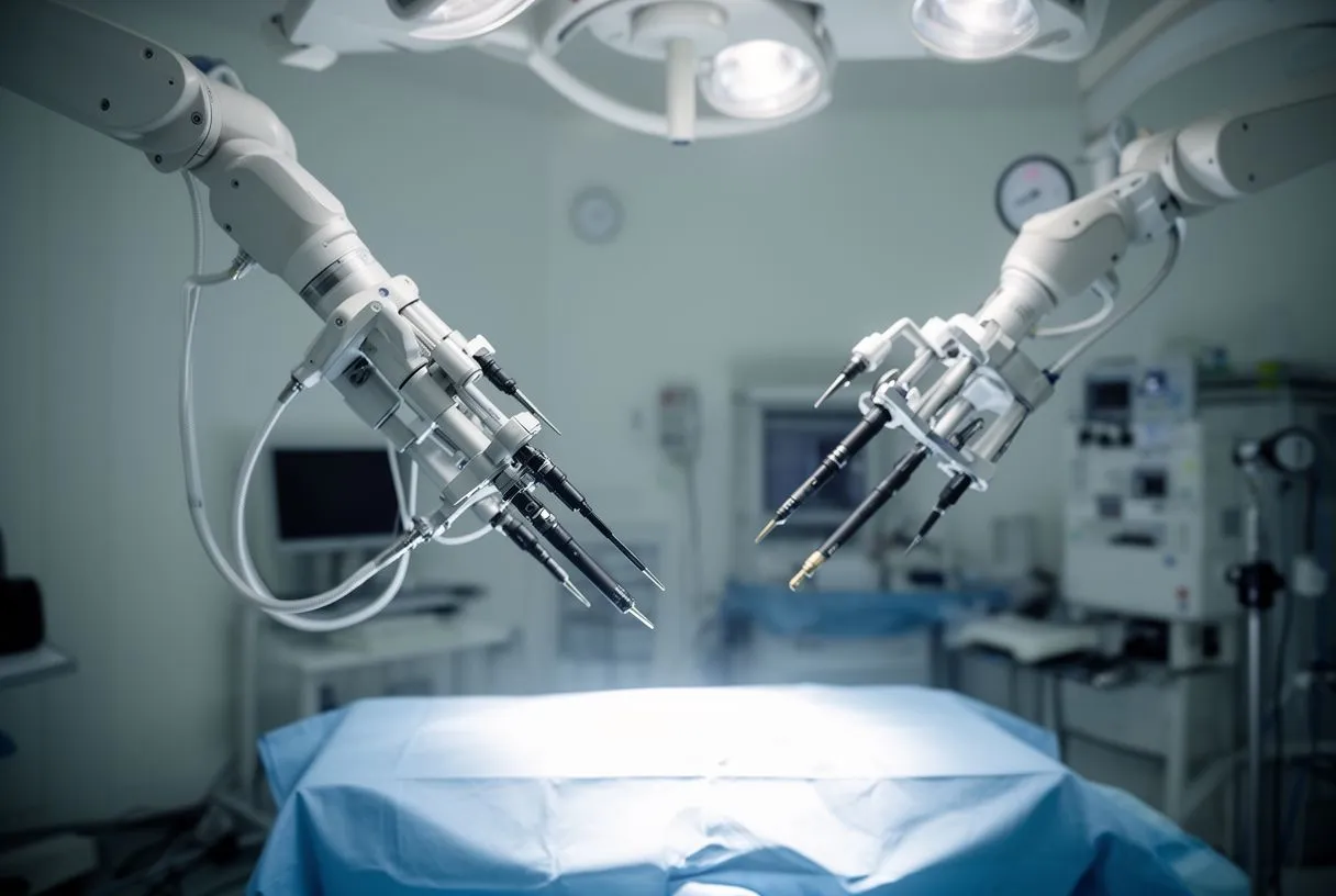
Robotic surgery adds precision. With the robotic system, the surgeon operates through tiny port incisions while seated at a console that translates hand movements into instruments inside the chest. We recommend the less invasive route only where the evidence supports it for that specific patient, which is a decision we make case by case.
How do you know which approach you will have? The choice depends on tumour size, location, and your overall health. This is exactly the kind of question a second opinion is designed to answer, and seeking one is a normal, sensible part of deciding about heart surgery.
What Is Cardiac Tumour Surgery Recovery Really Like?
Recovery after cardiac tumour surgery follows the same broad arc as other open-heart operations, and knowing the milestones in advance makes the whole process less intimidating.
The first days are in hospital. Most patients spend one to two nights in an intensive care unit and then a few more on a regular ward. You will be encouraged to sit up and walk short distances within the first day or two; early mobilisation genuinely speeds recovery and reduces complications. Breathing exercises and chest physiotherapy are part of the daily routine.
Weeks one to six are the healing phase. A sternotomy takes about six to eight weeks for the breastbone to knit, so during that time you should avoid lifting anything heavy, driving until your team clears you, and any activity that strains the chest. Patients who had minimally invasive surgery usually return to desk work and normal light activities sooner.
Follow-up is lifelong but light. Because myxomas can rarely recur, periodic echocardiograms are scheduled, typically annually at first and then less often. If the pathology shows a malignant tumour, you will also be referred to an oncology team. Follow-up after tumour removal usually includes echocardiograms at regular intervals, and it helps to know at discharge exactly when the next check will be.
Are There Risks of Malignant Cardiac Tumour Surgery?
Being honest about risk is part of good care, so let's set expectations clearly. For benign tumours such as myxoma, complete surgical removal is usually curative and complications are uncommon. For malignant tumours, the picture is more complicated.
Surgical risks are those of any open-heart operation. These include bleeding, infection, stroke, heart rhythm disturbances, and reactions to the bypass circuit. Overall risk depends heavily on your age, other health conditions, and the complexity of the operation, and your surgical team should quote figures from their own unit rather than generic averages.
For cardiac sarcoma, prognosis depends on type and spread. Cardiac sarcoma is a rare malignant tumour of the heart, and life expectancy varies widely depending on how far it has spread at diagnosis and how it responds to treatment. Questions like "what is the average life expectancy for someone with cardiac sarcoma?" have no single honest answer; outcomes range from months to years, and specialist oncology input shapes the treatment plan alongside surgery.
Chemotherapy and the heart interact. Some cancer drugs, notably anthracyclines and certain targeted therapies, can weaken the heart muscle over time, a condition called chemotherapy-related cardiotoxicity. This is why patients receiving these treatments have heart function monitored with echo before, during, and after therapy. The relationship between chemotherapy and heart problems is well documented in the oncology literature, and recognising it early allows medication to protect the heart to be started in time. Major medical publications such as The BMJ regularly cover this overlap between cancer care and cardiovascular medicine.
Cardiac tumour surgery should only be performed in centres with cardiac surgery capability, an intensive care unit, and a multidisciplinary team that includes cardiology, imaging, anaesthesia, and pathology. In Mexico, dedicated cardiac surgery programmes exist in major cities.
What drives the cost? The price of heart tumour removal depends on the surgical approach (open versus minimally invasive or robotic), the hospital tier, the length of stay, and whether valve repair is needed at the same time. A robotic case involves equipment costs that a standard operation does not. Any centre worth its name will give you a written, itemised estimate after reviewing your imaging, and beware of anyone who quotes a firm price without seeing your echo.
Questions to ask any team you are considering:
- Volume and approach: how many cardiac tumour removals does the team perform, and which approaches do they offer?
- Team structure: is your case reviewed by a multidisciplinary team against international guidelines before a recommendation is made?
- Transparency: will you receive a clear written proposal, including alternatives and the option of a second opinion?
Our team, led by Dr Alejandro Molina with consulting rooms in Guadalajara, Querétaro and Hermosillo, reviews every case against international practice guidelines before making a recommendation, and the final decision is always the patient's. The first contact is a free video consultation, where we get to know you, talk through your condition, and identify exactly which studies are still needed so no unnecessary ones are ordered; a face-to-face visit at the consulting rooms follows when you decide to go ahead. If you have been told you have a cardiac mass, do not wait for symptoms to decide for you. Bring your imaging, ask your questions, and let a qualified cardiac team help you weigh the options, starting with a consultation rather than a self-diagnosis.