

Echocardiogram
Learn what an echocardiogram shows, how it works, types, preparation, and costs.
What is an echocardiogram?
An echocardiogram is a non-invasive medical test that uses sound waves to produce detailed moving images of your heart. By bouncing high-frequency ultrasound waves off the structures inside your chest, this technology allows your cardiologist to see exactly how your heart muscle and valves are functioning in real time. It is a fundamental diagnostic step in evaluating overall cardiac wellness, providing crucial data without using radiation.
What does an echocardiogram show? The resulting images display the size and shape of your heart, the thickness of its muscular walls, and the internal chambers. Most importantly, it visualizes the four heart valves in motion, revealing whether they are opening fully and closing tightly to maintain proper blood flow. This clear visual mapping is essential for identifying structural heart disease before, during, or after surgical interventions.
For patients preparing for cardiac surgery, this diagnostic imaging provides your surgical team with a precise roadmap. It helps surgeons understand exactly what needs to be repaired or bypassed, ensuring the highest level of precision. By establishing a clear baseline of your cardiac anatomy, your medical team can safely plan the most effective path forward for your treatment.
How does an echocardiogram work?
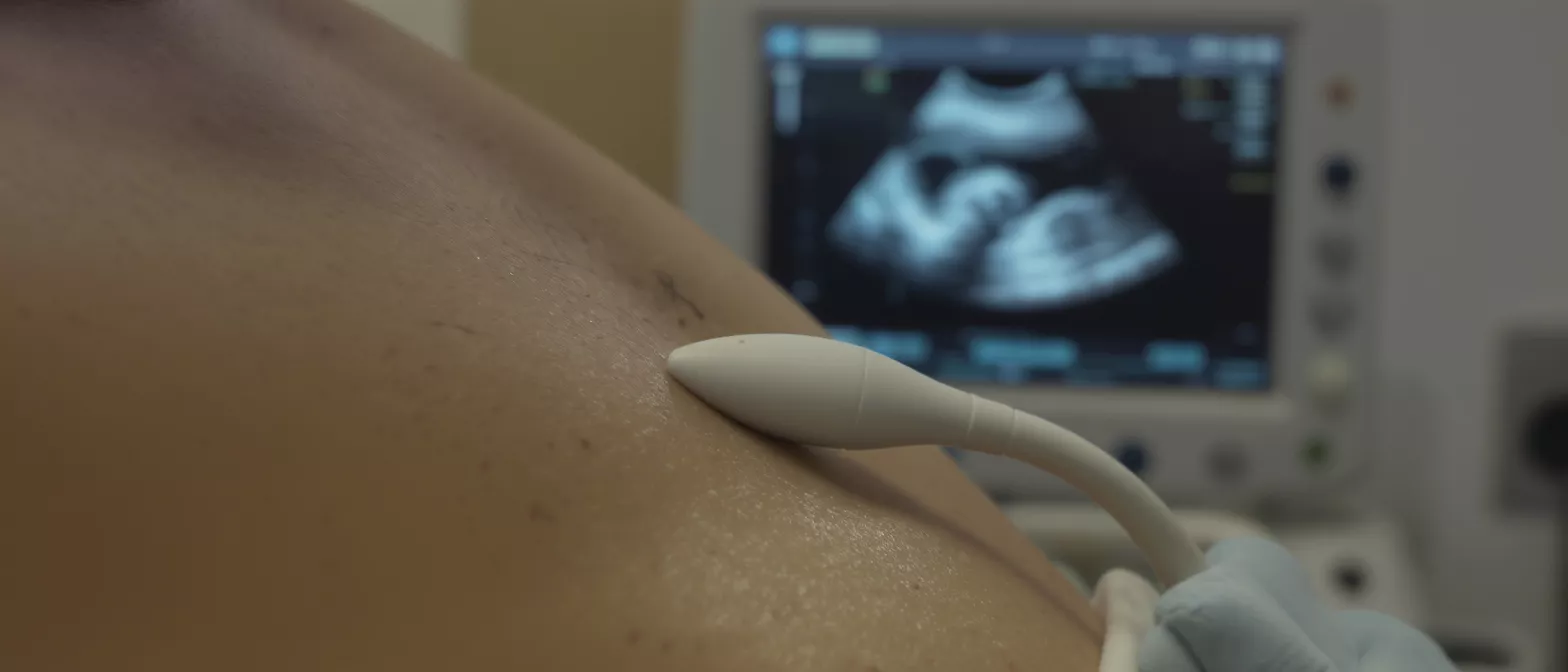
How does ultrasound create heart images? The echocardiogram procedure relies on a small handheld device called a transducer, which a technician glides across the skin of your chest. This probe emits brief pulses of sound waves that are far too high for the human ear to detect. As these harmless sound waves travel inward, they bounce off the solid structures of your heart and return to the transducer as echoes.
How are sound waves translated into video? A specialized computer inside the ultrasound machine measures the exact amount of time it takes for each sound wave to return, calculating the distance and density of the tissue it struck. This instant mathematical translation generates a detailed, real-time video feed on a monitor. The machine effectively maps a three-dimensional structure using the reflected acoustic data.
Because the heart is constantly in motion, the continuous stream of sound waves captures the rhythmic pumping, relaxing, and valve snapping with incredible accuracy. A Doppler echocardiogram enhances this process by tracking the speed and direction of blood flow, often displaying it as color overlays on the black-and-white image. This dynamic, functional view is what sets cardiac ultrasound apart from static imaging techniques.
Types of echocardiogram
What is a transthoracic echocardiogram? This is the most common and standard type of heart echo test. The technician places the transducer directly on the outside of the chest wall to capture images through the ribs. It is entirely painless, requires no needles, and typically takes about thirty to forty-five minutes to complete in a standard outpatient setting.
When a closer look is necessary, cardiologists may use a transesophageal echocardiogram (TEE). Because the esophagus sits directly behind the heart, passing a specialized, thin transducer down the throat provides an exceptionally clear, unobstructed view. This approach is frequently used to search for tiny blood clots or evaluate complex valve issues before surgery. A stress echocardiogram combines standard imaging with a physical or pharmacological stress test to see how the heart performs under exertion.
- Transthoracic: Standard, non-invasive imaging through the chest wall.
- Transesophageal: Detailed imaging via the esophagus for complex structures.
- Stress echo: Evaluates heart function during physical exertion or medication-induced stress.
Echocardiogram vs ECG
What is the difference between an echocardiogram and an ECG? While both are vital heart health checks, they measure entirely different things. An electrocardiogram (ECG or EKG) strictly records the electrical activity of the heart, tracking the timing and rhythm of your heartbeat via electrodes placed on the skin. It tells the doctor how the heart's internal wiring is functioning.
In contrast, an echocardiogram produces actual moving pictures of the heart's physical structures. While the ECG can tell a doctor that the heart is beating irregularly, the heart imaging test shows them why—perhaps revealing a damaged valve or a weakened muscle wall causing the electrical disruption. Both tests are highly complementary and are often used together to form a complete clinical picture.
Neither test replaces the other, as structure and electrical timing are two distinct aspects of cardiac wellness. Your cardiologist will interpret the electrical signals from the ECG alongside the structural video from the echo test. Together, these diagnostic metrics provide a comprehensive, multi-dimensional assessment of your heart health.
Why you might need a heart imaging test
What symptoms lead to a heart imaging test? Your primary care physician or cardiologist might request an echocardiogram if you experience unexplained shortness of breath, chronic chest pain, or an abnormal heart murmur. These symptoms often indicate that the heart muscle is struggling to pump efficiently or that a valve is leaking. The test provides immediate clarity, ruling out or confirming suspected cardiac conditions.
For a patient preparing for cardiac surgery, this diagnostic imaging is indispensable. It allows the surgical team to accurately measure the diameter of the heart chambers and assess the strength of the left ventricle's pumping motion. Knowing the exact ejection fraction—the percentage of blood pumped out with each heartbeat—helps the surgical team gauge the heart's resilience prior to operating.
Beyond pre-operative planning, this test is a cornerstone of post-surgical recovery monitoring. It allows your medical team to ensure that repaired valves are functioning perfectly and that overall heart function is improving over time.
Echocardiogram preparation guide
How do you prepare for a transthoracic echocardiogram? Preparation for a standard heart echo test is remarkably simple. Patients can eat and drink normally, take their prescribed medications, and go about their daily routine before the appointment. Unlike some gastrointestinal or cardiac procedures, you do not need to fast or alter your medication schedule for this specific diagnostic scan.
What should you wear to the appointment? Because the technician will need direct access to your chest, it is best to wear a two-piece outfit. A button-up shirt or a comfortable blouse allows you to easily undress from the waist up while keeping your lower body clothed. You will be given a medical gown to wear facing forward, ensuring you remain comfortable and maintaining your modesty throughout the process.
For a transesophageal echo or a stress echo, the preparation guidelines will differ significantly. You will typically be instructed to avoid eating or drinking for several hours before a TEE, and you may need to wear walking shoes if you are scheduled for a physical stress test. Always verify the specific instructions provided by your scheduling clinic to ensure a smooth experience.
What to expect during the echocardiogram procedure
When you enter the examination room, you will lie down on a specialized examination table on your left side. The cardiac sonographer will attach a few sticky electrodes to your chest to monitor your heart rhythm during the scan. They will then apply a warm, clear gel to the transducer, which helps conduct the sound waves smoothly through your skin and eliminates trapped air.
What will you feel during the scan? You will feel the firm pressure of the transducer pressing against your ribs as the technician repositions it to capture various anatomical angles. The process is painless, though the firm pressure required to get clear pictures between the rib bones can occasionally feel slightly uncomfortable. The technician may ask you to briefly hold your breath or change positions slightly to optimize the captured images.
Throughout the procedure, you might hear strange, pulsating audio sounds emanating from the ultrasound machine. This is the Doppler echocardiogram audibly translating the speed and direction of your blood flow into sound waves. The technician is listening to the pitch of these sounds to instantly assess whether blood is flowing smoothly or encountering turbulent resistance across a valve.
Understanding your echocardiogram results
How long do echocardiogram results take? The images captured during your scan are digitally recorded and sent to a specialized cardiologist for interpretation. Because heart anatomy is complex, the interpreting physician needs time to meticulously analyze the video frame by frame, measure the chamber sizes, and calculate the mechanical efficiency of the valves. Results are typically forwarded to your referring physician within one to two days.
The final echocardiogram diagnosis will be compiled into a clinical report detailing your heart's structural condition, valve integrity, and pumping strength. This report provides critical metrics like the ejection fraction, which indicates how forcefully the left ventricle pushes blood to the body. For surgical patients, these precise measurements are heavily relied upon to confirm whether a procedure is immediately necessary or can be delayed.
It is highly recommended to schedule a follow-up appointment with your cardiologist to review these results together in person. Seeing the moving images while hearing the clinical explanation makes the medical terminology much easier to grasp. Many patients find that wearing a post-operative cardiac monitoring wearable provides extra peace of mind by continuously tracking their heart rate and daily rhythm alongside their doctor's analysis.
Are echocardiograms safe?
Are there risks with an echocardiogram? The standard transthoracic echocardiogram is considered completely safe and carries virtually no risks for the patient. Unlike X-rays or CT scans, cardiac ultrasound does not use ionizing radiation. Instead, it relies entirely on harmless, high-frequency sound waves to penetrate the tissue and create the necessary images. This means the procedure can be safely repeated as often as necessary without causing cellular damage.
This inherent safety profile makes heart imaging tests an ideal, reliable tool for monitoring chronic cardiac conditions over many years. Your cardiology team can safely perform sequential scans to track the slow progression of a heart murmur or monitor the long-term durability of a previously repaired valve. It is a very low-risk way to monitor your heart health over time, giving both doctor and patient added confidence.
Because the test places no chemical stress on the kidneys or liver, it is uniquely suitable for patients with complex medical histories who cannot easily tolerate contrast dyes. When a higher-risk stress or transesophageal echo is required, the medical staff takes extensive safety precautions, including continuous monitoring of your vital signs, to mitigate any procedural risks.
Echocardiogram near me and cost considerations
Where can I get an echocardiogram near me? This vital heart health check is widely available at local hospitals, dedicated cardiology clinics, and specialized diagnostic imaging centers. Most medical networks require a referral from a primary care physician or a cardiologist to schedule the appointment, ensuring the correct medical protocol is followed and that the scan is medically justified.
When considering the echocardiogram cost, it is important to verify whether your specific health insurance policy covers the procedure. If you require a stress echo or a transesophageal echo, be aware that the clinical costs will be higher than a standard transthoracic scan due to the additional equipment, medical time, and specialized safety monitoring involved. Always check with your insurance provider to fully understand your coverage and any out-of-pocket expenses beforehand.
Understanding this comprehensive diagnostic process is a critical step in taking charge of your cardiac health, especially when preparing for or recovering from heart surgery. By providing clear, real-time pictures of your heart without using harmful radiation, the echocardiogram remains a safe, essential, and reliable tool in modern medicine. Always discuss your specific risk factors and concerns openly with your cardiology team to ensure the best possible surgical and long-term outcomes.






