

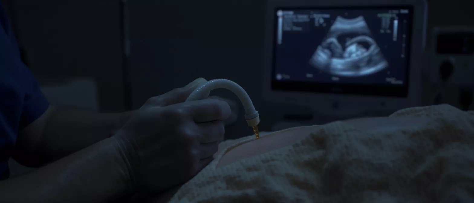
Cardiac Ultrasound
Cardiac ultrasound uses sound waves to create detailed heart images for surgery planning.
What is a cardiac ultrasound
A cardiac ultrasound is a safe, noninvasive imaging test that uses sound waves to produce detailed pictures of your heart. It allows your cardiologist to see your heart beating in real time. The test is commonly performed to evaluate how well the heart muscle pumps blood, to inspect the structure of the heart valves, and to assess the chambers and surrounding sac. Often referred to simply as an echocardiogram, this imaging method is one of the most frequently used diagnostic tools in cardiology.
Why cardiac ultrasound matters for heart surgery patients: If you or a loved one are preparing for heart surgery, this test provides your surgical team with a precise roadmap of your cardiac anatomy. It reveals the size and thickness of the heart walls, the strength of the muscle contractions, and the function of the valves. This information is crucial for planning interventions, determining the exact surgical approach, and establishing a baseline for your heart health before the operation.
Because it uses high-frequency sound waves rather than radiation, a cardiac ultrasound carries no known risks. It is painless, requires no needles, and can be repeated safely as often as necessary. For patients facing cardiac surgery, this test offers a clear picture of what is happening inside the chest without making a single incision, providing reassurance and clarity during a stressful time.
How does a heart ultrasound work
A heart ultrasound relies on a small device called a transducer, which is placed gently against the skin on the chest wall. The transducer emits short bursts of high-frequency sound waves that travel through the tissues of the chest. When these sound waves encounter boundaries between different structures, such as the heart muscle and the blood inside the chambers, they bounce back toward the transducer as echoes.
The ultrasound machine processes these returning echoes instantly, translating them into moving images on a screen. A technician, known as a sonographer, applies a clear gel to the chest to help the sound waves travel smoothly by eliminating air pockets between the transducer and the skin. By moving the transducer to different positions and angles across the chest, the sonographer captures multiple views of the heart, allowing every chamber, valve, and wall to be examined from several perspectives.
The entire process is completely passive from the patient perspective. You simply lie still on an examination table while the machine gathers data. The technology is similar to sonar used by ships to map the ocean floor, but here it maps the dynamic, moving structures within your heart, letting your doctor watch the valves open and close with every heartbeat.
Why is a cardiac ultrasound test needed
A cardiac ultrasound test is ordered when a doctor needs to evaluate the mechanical function of the heart. It is routinely used to diagnose and monitor a wide range of conditions, including heart valve disease, heart failure, cardiomyopathy, and congenital heart defects. If you are experiencing symptoms such as shortness of breath, chest pain, or an irregular heartbeat, this scan helps pinpoint the underlying cause.
Identifying valve disease: The test shows whether the valves are opening fully and closing tightly. It can reveal stenosis, where a valve is stiff and narrowed, or regurgitation, where a valve leaks and allows blood to flow backward. These findings are critical for determining whether a valve repair or replacement is necessary during heart surgery.
For surgical candidates, the heart ultrasound provides exact measurements of the heart chambers. If a chamber is enlarged, it suggests the heart has been working too hard against high pressure or a leaking valve. By assessing the pumping strength, the surgeon can decide whether the heart can tolerate a major operation or if a less invasive approach is required.
How to prepare for a cardiac ultrasound procedure
Preparing for a cardiac ultrasound procedure is simple and requires very few adjustments to your daily routine. Unlike some other imaging scans, this test requires no fasting, no intravenous lines, and no special dietary restrictions. You can eat, drink, and take your prescribed medications exactly as you normally would on the day of the appointment.
What to wear to the echo test: You should wear comfortable, loose-fitting clothing that allows easy access to your chest. Because you will need to undress from the waist up and wear a hospital gown for the scan, a front-opening shirt makes the process smoother. Leave valuables and jewelry at home, as necklaces and chains must be removed before the gel is applied.
When you arrive at the clinic, the sonographer will explain the process and answer any questions you have. The preparation is brief, usually taking only a few minutes to change into the gown and get comfortable on the examination table before the imaging begins. Staying relaxed and breathing normally helps ensure the clearest possible images.
What happens during an echo test
During an echo test, you will lie on your back or slightly turned onto your left side on a padded examination table. The sonographer will attach small, sticky patches called electrodes to your chest to monitor your heart rhythm during the scan. These electrodes connect to an electrocardiograph machine, which helps synchronize the moving images with your heartbeat.
The sonographer will then apply a warm, water-based gel to your chest and place the transducer against your skin. You will feel gentle pressure as the technician presses the wand against different areas of your chest to get the best views of your heart. You might hear a swooshing sound coming from the machine, which is simply the sound of the blood flowing through your heart chambers and valves.
The imaging itself usually takes between thirty and forty-five minutes. The room may be slightly darkened so the sonographer can see the screen clearly. You will occasionally be asked to hold your breath for a few seconds or change positions to capture specific angles, and you can speak up at any time if you feel uncomfortable.
What does a transthoracic echocardiogram show
A transthoracic echocardiogram is the standard, noninvasive type of cardiac ultrasound performed by moving the transducer across the outside of the chest. It provides a comprehensive view of the heart anatomy and function without requiring any internal probes. This test is the frontline imaging method for assessing overall cardiac health in surgical patients.
The images produced show the size and shape of the heart, the thickness of the muscle walls, and the pumping strength of the left ventricle. It clearly displays the motion of the heart walls with each beat, identifying areas of weakness that might indicate past heart attacks or muscle disease. The four heart valves are examined for structural damage, calcification, or restricted movement.
Blood flow and pressure evaluation: Using a feature called Doppler imaging, the ultrasound shows the direction and speed of blood flow inside the heart. It detects turbulent flow caused by narrowed or leaking valves and estimates the pressure inside the heart chambers. This functional data is essential for surgical planning, especially when deciding whether to repair a valve rather than replace it.
Understanding heart ultrasound results
Understanding heart ultrasound results requires comparing your heart measurements against established normal ranges. A cardiologist reviews the moving images and the Doppler data to generate a detailed report. This report details the size of the chambers, the thickness of the walls, the function of the valves, and the overall strength of the heart muscle.
What normal results mean: A normal report indicates that the heart valves are opening fully and closing tightly, the chambers are of standard size, and the heart muscle is pumping effectively without any signs of weakness or thickening. Normal results provide peace of mind and establish a healthy baseline for future comparisons.
Abnormal findings are carefully categorized. The report might note valve regurgitation, where blood leaks backward, or stenosis, where a valve is narrowed. It may identify a reduced ejection fraction, meaning the heart muscle is weaker than it should be. For surgical patients, these specific findings guide the surgical strategy and help predict postoperative recovery.
- Ejection Fraction: A measurement of the percentage of blood leaving the heart each time it contracts, showing muscle strength.
- Valve Grading: Assesses whether valve disease is mild, moderate, or severe based on blood flow turbulence.
- Chamber Dimensions: Records the exact size of the atria and ventricles to detect enlargement.
What affects echocardiogram cost
The echocardiogram cost can vary based on several factors, including the facility type, the complexity of the scan, and the geographic location of the clinic. A standard transthoracic ultrasound is generally less expensive than a transesophageal echocardiogram, which requires sedation and specialized probes. If Doppler imaging or contrast enhancement is added, the technical requirements increase, which can also affect the final price.
Insurance coverage plays a significant role in the out-of-pocket expense for patients. If the test is deemed medically necessary by a referring physician, health insurance plans typically cover a significant portion of the cost. It is always recommended to verify your coverage and any required pre-authorizations with your insurance provider prior to scheduling the appointment.
For patients without insurance or those seeking specialized care, many clinics offer self-pay rates or financial assistance programs. When evaluating the cost, consider that this single test provides comprehensive, real-time data about the heart that would otherwise require more invasive procedures to obtain. For those exploring diagnostic options, advanced cardiac imaging services provide detailed analysis.
Finding cardiac ultrasound near me
When searching for cardiac ultrasound near me, it is important to look for accredited imaging centers or hospital cardiology departments. Accreditation ensures that the facility meets strict standards for image quality, equipment maintenance, and technician training. The skill of the sonographer performing the scan directly impacts the quality of the images, which in turn affects the accuracy of the diagnosis.
Choosing the right facility: Look for a clinic with board-certified cardiologists who specialize in interpreting echocardiograms. The best facilities use modern ultrasound machines capable of producing high-resolution, three-dimensional images. For a patient facing potential heart surgery, having these high-quality images locally reduces travel stress and streamlines the surgical planning process.
In regions like Guadalajara, patients have access to high-quality diagnostic cardiac ultrasound staffed by experienced cardiac care teams. Choosing a specialized cardiac center ensures continuity of care from the initial diagnostic scan through to the surgical consultation and recovery. Patients looking for local heart imaging solutions might consider specialized heart diagnostic clinics for comprehensive evaluations.
Cardiac ultrasound and heart surgery recovery
Cardiac ultrasound plays a vital role not only before surgery but also during heart surgery recovery. After a procedure, the heart needs time to heal and adapt to the changes made by the surgeon. Follow-up echocardiograms allow the surgical team to monitor the repaired valves, assess the strength of the recovering heart muscle, and ensure there are no complications.
A postoperative echo test checks for fluid accumulation around the heart, known as a pericardial effusion, which can occur after surgery. It verifies that a newly repaired or replaced valve is functioning correctly without leaking. These routine scans are usually painless and provide immense reassurance that the surgery was successful and the heart is healing properly.
By comparing the postoperative images to the baseline cardiac ultrasound taken before the operation, the cardiologist can objectively measure the improvement in heart function. Ultimately, this noninvasive imaging tool is a pillar of lifelong cardiac care, ensuring the heart remains strong long after the surgery is over.
Conclusion
A cardiac ultrasound is a remarkable, noninvasive tool that uses simple sound waves to provide a window into the heart. It requires no radiation, involves minimal preparation, and delivers critical information about heart structure and function. From diagnosing valve disease to planning complex cardiac surgery and monitoring recovery, this imaging test is fundamental to modern cardiovascular care.
Understanding what to expect during the scan, how the results are interpreted, and why the test is necessary empowers patients and families facing heart treatment. By eliminating the unknowns, the heart ultrasound helps reduce anxiety and builds trust between the patient and the surgical team. It is a safe, essential step in the journey toward better heart health.






