

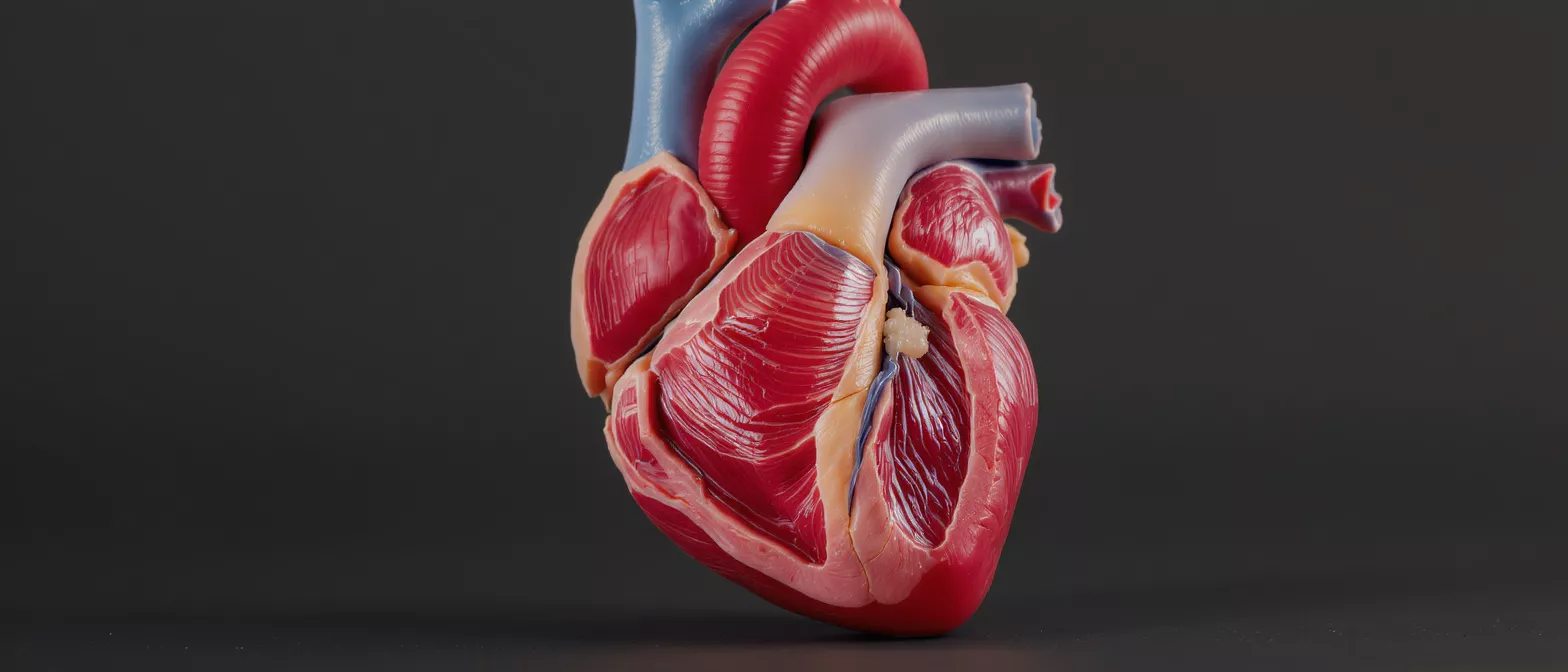
Cardiac Tumour
Learn about cardiac tumour types, symptoms, diagnosis and surgery at Cirugía de Corazón in Guadalajara.
What is a cardiac tumour?
A cardiac tumour is an abnormal growth of tissue within the heart muscle or the inner chambers of the heart. When patients first hear this term, it is entirely normal to feel a wave of fear or anxiety. However, it is deeply reassuring to understand that the vast majority of these growths are entirely benign, meaning they are not cancer. Even so, because the heart is a closed and mechanically active space, any mass requires careful evaluation by a specialist. Understanding the nature of the growth is the first step toward protecting your heart function.
Primary vs. secondary tumours: Medical professionals categorise these growths based on where they originate. A primary tumour begins directly inside the heart itself, while a secondary tumour has spread to the heart from another area of the body. Primary tumours are exceedingly rare, and secondary tumours are far more common, usually requiring an approach that addresses the original cancer site. Knowing this distinction helps your surgical team plan the most effective treatment strategy.
Whether the mass is a simple fluid-filled cyst, a small cluster of fat cells, or a solid benign structure, the structural integrity of the heart remains the top priority. Our team at Cirugía de Corazón focuses on precisely identifying the nature of the mass to ensure your cardiovascular system continues to function safely and efficiently.
How do cardiac tumour symptoms appear?
Cardiac tumour symptoms can be notoriously difficult to pinpoint because they often mimic other, more common heart conditions. The symptoms depend heavily on the size of the mass and its exact location within the heart chambers. A small growth might produce no symptoms at all, only being discovered incidentally during a routine echocardiogram, while a larger mass can physically obstruct normal blood flow or disrupt the heart's electrical pathways.
Positional shortness of breath: One of the most characteristic signs of an intracardiac tumour, particularly a myxoma, is difficulty breathing that changes depending on your physical position. A patient might feel perfectly fine while sitting upright but experience sudden shortness of breath when lying flat. This happens because gravity allows the tumour to shift, temporarily blocking the mitral valve and obstructing blood flow from the lungs to the heart.
Beyond respiratory changes, patients might experience palpitations, chest discomfort, or unexplained dizziness. It is vital to report any new or unusual cardiovascular sensations to your physician immediately. Early detection of these subtle changes provides the best opportunity for a straightforward and successful intervention.
What are the main heart tumour types?
Understanding the different heart tumour types helps demystify the diagnosis process. Medical science categorises these growths not only by whether they are benign or malignant, but also by the specific tissue type from which they arise. The vast majority of primary growths discovered in the heart are benign, meaning they do not spread to other organs, though they still require careful monitoring or removal due to their physical location.
The prevalence of myxomas: Cardiac myxomas account for roughly half of all primary heart tumours. These growths usually appear in the left atrium and are attached to the wall by a thin stalk. While benign, their movement within the chamber can cause dangerous obstructions.
Other benign varieties include lipomas, which are composed of fat cells, and fibromas, which consist of connective tissue. Malignant varieties, such as cardiac sarcomas, are significantly rarer and require a highly specialized, aggressive treatment approach. Identifying the exact cellular structure through imaging and biopsy is the cornerstone of creating an effective treatment plan.
- Myxomas: The most common benign primary tumour, typically found in the left atrium.
- Lipomas and Fibromas: Non-cancerous growths of fat and connective tissue, respectively.
- Sarcomas: Rare, malignant tumours that demand immediate, specialized oncological and surgical care.
How does cardiac tumour diagnosis work?
Accurate cardiac tumour diagnosis relies on advanced imaging technology to look inside the heart without needing to make a single incision. If a physician suspects a mass due to a heart murmur or unexplained symptoms, an echocardiogram is almost always the first diagnostic tool utilized. This safe, painless ultrasound test uses sound waves to create a moving picture of the heart, allowing the specialist to clearly visualize the size, shape, and movement of any intracardiac mass.
The role of MRI and CT scans: While echocardiograms are excellent for initial detection, a cardiac magnetic resonance imaging (MRI) scan provides unparalleled detail regarding the tissue composition of the tumour. A computed tomography (CT) scan may also be used to evaluate the surrounding blood vessels and check if the mass has affected any adjacent structures. These detailed images allow the surgical team to map out the exact surgical route before entering the operating room.
Once the imaging is complete, a multidisciplinary team reviews the results to confirm the tumour's size, its point of attachment, and its potential impact on heart valves. This comprehensive evaluation ensures that every surgical plan is customized to the unique anatomy of the individual patient, minimizing risk and maximizing long-term heart function.
What is a benign heart tumour?
A benign heart tumour is a non-cancerous growth that does not spread to other organs or tissues in the body. Despite being non-cancerous, the word benign can be slightly misleading in the context of the heart, as these growths can still pose serious medical risks. Because the heart is a rigid, highly pressurized organ, even a small, non-cancerous mass can disrupt delicate blood flow, damage valves, or break off and travel through the bloodstream.
Risks of embolization: One of the primary dangers of a benign mass is that a piece of the tumour, or a blood clot attached to it, can break away. This embolus can travel through the circulatory system and lodge in a critical vessel, potentially causing a stroke or a blockage in the lungs. This is precisely why surgeons often recommend surgical removal, even for asymptomatic benign tumours, as a preventative measure.
For patients preparing for the removal of a benign mass, maintaining optimal circulatory health is vital.
What is a malignant heart tumour?
A malignant heart tumour is a cancerous growth that originates in the heart's tissues, most commonly taking the form of a cardiac sarcoma. Unlike benign growths, malignant tumours are aggressive, grow rapidly, and can invade surrounding heart structures or spread to other parts of the body. These growths are incredibly rare, making specialized cardiac oncology expertise absolutely critical for effective treatment.
The challenge of sarcomas: Angiosarcomas, which develop in the lining of the heart's right atrium, and rhabdomyosarcomas, which arise from cardiac muscle cells, represent the most common malignant types. Because they grow aggressively and often present with non-specific symptoms, they are frequently discovered at an advanced stage. Treating these tumours requires a highly coordinated effort between cardiac surgeons and oncology specialists to combine surgical resection with targeted medical therapies.
Treatment for malignant cardiac masses may involve removing a portion of the heart tissue alongside the tumour. In complex cases, reconstructing the damaged heart chamber becomes a critical part of the procedure, ensuring the heart can continue to pump effectively after the cancerous tissue has been extracted.
How does cardiac tumour removal surgery work?
Cardiac tumour removal surgery is the primary treatment for most masses, aiming to completely extract the growth while preserving the heart's structural integrity. The operation is a major cardiovascular procedure that requires a highly skilled surgical team and a specialized operating environment. Patients can take comfort in knowing that modern surgical techniques have made these procedures remarkably successful, with excellent long-term outcomes for benign growths.
Cardiopulmonary bypass and reconstruction: To safely remove the tumour, the surgeon must temporarily stop the heart from beating. The patient is placed on a cardiopulmonary bypass machine, which takes over the job of pumping oxygen-rich blood to the body. With the heart still and empty of blood, the surgeon opens the chamber, carefully excises the tumour and its base of attachment, and reconstructs any damaged heart tissue or affected valves. Restoring structural stability to the repaired heart wall is essential, and utilizing myocardial tissue regeneration aids can support the heart's cellular repair mechanisms during recovery.
After the tumour is successfully removed, the heart is closed, and the bypass machine is gradually weaned off as the heart resumes its natural rhythm. The excised tissue is sent to a laboratory for thorough pathological analysis, providing the final confirmation of the tumour type and guiding any necessary follow-up care.
What is the cardiac tumour prognosis?
The cardiac tumour prognosis depends entirely on the type of tumour discovered, its size, and how early it was detected. For patients with benign tumours, particularly myxomas that are completely removed, the prognosis is excellent. The vast majority of these patients experience a full recovery, returning to their normal lives without the need for ongoing cancer treatments, though they will require routine echocardiograms to monitor for any rare signs of recurrence.
Navigating malignant tumour outcomes: The prognosis for malignant cardiac tumours is more guarded due to their aggressive nature and tendency to recur locally. Outcomes depend heavily on the success of the initial surgical resection and the patient's response to subsequent oncological therapies, such as chemotherapy or radiation. A proactive, multidisciplinary approach is the most effective way to manage these complex cases and optimize long-term survival.
Regardless of the tumour type, early detection and the expertise of the surgical team play the most significant roles in patient outcomes. Following the surgeon's guidelines for postoperative care and maintaining regular follow-up appointments are the best strategies for ensuring a healthy, resilient heart long after the surgery is complete.
When to see a heart tumour specialist in Mexico?
If you or a loved one has been diagnosed with a heart mass, or if you are experiencing unexplained cardiac symptoms, seeking out a qualified specialist is a crucial next step. For patients considering cardiac tumour surgery, Guadalajara offers world-class medical facilities and highly trained cardiovascular surgeons. Patients travel from across the globe to access state-of-the-art cardiac care that combines surgical excellence with compassionate, patient-focused recovery protocols.
Choosing Cirugía de Corazón: Finding a heart tumour specialist in Mexico means looking for a team with extensive experience in complex cardiac reconstruction and tumour resection. At Cirugía de Corazón, Dr Alejandro Molina and his team provide comprehensive evaluations using advanced imaging to determine the safest, most effective surgical approach. Our priority is ensuring you understand your condition and feel confident in your treatment plan.
Do not ignore persistent chest discomfort, unexplained shortness of breath, or unusual palpitations. Early consultation with a cardiovascular expert can provide peace of mind and timely intervention, ensuring your heart remains strong and functional for years to come.
Understanding cardiac mass evaluation
A cardiac mass evaluation is a systematic approach used by cardiologists to determine the exact nature of an abnormal structure seen on a heart scan. Not every mass discovered in the heart is a tumour. The evaluation process is designed to differentiate between true neoplasms, blood clots, and structural artifacts, ensuring patients do not undergo unnecessary invasive procedures.
Differentiating thrombi from tumours: One of the most common findings during a cardiac evaluation is a thrombus, or blood clot, rather than a true tumour. Clots often form in areas where blood flow is sluggish, particularly in patients with atrial fibrillation or severe heart failure. By utilizing advanced imaging techniques and contrast agents, specialists can clearly distinguish a clot from a solid tumour, directing the patient toward anticoagulation therapy instead of surgical extraction.
This detailed evaluation provides the foundation for the entire treatment plan. By definitively identifying the mass, the surgical team at Cirugía de Corazón ensures that every step taken, whether it is medical management or open-heart surgery, is precisely tailored to the patient's specific cardiovascular needs.






