Cardiac Magnetic Resonance Imaging: When It Is Used
Cardiac magnetic resonance imaging reveals detailed heart structure without radiation.
If your cardiologist has mentioned a cardiac magnetic resonance imaging scan — often shortened to cardiac MRI or CMR — you may be wondering what it is, why it was ordered, and what the appointment will actually involve. This test gives some of the most detailed pictures of the heart available today, showing the muscle, valves, and chambers in motion without radiation. In this guide we walk through when doctors order a heart MRI test, how it compares with an echocardiogram and a CT scan of the heart, what preparation involves, and how the results fit into decisions about treatment — including surgery.
What Is Cardiac Magnetic Resonance Imaging?
A scanner that uses magnets, not radiation. Cardiac magnetic resonance imaging uses a powerful magnetic field and radio waves to build detailed pictures of the heart while it beats. Unlike a CT scan, it involves no ionising radiation, which makes it especially valuable for younger patients and for anyone who may need repeated imaging over the years. The images are far more detailed than a standard X-ray and can distinguish between healthy muscle, thickened muscle, and scarred tissue in ways other tests cannot.
The difference between an MRI and a CMR scan. People often ask what separates a general MRI from a CMR scan. The machine itself is similar; the difference lies in the technique. A cardiac study uses special sequences timed to your heartbeat and breathing, often with electrocardiogram (ECG) leads attached to your chest, so each picture is captured at a precise moment in the cardiac cycle. Without that timing, a beating heart would appear as a blur.
What does a cardiac MRI show? A single study can answer several questions at once: how well the heart muscle pumps, whether there is scar tissue from a previous heart attack, how the valves open and close, whether the heart muscle is inflamed, and whether the aorta — the main artery leaving the heart — is enlarged. This breadth is why cardiologists reach for magnetic resonance imaging of the heart when simpler tests leave questions unanswered.
How Does a Cardiac MRI Scan Work Inside the Scanner?
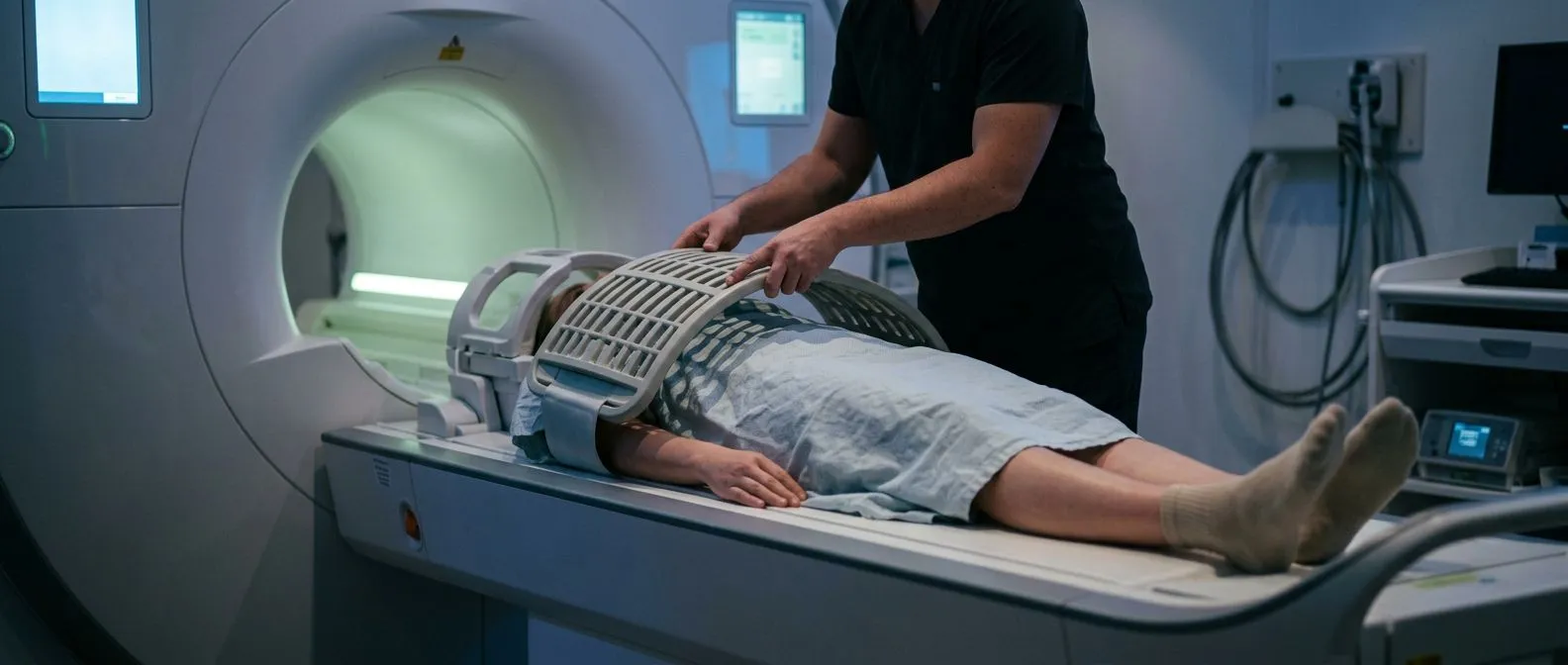
What the appointment actually involves. Most cardiac MRI scans take between 45 and 90 minutes. You lie on a table that slides into a wide, tunnel-shaped scanner. Small pads placed on your chest detect your heartbeat, and you will be asked to hold your breath for short bursts of several seconds while images are taken. The machine makes loud tapping and thumping sounds, so you will be given earplugs or headphones. The team operating the scanner can see and speak to you throughout.
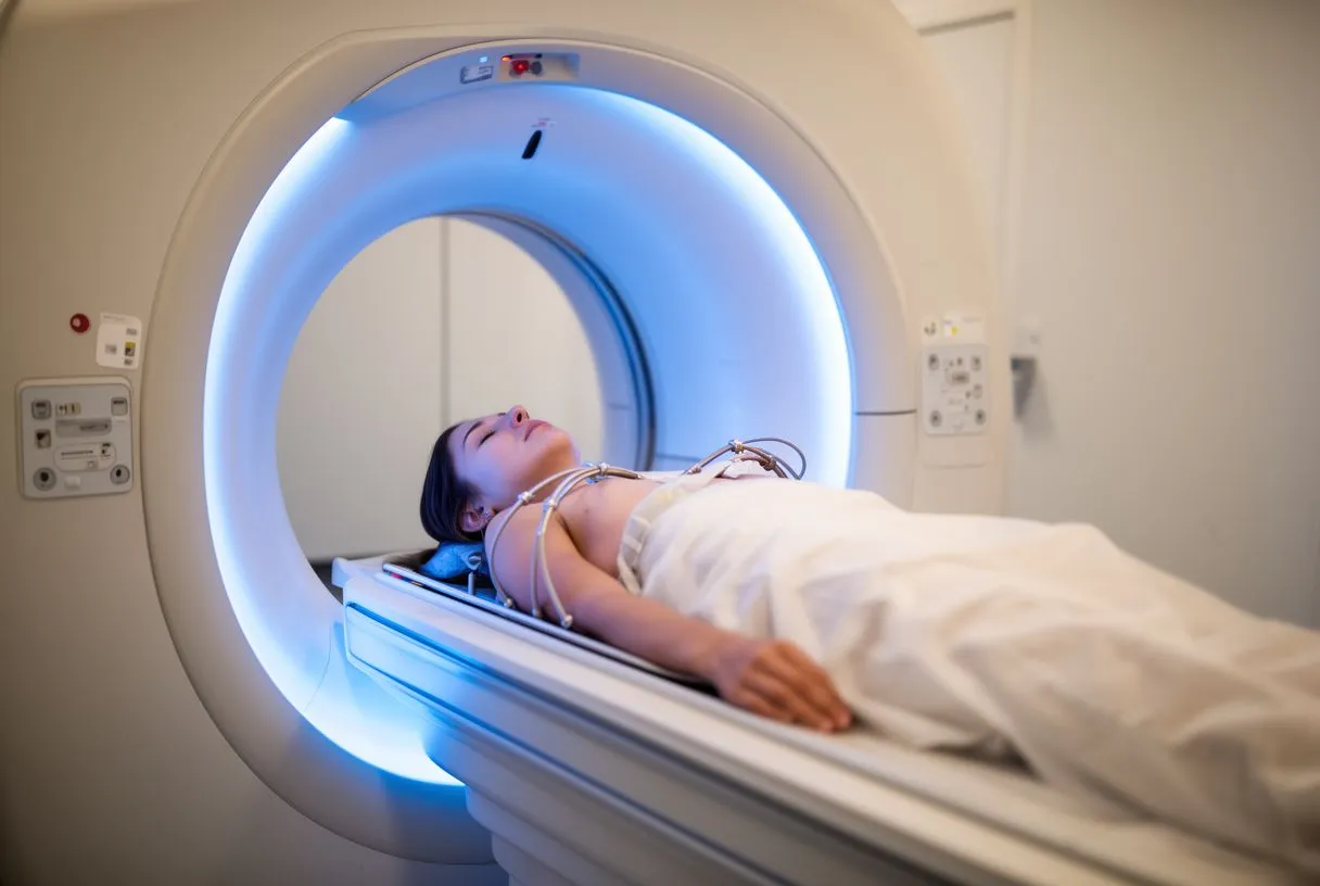
Staying still matters more than anything else. Movement blurs the images, so the most important thing you can do during a cardiac MRI scan is lie still and follow the breathing instructions. Some centres ask you to breathe in a specific rhythm for certain sequences. If you feel anxious in enclosed spaces, mention this when the appointment is booked — options such as a wider-bore scanner, music through the headphones, or in some cases mild sedation can be arranged.
Contrast dye, if needed. Many studies include a contrast agent called gadolinium, injected through a small needle in your arm partway through the scan. This dye highlights blood flow and allows the radiologist to see scar tissue or inflammation clearly. The injection is quick and usually painless, apart from a brief cold sensation in the arm.
Why Would Someone Need a Cardiac MRI?
When an echocardiogram is not enough. Most people start with an echocardiogram, an ultrasound scan of the heart performed in the clinic. It is quick and answers many questions, but it cannot see through lung or bone well in some patients, and it cannot reliably detect scar tissue. A cardiac MRI is ordered when the echocardiogram is unclear or when a specific question — such as whether chest pain came from a small heart attack or from inflamed muscle — needs a sharper answer.
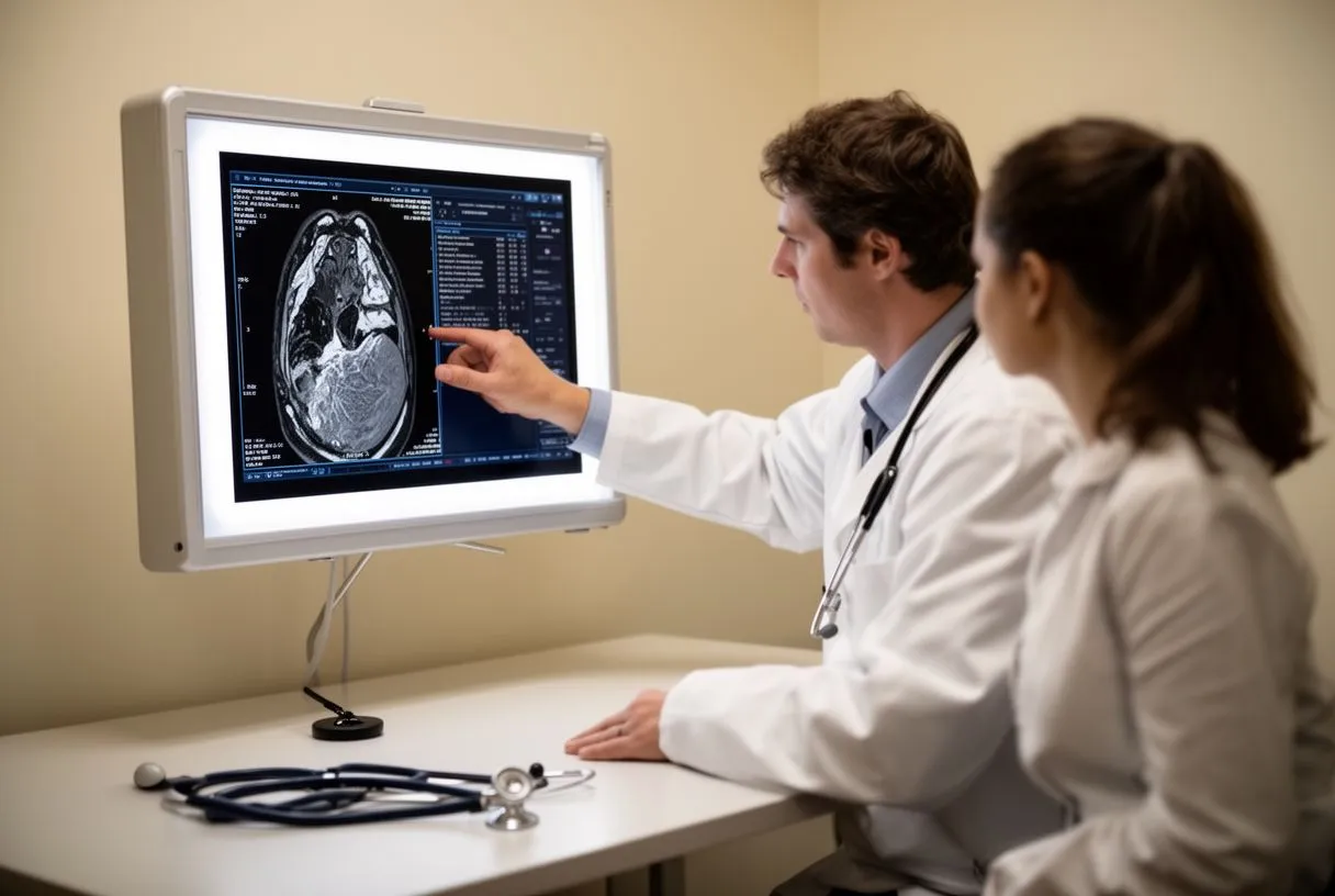
Common reasons doctors order one. In our consulting rooms we explain the reason for every study before it is ordered, because a test should always answer a question that changes the plan. The most frequent indications include:
- Suspected cardiomyopathy: when the heart muscle appears thickened, stretched, or weakened and the cause is unclear.
- Assessing heart failure: to measure pumping function precisely and look for scar or inflammation.
- Chest pain without blocked arteries: to detect microvascular problems or myocarditis, an inflammation of the heart muscle often seen after viral illness.
- Aortic disease: to measure an enlarged aorta, or aneurysm, and follow its size over time.
- Congenital heart disease: to map the anatomy of a heart the patient was born with, before or after corrective surgery.
Before heart surgery. For patients being considered for an operation, magnetic resonance imaging of the heart often completes the picture. We review every case as a multidisciplinary team against international practice guidelines before recommending surgery, and a detailed CMR frequently forms part of that review — for example, when planning a valve repair or assessing whether a stiff, thickened heart muscle explains a patient's breathlessness.
Cardiac MRI vs Echocardiogram: Which Test, and When?
Two tests, different strengths. The question of cardiac MRI versus echocardiogram comes up constantly, and the honest answer is that they complement each other rather than compete. An echocardiogram takes minutes, costs less, and shows the heart moving in real time at the bedside. A cardiac MRI takes longer but produces measurements that are more repeatable between different operators and different machines, and it can see tissue characteristics — scar, inflammation, iron deposits — that ultrasound simply cannot.
When each one is chosen. As a practical rule of thumb:
- Echocardiogram first: for initial assessment of murmurs, breathlessness, and most valve questions.
- Cardiac MRI next: when precise measurement is needed, when ultrasound windows are poor, or when tissue characterisation will change the diagnosis or the surgical plan.
- CT scan of the heart sits alongside these: it is faster and better for the coronary arteries, while MRI is better for the muscle and soft tissue.
How this plays out in practice. In our experience, patients who arrive having had only an echocardiogram often need one more detailed study before a surgical recommendation can be made responsibly. We take the time in consultation to explain what the studies actually show, in plain words, so that the patient understands why each test was chosen and what it means for the options ahead.
Cardiac MRI With Contrast: What Does It Add?
Seeing scar tissue. A cardiac MRI with contrast uses a gadolinium-based dye that briefly changes the magnetic properties of tissue. Areas of dead or scarred muscle hold onto the dye longer than healthy muscle, so late images taken after injection reveal exactly where and how much of the heart muscle has been damaged. This technique, called late gadolinium enhancement, is one of the most powerful tools in modern cardiology: it can distinguish a scar from a previous heart attack from scarring patterns seen in cardiomyopathy, which changes the diagnosis entirely.
Is a contrast study always needed? Not always. A cardiac MRI without contrast can measure the pumping function of the ventricles, the size of the chambers, valve flows, and the aorta very well. Contrast is added when the question involves scar, inflammation, tumours, or the precise cause of a weakened heart muscle. Your cardiologist decides based on the clinical question, not automatically.
Gadolinium safety in plain terms. The small doses used today, especially the newer agents, are considered safe for most people, and the dye leaves the body through the kidneys. If you have significant kidney disease, tell the imaging team in advance; they will check a recent blood test and may perform the study without contrast or use an alternative approach. Patients with severe kidney impairment are generally advised against gadolinium.
What Should You Not Do Before a Cardiac MRI?
Food, drink, and daily habits. Preparation for a cardiac MRI is refreshingly simple compared with surgery. You can usually eat and drink normally — some centres ask you to avoid caffeine for a few hours beforehand because it can slightly increase the heart rate and affect some sequences. Continue your regular medications unless told otherwise. Avoid wearing jewellery, watches, hair products, and clothing with metal, since metal interferes with the magnetic field and can heat up or create image artefacts.
Metal implants and devices. Before the appointment you will complete a safety questionnaire. Most modern pacemakers and implantable defibrillators are MRI-conditional, meaning the scan can be done with specific protocols and device settings; older devices or certain metallic implants — some aneurysm clips, cochlear implants, metal fragments in the eyes — may rule the study out. Bring a list of your implants and device card if you have one.
Practical tips for the day itself. Wear comfortable clothing without metal fasteners, arrive with time to spare for the safety checks, and use the bathroom before the scan starts, since you may be asked to lie still for extended periods. If you are claustrophobic, this is the moment to say so — the imaging team can plan accordingly, and knowing what to expect removes much of the worry.
Is Cardiac MRI Safe? Risks and Who Should Avoid It
No radiation, few risks. For most patients the answer to "is cardiac MRI safe?" is a clear yes. The scan uses no ionising radiation, and the magnetic field at clinically approved strengths has no known harmful effects. The most common complaints are the noise, the enclosed space, and the need to lie still — discomforts rather than dangers. Serious reactions to gadolinium contrast are rare with modern agents.
Cardiac magnetic resonance is an established, well-studied technique; major references such as the European PubMed Central archive contain thousands of peer-reviewed studies documenting its accuracy across heart muscle disease, valve disease, and congenital conditions. That depth of evidence is part of why it has become a standard tool before major cardiac decisions.
Who should not have one. The absolute contraindications are few but important: certain older metallic implants, some types of aneurysm clips, metal fragments in the eye, and, as noted, severe kidney disease when contrast is required. A detailed screening questionnaire covers all of this before you enter the scanner room. We check each patient's implant list and imaging history carefully before recommending the study, and if an alternative test serves the question better, we say so plainly — no unnecessary studies are ordered.
Heart MRI for Cardiomyopathy and Heart Failure
Finding the cause, not just the symptom. A heart MRI for cardiomyopathy does something no other test can do as well: it identifies which type of muscle disease a patient has. Hypertrophic cardiomyopathy — thickened muscle, often inherited — shows a distinctive pattern. Dilated cardiomyopathy — a stretched, weak-pumping heart — may show scar that points to inflammation or previous infection. Amyloidosis, a condition where abnormal protein builds up in the heart, has its own signature. Each pattern leads to a different treatment, which is why the scan matters so much.
Research on this question is extensive. Studies available through cardiac magnetic resonance imaging findings in specific heart conditions show how CMR evaluates heart function in patients with structural heart disease, confirming the test's role in distinguishing conditions that look similar on an echocardiogram.
Monitoring over time. For patients with established heart failure, cardiac MRI provides the most reliable way to track the heart's pumping function between treatments. Doctors call this Surveillance Imaging — repeat scans at intervals to see whether the muscle is recovering, stable, or deteriorating. Because the test involves no radiation, repeating it carries no cumulative risk, which matters for younger patients who may need decades of follow-up.
Cardiac MRI for Valve Disease and Congenital Heart Disease
Measuring valve leaks precisely. When an echocardiogram shows a leaky mitral or aortic valve, the next question is how severe the leak is and what it is doing to the heart's size and pumping strength. A cardiac MRI for valve disease answers this with measurements that guide the timing of surgery. Choosing the right moment to operate — before the heart muscle is permanently damaged, but not so early that the operation carries more risk than benefit — is one of the most important decisions in cardiac surgery, and precise imaging sits at the centre of it.
Congenital heart disease in adults. Adults born with heart defects — repaired in childhood or never repaired — need imaging that maps complex anatomy. The depth of published work in this area is remarkable; detailed reviews such as magnetic resonance imaging in structural heart evaluation describe how MRI delineates complex cardiac and extracardiac anatomy that ultrasound cannot fully capture. CMR measures blood flow through abnormal connections, quantifies volumes of the right ventricle — the chamber hardest to see with ultrasound — and plans surgery or catheter-based treatment.
How imaging feeds into our recommendations. Our team performs conventional, minimally invasive, robotic, and transcatheter procedures, including TAVR — transcatheter aortic valve replacement, where a new aortic valve is placed via a catheter through the groin rather than through open surgery. The choice between an open operation and a catheter-based approach depends on the patient's anatomy, age, and overall condition, and detailed imaging studies are part of every case we review together as a team. The final decision always rests with the patient, with every alternative explained.
What a cardiac MRI costs in Mexico. The question "how much does a heart MRI cost?" has no single answer. In Mexico, prices vary widely by city, hospital, and whether contrast is used, and patients from abroad should ask whether the quoted price covers the radiologist's report and any follow-up consultation. Rather than quote figures here — which change constantly — we suggest asking any imaging centre for a written, itemised quote. For patients planning a consultation with us in Guadalajara, Querétaro, or Hermosillo, the first contact is a free video consultation, and we tell you honestly which studies are still needed so no unnecessary ones are ordered.
Getting the most from your report. A cardiac MRI report contains technical measurements that can look like a wall of numbers: ejection fraction (the percentage of blood the heart pumps out with each beat), ventricular volumes, mass of the muscle, and descriptions of scar. Bring the full report — not just a summary — to your consultation. Ask which numbers matter for your specific situation and what they change about the plan.

From images to decisions. The scan itself never decides anything; it informs a conversation. When we sit with a patient and their family, we go through the images and the measurements together, explain what the studies actually show in plain words, and answer every question before any recommendation is made. Where surgery is being considered, we encourage patients to seek a second opinion and treat that as a normal, healthy part of deciding about heart surgery — a confident decision is built on understanding, not on pressure.
Wrapping up. Cardiac magnetic resonance imaging is the most detailed, radiation-free view of the heart available, and it earns its place when an echocardiogram leaves questions unanswered or when a surgical decision demands precision. It is safe for most people, the preparation is simple, and the results can clarify diagnoses that would otherwise remain uncertain. If your doctor has suggested this test — or if you have symptoms such as breathlessness, chest discomfort, or fainting that have never been fully explained — the next step is a conversation with a qualified cardiac team who can tell you which study answers your specific question. Dr Alejandro Molina and our multidisciplinary team in Guadalajara, Querétaro, and Hermosillo review every case against international guidelines and will gladly talk through your situation in a first video consultation. Never self-diagnose from this article; book a consultation and let the right test be chosen for the right reason.