Right Heart Catheterisation: Why and How It Is Done
Learn the right heart catheterisation purpose: how doctors measure heart and lung pressures directly to guide diagnosis and treatment.
If your cardiologist has mentioned a right heart catheterisation, you may be wondering what the test actually measures and why it cannot simply be replaced by an ultrasound or an X-ray. The purpose of right heart catheterisation is to measure the pressures inside the right-sided chambers of your heart and the arteries of your lungs directly — something no scan can do with the same accuracy. In this guide we explain, in plain language, what happens during the test, why doctors order it, how it differs from other cardiac catheterisation procedures, and what recovery really looks like.
What Is Right Heart Catheterisation?
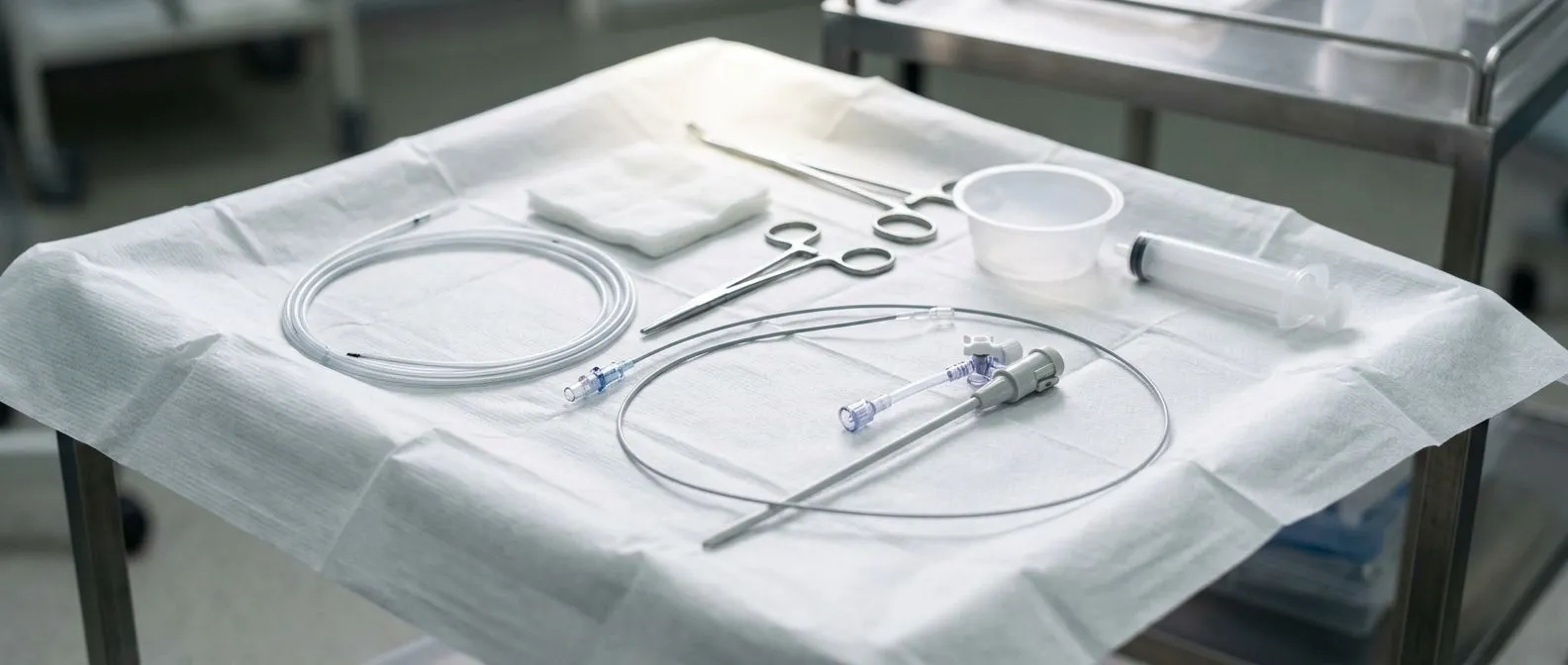
A thin, flexible tube measures pressures directly. Right heart catheterisation (often abbreviated RHC) is a diagnostic test in which a doctor guides a very thin, soft tube called a catheter through a vein into the right side of your heart. The catheter carries a tiny pressure sensor at its tip. As it is advanced through the Right Atrium (the upper receiving chamber), the Right Ventricle (the lower pumping chamber), and into the pulmonary artery — the vessel that carries blood to the lungs — it records the pressure at each point.
The Swan-Ganz catheter is the classic tool. The balloon-tipped catheter most often used for this test is commonly called a Swan-Ganz catheter, named after the doctors who popularised it. Its pulmonary artery catheter purpose is specifically haemodynamic assessment: measuring blood flow and pressures rather than taking pictures. Because the catheter is soft and floats with the blood flow, it typically settles in a branch of the pulmonary artery without needing to be steered aggressively.
It is a measurement test, not a treatment. Unlike procedures that open blocked arteries or replace valves, a right heart cath usually involves no balloons, stents or implants. Its entire job is to produce precise numbers. Those numbers — pressures, cardiac output (how much blood your heart pumps each minute), and the resistance in the lung vessels — often decide which treatment you are offered next. In our practice, every case is reviewed by the multidisciplinary team against international guidelines before any recommendation is made, and pressure data from this test is frequently the evidence that drives that discussion.
What Is the Purpose of Right Heart Catheterisation?
Diagnosing pulmonary hypertension accurately. The single most important purpose of right heart catheterisation today is confirming or excluding pulmonary hypertension — abnormally high pressure in the arteries between the heart and the lungs. An ultrasound of the heart can estimate these pressures, but it can be wrong in either direction. The 2015 ESC/ERS Guidelines for the diagnosis and treatment of pulmonary hypertension 2015 ESC/ERS Guidelines for the diagnosis and treatment of pulmonary hypertension establish that pulmonary hypertension should be confirmed by right heart catheterisation before disease-specific therapy is started, because treatment drugs are strong, expensive and not harmless.
Distinguishing between different causes. During the test, the doctor can calculate pulmonary vascular resistance and measure the pressure of blood filling the left side of the heart (the "wedge" pressure). This tells the team whether high lung pressures come from the lung arteries themselves, or from back-pressure caused by a leaky or narrowed mitral valve, or from left-heart failure. The treatment for these conditions is completely different, and this test is often what tells them apart.
Guiding treatment and tracking response. For patients already on therapy for pulmonary hypertension, repeat right heart catheterisation can show whether the treatment is working. It is also used before major interventions — for example, assessing whether a patient's lung pressures make a valve procedure or heart surgery safe, or evaluating candidates for heart transplantation. We encourage patients to ask for the actual numbers from their catheterisation and to have them explained in plain words; in our consultations we take the time to do exactly that before any decision is discussed.
When Is Right Heart Catheterisation Needed?
Common right heart catheterisation indications. Doctors order this test in a fairly defined set of situations. The most frequent ones include:
- Confirmed or suspected pulmonary hypertension: when an ultrasound shows high lung pressures, or when you have unexplained breathlessness that scans cannot explain.
- Unclear heart failure: when it is uncertain whether symptoms come from the left side of the heart, the lungs, or both.
- Before major decisions: assessing risk before valve surgery, heart transplantation evaluation, or complex congenital heart disease repair.
- Monitoring therapy: checking whether pulmonary hypertension medication is improving the pressures over time.
Why a scan alone is not enough. Echocardiography (heart ultrasound) estimates lung pressures from the speed of blood leaking through valves. That estimate can be inaccurate, especially in patients with lung disease, obesity or valve problems. Because the diagnosis of pulmonary hypertension changes a person's life — from insurance and travel to lifelong medication — most specialists insist on direct measurement before making it. The Serum Pentraxin 3 and hs-CRP Levels in Children with Severe Pulmonary Hypertension Serum Pentraxin 3 and hs-CRP Levels in Children with Severe Pulmonary Hypertension research reflects the broader effort to find blood markers that track this disease, but no blood test replaces the catheter measurement.
When the test is not needed. A right heart cath is not a screening test for the general population, and it is not the first step for ordinary chest pain or suspected coronary blockages — those are investigated with other tools. If your team orders one, there is almost always a specific question they need the pressure numbers to answer. I tell my own patients that a test which does not change the treatment plan is not worth the risk of any needle stick, and we only order studies when the answer will genuinely change what we offer.
How Is Right Heart Catheterisation Done?
Where the catheter enters. The catheter is introduced through a vein — most commonly the internal jugular vein in the neck, the femoral vein in the groin, or increasingly a vein in the arm (the basilic or brachial vein). The entry site is numbed with local anaesthetic, and a small needle is used to place a thin sheath, a short plastic tube through which the catheter slides. No general anaesthesia is needed; you remain awake and able to talk to the team.
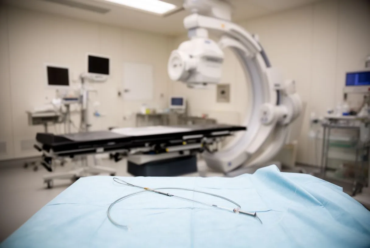
The route through the heart. From the entry vein, the catheter travels with the blood: into the Right Atrium, across the tricuspid valve into the Right Ventricle, and out through the pulmonary valve into the pulmonary artery. Modern fluoroscopy (a kind of live X-ray) lets the doctor watch the catheter's progress on a screen at all times. In some cases the catheter is advanced into the small lung artery branches until the tip "wedges", which allows measurement of the filling pressure on the left side of the heart.
What is measured. Once positioned, the team records a series of pressures and calculations. The procedure is performed in the Catheterisation Laboratory — the dedicated room where these procedures are performed — and each pressure tracing is checked carefully, because a technically poor tracing can mislead the whole diagnosis. The procedure usually takes between 30 and 60 minutes, and longer if it is combined with other tests.
Right Heart Catheterisation vs Left Heart Catheterisation: What Is the Difference?
They study different sides of the heart through different vessels. Left Heart Catheterisation enters an artery — usually at the wrist or groin — and travels backwards into the left ventricle and the coronary arteries, which supply the heart muscle itself. It is the test used to look for coronary artery narrowings and to measure pressures on the left side. Right heart catheterisation enters a vein and studies the right chambers and the pulmonary arteries instead.
Can you do a left and right heart catheterisation at the same time? Yes — and this is common. When a patient has both suspected coronary disease and suspected pulmonary hypertension, or when valve surgery is being planned, both catheters can be placed in one session through the vein and the artery at the same entry site or nearby sites. Doing both at once means one preparation, one recovery, and pressures measured simultaneously, which is more accurate for some calculations.
Which one you need depends on the question. Chest pain and suspected blocked arteries point to the left-sided test. Breathlessness, suspected lung vessel disease, or assessment before heart failure therapies point to the right-sided test. Many patients with complex valve disease actually need both. Our team's practice is to recommend the least invasive combination that answers the clinical question for that specific patient — and to explain clearly why each component is being proposed.
What Happens During the Procedure, Step by Step?
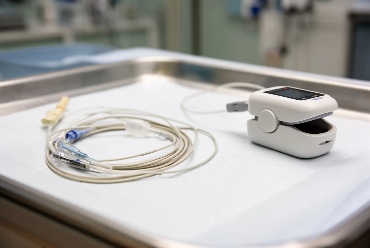
Arrival and preparation. You will be asked not to eat for several hours beforehand and to arrange someone to drive you home. On the day, staff check your blood tests, medications and allergies, place an intravenous line, and clean and drape the entry site. You will be on a table under an X-ray machine, connected to monitors showing your heart rhythm and oxygen levels.
Insertion and measurement. After local anaesthetic numbs the skin, the sheath is placed and the catheter is advanced while the doctor watches the pressures change on the monitor — each chamber has a characteristic pressure shape, confirming where the tip is. You may feel a sensation of pressure or a brief flutter as the catheter crosses valves; many patients describe it as odd but not painful. At the end, the catheter and sheath are removed and firm pressure or a small closure device is applied to the vein entry site.
Right after the test. You will rest lying flat for a short period — typically one to a few hours depending on the entry site — while nurses check for bleeding or swelling. Most people go home the same day. The results are usually available immediately in raw form, and a full report follows within days.
How Painful Is Right Heart Catheterisation?
Most patients are surprised by how manageable it is. The genuinely uncomfortable moments are few: the sting of the local anaesthetic injection, the pressure sensation when the sheath enters the vein, and the odd internal flutter as the catheter crosses valves. Once the catheter is in place, most people feel nothing from it at all. Sedation can be offered to help you relax, though many patients choose to stay fully awake and chat with the team.
The entry site matters. Arm (basilic) and wrist approaches tend to be the easiest to tolerate and the quickest to recover, because you can sit up sooner. Neck and groin sites require lying flat a little longer, and a bruise at the groin is common and harmless. If a site is sore afterwards, simple pain relief such as paracetamol is usually enough.
What patients tell us afterwards. The anticipation is almost always worse than the procedure itself; many patients say afterwards that they had built it up far more than it deserved. Tell the team immediately if anything hurts so they can add anaesthetic or sedation — that is exactly what the team is there for.
Are Right Heart Catheterisation Risks Serious?
It is a low-risk test in experienced hands. Right heart catheterisation is one of the most common procedures performed in cardiac units worldwide, and serious complications are uncommon. The risks that do exist are worth knowing about honestly, because being prepared is part of making an informed decision:
- Bruising or bleeding at the entry site: the most frequent issue, usually minor and self-limiting.
- Irregular heartbeat during passage: the catheter can briefly trigger extra beats or short runs of arrhythmia as it crosses the Right Ventricle; these usually stop when the catheter moves on, and the team monitors continuously.
- Rare lung complications: very rarely, the catheter can injure a pulmonary artery branch, or a vagal reaction can drop the blood pressure temporarily.
- Infection or vein irritation: uncommon, and minimised by sterile technique.
How risk is kept down. Continuous ECG and pressure monitoring throughout, gentle catheter handling, and appropriate choice of entry site all reduce complications. Serious events are rare enough that the test is routinely performed as an outpatient procedure. Harvard Health Publishing notes that cardiac catheterisation procedures, when appropriately indicated and performed by experienced teams, carry low complication rates relative to the information they provide Harvard Health Publishing.
Being honest about your own risk. Risk is not identical for everyone — patients with severe lung disease, very high lung pressures, or bleeding tendencies need extra care, and the team should discuss this with you beforehand. In our team every proposed procedure is reviewed against international practice guidelines, and we present you with the alternatives as well as the recommendation, so the final decision about whether to proceed is always yours.
How Long Does Recovery Take After a Right Heart Cath?
Same-day recovery for most patients. How long it takes to recover from a right heart catheterisation is one of the most reassuring answers in cardiology: for most people, it is hours, not weeks. After the sheath is removed, you rest flat for a period — the exact time depends on the entry site — then sit up gradually, have a drink and something to eat, and usually go home the same afternoon. The entry site may be tender, and a small bruise is normal.
First 24 to 48 hours. Avoid heavy lifting, vigorous exercise, and submerging the entry site in baths or pools for a day or two. Most people return to office work and light activity the next day. Watch for warning signs: spreading swelling, active bleeding, increasing pain, fever, or new breathlessness — any of these deserve a call to the team.
Getting your results. Because the catheter has no lasting physical effect on your heart, the "recovery" that matters most is understanding what the numbers mean. We make a point of reviewing the results with patients in plain words — what each pressure means, what it changes about the plan, and what happens next — before any treatment decision is proposed. If a result is normal, that too is valuable information: it can save you from years of unnecessary medication.
How to Prepare and What Questions to Ask Your Team
Practical preparation. Preparation is simple but worth doing properly: follow your team's instructions about fasting, bring a full list of your medications (especially blood thinners, which may need pausing), tell them about any allergies and kidney problems, and arrange a lift home. Wear comfortable clothing and leave valuables at home. If you are anxious about the procedure, say so — teams can offer sedation, and knowing what to expect removes much of the fear.
Questions that get you better answers. Before the test, consider asking your team these questions — good doctors welcome all of them:
- What specific question will this test answer for me? If the answer would not change your treatment, ask whether it is truly needed.
- Where will the catheter enter, and why that site? Arm, neck and groin sites have different comfort and recovery profiles.
- Will other tests be done at the same time? Combining tests can save a repeat procedure later.
- Who will explain the results to me, and when? You deserve the numbers and what they mean, not just "it was fine".
A final word. Right heart catheterisation is a well-established, low-risk test that answers questions no scan can, and for many patients it is the step that finally turns years of unexplained breathlessness into a clear diagnosis and a real treatment plan. If you have been told you may need this test — or you have breathlessness that no one has fully explained — the right next step is a conversation with a qualified cardiac team, not self-diagnosis. Our multidisciplinary team, led by Dr Alejandro Molina, offers a first free video consultation in which we review your situation, explain which studies are genuinely needed, and put you in control of every decision that follows. Consulting rooms are available in Guadalajara, Querétaro and Hermosillo, and a second opinion is always welcome.