Transvalvular Gradient in Aortic Stenosis Explained
Transvalvular gradient aortic stenosis explained: learn what your echo numbers mean, when severity changes treatment.
If you or a family member have been told you have aortic stenosis, you have probably seen numbers on an echocardiogram report that nobody fully explained. One of the most important is the transvalvular gradient in aortic stenosis — in simple words, the pressure difference the heart has to overcome to push blood through a narrowed aortic valve. Understanding this number helps you understand why your cardiology team is recommending monitoring, medication, or treatment, and what actually changes after a valve procedure. This guide walks through what the gradient means, how it is measured, what the cut-off values are, and what the numbers mean for your decisions.
What Is the Transvalvular Gradient in Aortic Stenosis?
The basic idea. The aortic valve is the one-way door between the heart's main pumping chamber (the left ventricle) and the aorta, the large artery that carries blood to the whole body. When the valve becomes stiff and narrowed — aortic stenosis — the heart must squeeze harder to force blood through the smaller opening. The transvalvular gradient is the pressure drop across that narrowed valve, measured in millimetres of mercury (mmHg).
Think of it like a garden hose. If you pinch the nozzle, pressure builds up behind the pinch. The tighter the pinch, the bigger the difference in pressure before and after it. In the same way, a tighter valve produces a larger pressure gradient, and that number tells your cardiologists how much work the valve is forcing the heart to do.
It is worth knowing that the gradient is one of three measurements used together. The others are the valve opening area (measured in square centimetres) and how well the heart muscle itself is pumping. A full picture of aortic stenosis hemodynamics — how blood and pressures behave — always combines all three, because each one tells part of the story.
What Is the Mean Gradient on an Echocardiogram?
Peak versus mean. When you read an echo report, you will usually see two gradient numbers: peak and mean. The peak gradient is the highest instantaneous pressure difference measured during a heartbeat. The mean transvalvular gradient is the average pressure difference across the whole period when blood is actually flowing through the valve. Guidelines and treatment decisions rely mainly on the mean gradient, so when your team talks about "the gradient," that is usually the number they mean.
Where the number comes from. Echocardiography does not measure pressure directly. It measures the speed of blood jetting through the valve using ultrasound (the Doppler principle), and then converts that velocity into a pressure estimate. A faster jet means a bigger pressure drop. This is why the same report also lists a "peak velocity" in metres per second — it is the raw measurement behind the gradient figure.
Because it is a derived estimate rather than a direct measurement, small variations between studies are normal. Trending the number over time at the same lab, with the same equipment, is more meaningful than comparing a single reading against a report from another centre.
How Is Transvalvular Gradient Measured?
Doppler echocardiography first. In practice, transvalvular gradient measurement starts with a standard transthoracic echocardiogram — the ultrasound probe on the chest. The sonographer aims the beam along the narrowed jet of blood and records the velocity curve. The Doppler transvalvular gradient is then calculated from that curve, with the mean gradient derived by averaging the instantaneous pressures during systole.
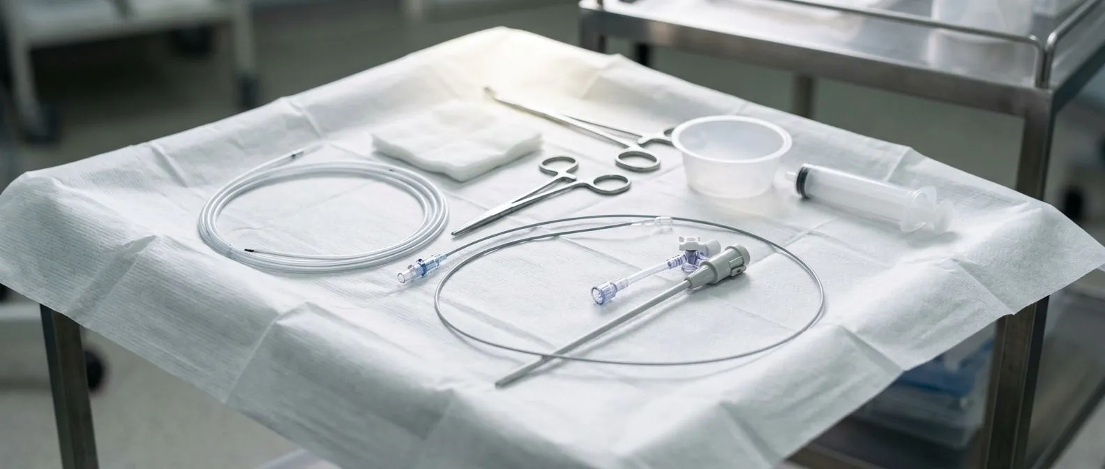
Catheter confirmation. Sometimes there is doubt about the echo numbers — perhaps the patient's symptoms do not match the reported gradient, or the technical quality of the study is limited. In those cases, a cardiac catheterisation can measure pressures directly on either side of the valve. This is the reference standard for transvalvular pressure gradient measurement, though it is an invasive test reserved for genuine uncertainty rather than routine use.
The catheter effect. One practical detail worth knowing: when a pressure-measuring catheter sits inside the narrowed valve during catheterisation, it can itself distort the reading and make the gradient look slightly higher than it really is. Echocardiography and catheterisation also measure pressure at slightly different points, so the two numbers are expected to differ a little — this is understood physics, not an error in your test.
In our consultation rooms we take the time to go through the actual echo images and measurements with you, in plain words, so the numbers on the report stop being a mystery before any decision is made. In the consultation we take the time to explain what the studies actually show, in plain words, and to answer every question before any decision.
What Is the Normal Range for the Aortic Valve Gradient?
What a healthy valve looks like. A normal aortic valve opens widely, so very little energy is lost pushing blood through it. The mean gradient of a healthy valve is typically close to zero — only a few mmHg. There is no single "normal" figure with a hard boundary, but a mean gradient that stays in the low single digits, with a valve area around 3–4 cm², is reassuring.
Mild, moderate and severe bands. As the valve narrows, the gradient climbs. International guidelines group aortic stenosis into stages using the mean transvalvular gradient, together with valve area and blood flow velocity. A commonly used framework looks like this:
- Normal valve: mean gradient under about 20 mmHg, no significant narrowing.
- Mild stenosis: mean gradient generally below 20 mmHg, valve area above 1.5 cm².
- Moderate stenosis: mean gradient roughly 20–39 mmHg, valve area 1.0–1.5 cm².
- Severe stenosis: mean gradient 40 mmHg or more, valve area 1.0 cm² or less, jet velocity 4 m/s or more.
These bands are guides, not verdicts. A patient with a weak heart muscle can have a severe valve with a lower gradient, which is exactly why the full hemodynamic picture matters and why the numbers should always be interpreted by a team, not read in isolation.
What Are the Numbers for Severe Aortic Stenosis?
The classic severe thresholds. When people ask what level of aortic stenosis requires surgery, the answer combines three numbers. Severe aortic stenosis is generally defined as a valve area of 1.0 cm² or less, a mean transvalvular gradient of at least 40 mmHg, and a peak jet velocity of 4.0 metres per second or more. Any one of these alone is not enough — the diagnosis of severe stenosis rests on the combination, confirmed in a patient who has symptoms or clear progression.
Why symptoms drive the decision. Among patients with severe stenosis, the presence of symptoms — breathlessness on exertion, chest tightness, dizziness or fainting — is the strongest signal that treatment should be considered. Guidelines recommend intervention for severe symptomatic aortic stenosis because the outlook changes significantly once symptoms appear. Asymptomatic patients are usually monitored closely at intervals, with treatment discussed earlier if tests show the heart is already being affected.
When numbers and symptoms disagree. Sometimes a patient reports real symptoms but the gradient looks only moderate. That is when the team looks deeper: exercise testing, additional imaging of the valve's calcium burden, or a low-flow state may explain the mismatch. Every case is different, and a rigid reading of a single threshold can mislead. Every case we review is discussed by the full multidisciplinary team against international practice guidelines before any recommendation is made, and the final decision always rests with the patient.
What Does a High Gradient Mean for Treatment?
Why gradient predicts outcome. A high gradient severe aortic stenosis pattern — the classic combination of a small valve area and a mean gradient of 40 mmHg or more — tells the team that the valve is truly tight and the heart is working hard against it. Research following patients through valve replacement has shown that the pre-procedure gradient carries real information about outcomes after treatment, which is one reason the number features so prominently in decision-making.

Treatment options once the gradient is severe. For high-gradient severe stenosis with symptoms, replacing the valve is the established treatment. The two main routes are surgical aortic valve replacement, performed through the chest with the heart stopped, and transcatheter aortic valve implantation — usually called TAVR — where a new valve is delivered through a catheter, most often from an artery in the groin. Neither is universally "better": the choice depends on age, surgical risk, valve anatomy and the patient's own priorities.
How we weigh the trade-offs. We perform conventional, minimally invasive, robotic and transcatheter procedures, and we recommend the less invasive route only where the evidence supports it for that particular patient. That honesty matters, because a gradient number alone never decides the procedure — the whole patient does. If you are weighing options, seeking a second opinion is a normal and sensible part of deciding about heart surgery, and a good team will welcome it rather than resist it.
What Is Low-Flow Low-Gradient Aortic Stenosis?
A subtler pattern. Not every severe valve announces itself with a high number. In low-flow low-gradient aortic stenosis, the valve area measures severe (1.0 cm² or less) but the mean gradient sits below 40 mmHg because the heart is not generating much flow across it. This happens most often in two situations: when the heart muscle is weak (classically called "low-flow, low-gradient with reduced ejection fraction"), or in older patients with a stiff, small ventricle that fills poorly even though pumping looks normal on standard tests.
Why it needs careful work-up. Because the gradient is low, it can be mistaken for only moderate stenosis. The work-up usually includes dobutamine stress echocardiography — a medication given during the echo to make the heart pump harder — to see whether the gradient rises and the valve behaves like a genuinely tight one. Additional imaging of valve calcium and flow measurements help confirm the diagnosis.
What it means for treatment. When low-flow low-gradient severe stenosis is confirmed and the patient has symptoms, valve replacement can still be offered and can help, but the decision is more nuanced and the evidence base is subtler than for high-gradient disease. This is precisely the kind of situation where a multidisciplinary heart team discussion earns its place.
What Does an Elevated LVOT Gradient Mean?
A different measurement entirely. The LVOT — the left ventricular outflow tract — is the channel just below the aortic valve. A gradient measured there is not the same as the transvalvular gradient across the valve itself. An elevated LVOT gradient usually points to dynamic obstruction inside the pumping chamber, most commonly seen in hypertrophic cardiomyopathy, where thickened heart muscle bunches up and blocks outflow during contraction.
Why the distinction matters. Mixing up these two gradients can lead to confusion: a report showing a high LVOT gradient does not mean the aortic valve is narrowed, and vice versa. The two conditions are managed very differently — hypertrophic obstructive physiology is usually treated first with medication, while severe aortic stenosis with symptoms is treated by replacing the valve.
When both can coexist. In some older patients with long-standing high blood pressure and a thickened septum, both a valve problem and dynamic outflow obstruction can be present. Untangling which is causing the symptoms requires careful Doppler mapping, and it is one of the reasons a specialist structural heart assessment is worthwhile before committing to any intervention.
How Does the Gradient Change After TAVR and Valve Surgery?
What to expect immediately afterwards. One of the satisfying things about valve replacement — whether surgical or transcatheter — is how quickly the transvalvular gradient falls. The tight, calcified valve is replaced by an opening with far less resistance, and the mean gradient typically drops from the severe range down into single digits or the low teens. The heart no longer has to squeeze against a pinch point, and most patients notice the difference in their breathing and energy within weeks.
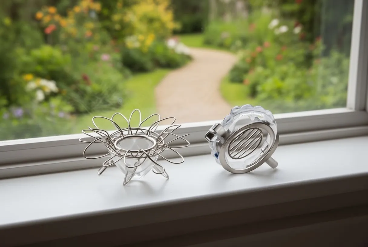
Watching the new valve over time. Follow-up echocardiograms after TAVR or surgical replacement check the new valve's function, including its own gradient. A mildly elevated gradient across a new valve can be normal, especially with a smaller valve size. What matters is the trend: a stable gradient is reassuring, while a rising gradient over successive studies raises the question of valve deterioration or other dysfunction and prompts closer review.
Research on gradients and long-term outcome. Studies have examined how the gradient measured after valve procedures relates to outcomes, and elevated residual or early post-procedure gradients have been associated with differences in longer-term results. This is an active research area, and it reinforces the same practical message: the gradient is a number to be tracked over time, not a one-off verdict. Follow-up imaging is reviewed against international guideline criteria, so any change in the new valve can be picked up early.
When to Talk to a Cardiac Team About Your Gradient
Warning signs that should not wait. The gradient itself is a number on a report, but your symptoms are the alarm bell. Seek medical attention promptly if you experience fainting or near-fainting, new or worsening breathlessness on modest activity, chest pressure with exertion, or palpitations with light-headedness. In established aortic stenosis, these symptoms mark a change in stage and deserve urgent assessment rather than watchful waiting.
Making sense of your report before the appointment. It helps to arrive at any consultation with your echo report and know three numbers: the mean gradient, the valve area, and the peak velocity. It also helps to know when the study was done, because stenosis progresses over years. A gradient in the moderate range today may be severe within a couple of years, which is why serial monitoring — usually yearly echo, or more often if values are drifting — is part of every stenosis patient's care.
- Bring the actual images or report, not just a verbal summary — trends matter more than single readings.
- Describe your symptoms in your own words: what you could do six months ago that you cannot do now is more informative than any single number.
- Ask whether the picture is complete: if symptoms and numbers disagree, ask what further testing would resolve it.
Getting a proper evaluation. If your numbers or symptoms suggest aortic stenosis, the next step is a conversation with a cardiac team that can look at the whole picture. Our first contact is a free video consultation: we get to know you, talk through your condition, and tell you exactly which studies are still needed so that no unnecessary ones are ordered. A face-to-face visit at one of our consulting rooms in Guadalajara, Querétaro or Hermosillo follows only when you decide to move forward.

The bottom line. The transvalvular gradient in aortic stenosis is the pressure price your heart pays to push blood through a narrowed valve. A mean gradient of 40 mmHg or more, with a valve area of 1.0 cm² or less and symptoms, is the classic picture that leads teams to recommend valve replacement — while low-gradient patterns need a more careful, individualised work-up. Numbers guide decisions; they never replace the judgement of an experienced team or the voice of the patient. If you have a report with a gradient you do not fully understand, do not self-diagnose from it: bring it to a qualified cardiac team, ask your questions, and let the decision be made with you, not for you. Your heart — and your peace of mind — deserve nothing less.