Confirming a Heart Diagnosis Before Surgery
Confirming a heart diagnosis before surgery: the tests and steps specialists use to verify your condition before any operation is planned.
Hearing that you may need heart surgery is overwhelming. Before any operation happens, your medical team needs to be certain about what is actually wrong — this process of heart diagnosis confirmation before surgery protects you from undergoing a procedure you may not need, or from having a procedure planned on incomplete information. This guide walks through every test and step specialists use to verify a heart condition before an operation, in plain language, so you know what to expect and what questions to ask.
What Is Heart Diagnosis Confirmation Before Surgery?
The definition in plain terms. Heart diagnosis confirmation before surgery means proving, with objective tests, that the condition your cardiologist suspects is really present — and that it is the actual cause of your symptoms. A suspicion based on symptoms alone is a starting point, never a reason to open the chest. Confirmation usually involves several complementary tests, because each one looks at a different part of the heart's anatomy and function.
Why one test is never enough. An electrocardiogram (a tracing of the heart's electrical activity) can suggest a problem, but it cannot show a blocked artery or a leaking valve directly. An echocardiogram — an ultrasound scan of the heart — shows the valves and pumping chambers beautifully, but it may underestimate blockages in the coronary arteries, the vessels that feed the heart muscle. That is why specialists build a diagnostic picture layer by layer, and why a complete confirmation process typically takes days or weeks, not a single afternoon.
What confirmation changes. Once the diagnosis is confirmed, everything downstream changes: which operation is offered, whether a minimally invasive approach is possible, and what your realistic outcome looks like. At our practice, every case is reviewed by the multidisciplinary team against international practice guidelines before any recommendation is given, so the diagnosis itself is examined by several specialists, not just one.
Why Is Confirming a Heart Condition Before an Operation So Important?
The cost of operating on a wrong or incomplete diagnosis. Heart surgery carries real risks, and those risks are only justified when the operation clearly solves a confirmed problem. Operating on a valve that is only mildly diseased, or bypassing arteries that are not the true source of symptoms, exposes the patient to serious risk without a matching benefit. Confirmation protects the patient first and foremost.
The problem of "symptom overlap." Chest discomfort, breathlessness, and fatigue are shared by many conditions: coronary artery disease, valve disease, anemia, thyroid problems, lung disease, and acid reflux can all mimic one another. This is why the workup often includes blood tests and lung assessments alongside heart imaging — the team must exclude other explanations before attributing symptoms to the heart.
Confirmation also shapes the type of surgery. The same confirmed diagnosis can often be treated in more than one way. A narrowed aortic valve (the main outflow valve of the heart) might be replaceable through open surgery, through a small incision, or through a catheter-based procedure called TAVR — transcatheter aortic valve replacement — where a new valve is delivered through an artery in the groin without opening the chest. Which route is right depends on the confirmed anatomy and the patient's overall condition. We perform conventional, minimally invasive, robotic and transcatheter procedures, and we recommend the less invasive route only where the evidence supports it for that specific patient.
What Does an Echocardiogram Before a Heart Operation Show?
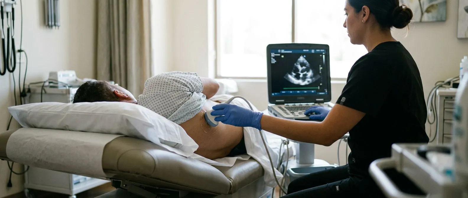
The cornerstone of cardiac imaging. An echocardiogram is an ultrasound scan of the heart, the same painless technology used to see babies before birth. It shows the four chambers, the four valves, the thickness of the heart muscle, and how well the heart pumps with each beat. For most valve problems, the echocardiogram is the single most important test, and the diagnosis of a leaking or narrowed valve is essentially confirmed on it.
Key measurements your team will discuss. The report will include numbers that sound technical but matter enormously:
- Ejection fraction: the percentage of blood the heart pumps out with each beat — a direct measure of pumping strength.
- Valve area and gradient: for a narrowed valve, how tight the narrowing is and how much pressure the heart must generate to push blood through it.
- Chamber sizes: enlarged chambers can show that the heart has been straining under an untreated problem, sometimes for years.
- Leak severity: for a leaking (regurgitant) valve, how much blood flows backward with each beat.
When a standard echo is not enough. Sometimes images are unclear — because of body habitus, lung disease, or prosthetic material — and a transesophageal echocardiogram is ordered. Here the ultrasound probe is swallowed as a thin, sedated tube, positioning it right behind the heart for far sharper images. In our consultations we take the time to explain what these studies actually show, in plain words, before any decision is made, because a patient who understands their echo understands their own diagnosis.
When Is Cardiac Catheterization Needed Before Surgery?
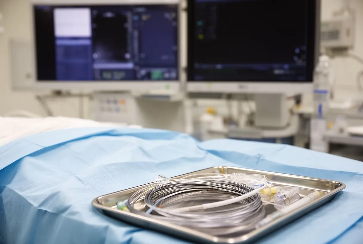
What the procedure actually involves. Cardiac catheterization — often called an angiogram — is a test in which a thin, flexible tube (catheter) is threaded through an artery, usually in the wrist or groin, up to the heart. Contrast dye is injected into the coronary arteries and X-ray video shows exactly where and how severely they are narrowed. Dye can also be injected into the heart chambers and across the valves to measure pressures directly.
Why nearly every open-heart operation requires it. Before bypass surgery, the surgeon must know precisely which arteries are blocked, how far the blockages extend, and which vessels are suitable targets for grafts. Before valve surgery, catheterization rules out co-existing coronary disease that should be treated in the same operation — discovering it after surgery would mean a second procedure. This is why catheterization is standard in the preoperative workup for most planned cardiac operations.
What it feels like and what the risks are. The test is done under local anesthetic and light sedation; most patients are comfortably awake and go home the same day. You may feel brief warmth when the dye is injected. Serious complications are uncommon but real — bleeding at the puncture site, kidney effects from the dye, and rarely injury to an artery — and your team should discuss them candidly beforehand rather than wave them away.
How Does a Stress Test Before a Heart Procedure Help?
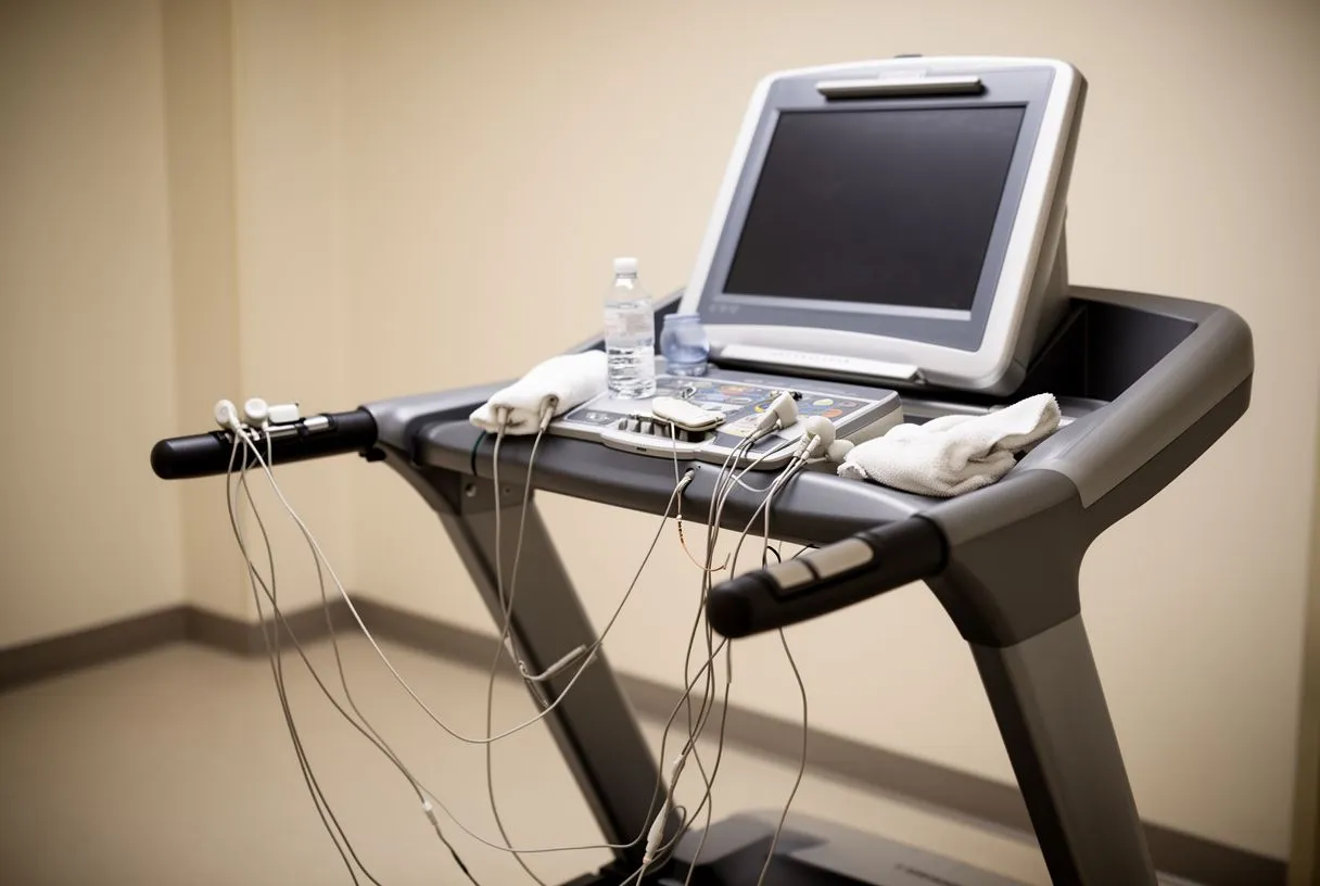
Making the heart reveal its limits. Some blockages do not restrict blood flow at rest, so they can hide on resting tests. A stress test — on a treadmill, on a bicycle, or with medication that simulates exercise for patients who cannot walk — forces the heart to work harder. If a narrowed artery cannot keep up, the heart muscle beyond it shows characteristic changes on the monitoring, revealing territory at risk that a resting test would have missed.
The varieties you may encounter. A plain exercise electrocardiogram is the simplest form. A stress echocardiogram adds ultrasound images taken before and immediately after exertion, showing wall segments that stop contracting properly. A nuclear perfusion scan uses a small tracer dose to create pictures of blood distribution across the heart muscle. Each has strengths, and the choice depends on the question being asked and the patient's ability to exercise.
What the result changes. A stress test does not usually confirm a diagnosis on its own, but it answers a crucial surgical question: which parts of the heart muscle are jeopardized, and how much. That information helps determine whether bypass surgery will genuinely relieve symptoms and improve outlook, or whether medication alone is the better path. We say honestly which studies are still needed and which are not, so no unnecessary ones are ordered.
When Is a Cardiac MRI Ordered Before Surgery?
The most detailed picture of heart muscle. A cardiac MRI uses a powerful magnetic field — no radiation — to photograph the heart in fine detail. Its unique strength is tissue characterization: it can distinguish scarred muscle from stunned but living muscle, identify inflammation, and measure precisely how much of the heart muscle has been damaged by a previous infarction. That distinction is decisive in planning surgery, because operating offers little benefit to muscle that has already died, while living but under-supplied muscle is exactly what bypass is meant to rescue.
Where MRI settles diagnostic uncertainty. When an echocardiogram suggests an uncommon condition — thickened or restrictive heart muscle, congenital abnormalities, or ambiguous masses — MRI often provides the definitive answer. It is also the reference test for quantifying right ventricular function, which matters in certain congenital and valve conditions.
Practical realities. The scan takes 45 to 90 minutes inside a narrow, noisy tube; patients with certain pacemakers or implanted devices may need modified protocols or an alternative test. If your team orders an MRI, it is worth asking why — a good answer will connect the scan directly to a surgical decision, not to general curiosity.
How Do Specialists Verify Heart Blockage Before Bypass Surgery?
Matching the angiogram to the symptoms. Confirming that a blockage explains the symptoms is a two-step logic. First, the angiogram must show a narrowing in a territory that matches the symptoms — chest pressure with a critical blockage of the artery supplying the front of the heart, for example. Second, tests must show that the muscle supplied by that artery is still alive and under threat. Both pieces together justify bypass surgery.
Pressure measurements inside the artery. Modern catheterization can go further than pictures: a thin pressure wire measures blood flow across a borderline narrowing during catheterization. If the measured pressure drop falls below a validated threshold, the blockage is proven to be limiting flow and deserves treatment; if not, medication is usually the better first step. This technology has prevented many unnecessary stents and operations.
Coronary anatomy drives the surgical plan. Once blockages are verified, the surgeon maps which arteries will be grafted, which conduit will be used (a vein from the leg, or the internal mammary artery from the chest wall, which has the best long-term durability), and whether the operation can be done on or off the heart-lung machine. Walking patients through their own angiogram images, pointing at the blockages on the screen, can turn an abstract fear into a concrete, understandable plan.
What Does Heart Valve Diagnosis Verification Involve?
Confirming severity against guideline thresholds. Valve disease is graded: mild, moderate, severe. Severe aortic stenosis (a tightly narrowed aortic valve) is typically defined by a valve area around 1.0 square centimeter or less with characteristic pressure measurements on echo. Aortic aneurysms — bulges of the body's main artery — carry a widely used threshold too: repair is generally considered when the aortic diameter exceeds 5.5 centimeters, or at smaller sizes in certain inherited conditions. These numbers, not anyone's impression, drive the recommendation.
When symptoms and measurements disagree. Some patients have severe valve disease on paper but few symptoms; others have disabling symptoms with borderline measurements. In these gray zones, extra tests — exercise echo, cardiac MRI, or BNP blood tests (a hormone the stretched heart releases) — help confirm whether the valve truly needs treating now. This is exactly where a second look by a different specialist adds real value, and we treat seeking a second opinion as a normal, sensible part of deciding about heart surgery.
A valve judged "borderline" on one set of images can look clearly severe, or clearly mild, once a multidisciplinary team reviews the echo, the catheterization and the exercise data together.
What Is a Cardiac Risk Assessment Before an Operation?
Risk scores put numbers on the decision. Before offering any operation, teams estimate the individual patient's operative risk using validated scoring tools such as EuroSCORE II, which weighs age, kidney function, lung function, prior strokes, the urgency of the case, and the type of operation. The score does not predict any single person's outcome with certainty, but it anchors an honest conversation: it tells the patient what the average risk looks like for someone with their profile.
Optimization before the operation. A confirmed diagnosis also allows time to prepare the body. Uncontrolled diabetes, anemia, active smoking, dental infections, and untreated atrial fibrillation (a common rhythm disturbance) all raise operative risk; correcting what can be corrected before surgery measurably improves recovery. In patients with significant pumping weakness, the heart failure evaluation that precedes surgery follows established guideline principles for staging and medical therapy — principles set out in documents such as the 2016 ESC Guidelines for the diagnosis and treatment of acute and chronic heart failure.
What a realistic risk conversation sounds like. Beware of any team that quotes a risk of zero. An honest assessment names the specific complications relevant to the planned operation — bleeding, stroke, kidney injury, rhythm problems, wound infection — alongside their approximate frequencies for your risk category. At our consulting rooms in Guadalajara, Querétaro and Hermosillo, every proposal to a patient includes the alternatives and their trade-offs, and the final decision is always the patient's.
Why Get a Heart Surgeon's Diagnosis Review or Second Opinion?
A surgeon sees the diagnosis differently. A cardiologist confirms what is wrong; a cardiac surgeon evaluates whether an operation is the right answer, which approach fits, and how the procedure would technically be performed. When a heart surgeon reviews the diagnosis before surgery, they may see an option that was never mentioned — a robotic approach through small incisions, or a TAVR route that avoids open surgery entirely — or they may confirm that the original plan was already the best one, which is equally valuable.
The evidence that second opinions change plans. Studies of surgical second opinions consistently show that a meaningful share of recommended operations are revised after review — sometimes the operation changes technique, sometimes the urgency changes, and sometimes the diagnosis itself is reconsidered. Bringing your images, reports and medication list makes that review far more productive.
How a proper remote review works. The first contact with our team is a free video consultation: we get to know the patient, talk through the condition, and say which studies are still needed so no unnecessary ones are ordered. A face-to-face visit at the consulting rooms follows when the patient decides to go ahead. If you are weighing a major heart operation, the most useful next step is not more internet research — it is sitting down with a qualified cardiac team that will confirm your diagnosis, explain every option, and respect your right to decide. Dr Molina trained in cardiothoracic surgery at the Universidad de Guadalajara, in cardiac, minimally invasive, robotic and structural surgery at the University of Montreal, and in structural cardiology and TAVR at CHU Bordeaux, and our team reviews every case together before recommending anything.
Heart diagnosis confirmation before surgery is not bureaucratic caution — it is the process that helps ensure the operation you undergo matches the problem you actually have. Build your questions around the tests covered here: what does each study show, which ones are still missing, and what happens if the findings disagree? Any trustworthy cardiac team will welcome those questions, encourage a second opinion, and only recommend surgery once the diagnosis is confirmed and the alternatives have been laid out clearly in front of you.