

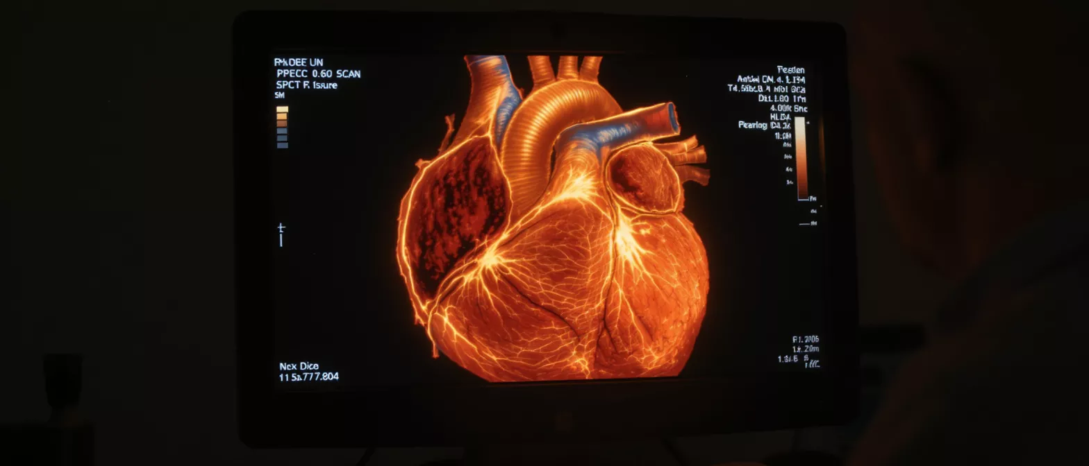
Nuclear Perfusion Scan
Learn how a nuclear perfusion scan evaluates heart blood flow, what to expect, and how to prepare.
What is a nuclear perfusion scan
When your cardiologist needs to see how well blood is flowing to your heart muscle, they may order a nuclear perfusion scan. This non-invasive diagnostic test uses a small amount of radioactive tracer to create detailed pictures of your heart. It helps your medical team determine if specific areas of the heart are receiving enough oxygen-rich blood, both while you are resting and under stress.
Unlike a standard x-ray that just shows the structure of your bones and organs, this test reveals how your heart muscle actually functions. It allows doctors to see the direct impact of any potential blockages in your coronary arteries. Understanding this blood flow is a critical step in deciding whether you need medication, a stent, or heart surgery.
The term itself can sound intimidating, especially when you are already facing cardiac concerns. However, "perfusion" simply means the passage of fluid through the circulatory system. In this case, it refers to how effectively your heart tissue is being nourished. The scan provides a clear map of this process so your surgical team can plan the safest route forward.
Why do I need a myocardial perfusion scan
Your doctor might recommend a myocardial perfusion scan if you are experiencing symptoms like unexplained chest pain, shortness of breath, or uncharacteristic fatigue. It is a powerful tool for diagnosing coronary artery disease early. By comparing blood flow at rest and during exertion, the test shows exactly which parts of the heart are starving for oxygen.
Distinguishing between old and new blockages: If you have a history of heart issues, this test helps your care team see if a previously treated area has narrowed again. It provides a clear picture of whether your current medications are working or if your treatment plan needs to be adjusted before any surgical intervention is considered.
Assessing surgical risk before an operation: If you are preparing for a major heart surgery like a bypass or valve replacement, your surgeon needs to confirm the underlying health of your heart muscle. A scan that shows poor blood flow in a specific region will guide the surgeon to that exact area, ensuring the procedure addresses the root cause of the problem.
How does cardiac perfusion imaging work
Cardiac perfusion imaging relies on a specially designed radioactive tracer, which is essentially a short-lived, safe dye. This substance is injected into your bloodstream through an IV line. As the tracer circulates, it is absorbed by your heart muscle in direct proportion to the amount of blood flow that area receives. Healthy tissue absorbs the tracer normally, while areas with restricted blood flow appear as dark spots.
A specialized camera, usually positioned just above your chest, detects the energy emitted by the tracer. This camera rotates around your body to capture multiple angles. A computer then compiles these images into detailed cross-sections and three-dimensional models of your heart. This process is known as a cardiac nuclear medicine study and is standard practice in modern cardiology.
By taking two sets of pictures—one while you are resting and another after your heart has been stressed—the technology reveals the difference in blood flow. If blood flow is normal at rest but reduced during stress, it usually indicates a partial blockage. If the blood flow is reduced both at rest and during stress, it may suggest a more severe blockage or previous tissue damage.
What to expect during a nuclear stress test perfusion
Understanding the procedure can ease much of the anxiety associated with a nuclear stress test perfusion. The test is typically divided into two phases: the resting phase and the stress phase. You will be closely monitored by a technician throughout the entire process, and the environment is designed for patient safety and comfort.
During the stress portion of the test, your heart rate needs to be elevated. If you are physically able, you will walk on a treadmill. If you cannot exercise, you will be given a medication that safely mimics the effects of exercise on your heart. Once your heart is working harder, the tracer is injected and the first set of images is taken.
- The Resting Phase: An IV is placed, and the tracer is injected. You wait quietly for about 30 minutes before the first set of images is captured.
- The Stress Phase: Your heart is stressed via treadmill or medication. Your heart rate, blood pressure, and rhythm are continuously monitored.
- Image Capture: You lie flat under a gamma camera for 15 to 20 minutes while it captures the detailed blood flow images.
How to prepare for a heart blood flow test
Preparation for a heart blood flow test is straightforward, but following your doctor's instructions carefully ensures accurate results. You will typically be asked to avoid caffeine for 24 hours before the test, as caffeine can artificially alter your heart rate and interfere with certain stress medications. This includes coffee, tea, chocolate, and some sodas.
Medication adjustments: Your cardiologist will review your current prescriptions and let you know which ones to take the morning of the test and which to skip. Certain medications, like beta-blockers, might need to be paused so your heart rate can rise appropriately during the stress phase. Never stop taking medication without specific instructions from your surgical team.
You should wear loose, comfortable clothing and walking shoes, as you may be exercising. It is best to eat a light meal a few hours before the appointment, unless instructed otherwise, and drink plenty of water to help flush the tracer from your system afterward. Remember to bring a list of your current medications and a form of identification to the clinic.
What do perfusion scan results explained mean
Waiting for test results can be stressful, but having perfusion scan results explained clearly by your doctor helps demystify the process. A normal result means your heart muscle is receiving an adequate and even supply of blood, both at rest and under stress. This usually indicates that major coronary arteries are clear of significant blockages.
An abnormal result does not necessarily mean you need immediate surgery. It simply shows that there is an area of the heart where blood flow is compromised. Your doctor will look at the specific pattern of the tracer to determine if the issue is a temporary shortage of blood, known as ischemia, or permanent damage from a previous heart attack. They may then recommend a myocardial perfusion imaging test to monitor changes over time.
These results serve as a roadmap for your care. If a reversible defect is found, it suggests a blockage that might be treated with medication or a stent. If a fixed defect is present, it means the tissue is scarred, and the focus shifts to preventing further damage. Your surgical team will use this data to decide if a bypass operation is necessary.
Are there risks with a cardiac SPECT scan
Many patients worry about radiation, but the risks associated with a cardiac SPECT scan are very low. The amount of radioactive tracer used is carefully measured to be just enough for imaging while remaining completely safe for your body. The tracer loses its radioactivity very quickly and is eliminated naturally through your urine over the course of a day.
Allergic reactions: Severe allergic reactions to the tracer are exceptionally rare. The compound is designed to pass through your system without interacting with your body's chemistry. If you have a history of severe allergies to contrast dyes or medications, inform your technician beforehand.
Stress test safety: Because the test includes a stress component, your heart rate and blood pressure will increase. You will be continuously monitored by medical professionals equipped to handle any rare adverse events. If you feel chest pain, dizziness, or extreme shortness of breath during the test, the technicians will stop immediately and attend to you.
How does nuclear cardiology diagnostic test differ from others
A nuclear cardiology diagnostic test offers unique advantages compared to other heart tests. A standard electrocardiogram (ECG) records the electrical activity of your heart but does not show the actual blood flow or the extent of structural damage. An echocardiogram uses sound waves to show the heart's pumping motion, but it can miss subtle regional differences in blood supply.
Perfusion scanning bridges this gap by showing function down to the tissue level. It reveals exactly how well the heart muscle is being fed, making it a highly sensitive tool for detecting ischemia. Because it highlights areas that might still be salvageable before permanent damage occurs, it is an essential tool for planning interventions. Patients often appreciate that they can consult with a nuclear cardiology specialist to review these detailed findings.
While an angiogram provides a detailed look at the plumbing of the arteries, a nuclear scan reveals how that plumbing affects the living tissue. This functional information is crucial. It tells your surgeon not just where the blockage is, but whether fixing it will actually restore health to that part of the heart.
Recovering after a heart perfusion study
Recovery from a heart perfusion study is generally immediate and requires no downtime. Because it is a non-invasive diagnostic test, you will not have incisions or require anesthesia. Once the imaging is complete, the IV is removed, and you can usually return to your normal daily activities immediately.
Hydration and rest: The most important step after the test is to drink plenty of water. Staying hydrated helps your kidneys flush the remaining radioactive tracer out of your body efficiently. You may resume your normal diet and medications unless your doctor has specifically instructed otherwise based on the test findings.
If you underwent the pharmacological stress portion of the test, you might experience a mild headache, flushing, or a slightly elevated heart rate for a few minutes after the medication is stopped. These sensations are normal and fade quickly. Your care team will ensure you feel stable and well before you leave the facility.
Questions to ask about your coronary blood flow scan
Being an active participant in your care leads to better outcomes. When discussing the results of your coronary blood flow scan, having a list of questions prepared can help you understand your condition and your surgical options. Do not hesitate to ask your surgeon or cardiologist to clarify anything that seems confusing.
It is helpful to know exactly what the images showed. Ask if there were any areas of reduced blood flow and whether those areas are considered reversible or permanent. Inquire about the next steps: do these results change your medication, or do they indicate that a surgical procedure like a bypass is necessary?
Finally, discuss your overall risk factors. The scan provides a snapshot of your heart's health today, but managing the underlying cause of any blockages is a lifelong process. Ask what lifestyle changes, dietary adjustments, or exercise routines will best support your heart health in the long term. Your surgical team is there to guide you through both the test results and the path to recovery.






