

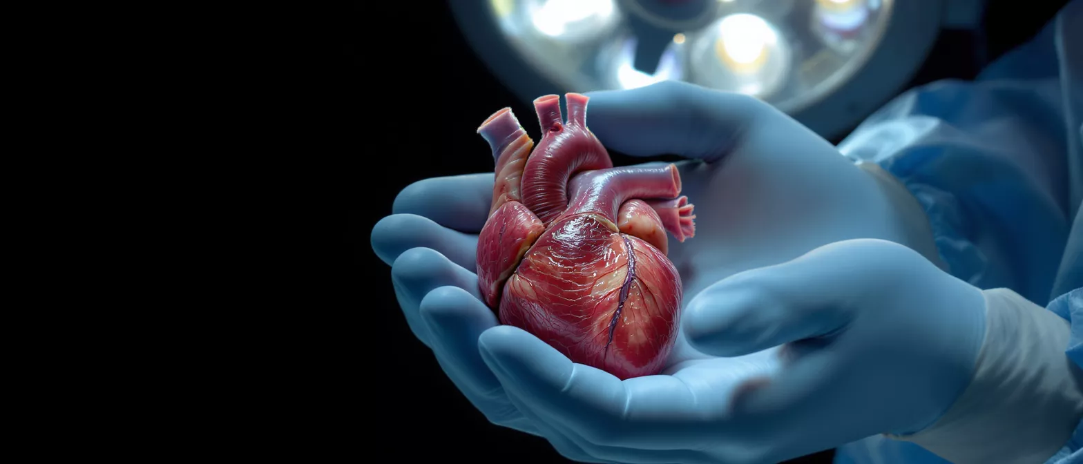
Heart
Explore our heart glossary to learn about heart anatomy, function, valves, and cardiac conditions.
What is the human heart?
The human heart is a muscular organ roughly the size of a closed fist, located slightly to the left of the center of the chest. Its primary purpose is to act as a tireless pump, circulating blood throughout the body to deliver oxygen and vital nutrients to tissues and organs. For a patient preparing for cardiac surgery, understanding this organ demystifies the surgical process and highlights why the planned procedure is necessary.
Every minute of every day, this organ contracts and relaxes in a coordinated rhythm, moving blood through two distinct phases: systole, when it pushes blood out, and diastole, when it fills with blood again. This continuous cycle ensures that your brain, kidneys, liver, and every other organ receive the sustenance they need to keep you alive and healthy.
When disease affects this crucial pump, symptoms like fatigue, shortness of breath, or chest pain often develop. These signs indicate that the heart's structure or electrical system needs medical attention, which is precisely where surgical intervention can repair damage and restore normal function.
How the heart works in the cardiovascular system
The heart functions as the central hub of the entire cardiovascular system, working in tandem with a vast network of arteries, veins, and capillaries. It manages two separate but simultaneous circuits: the pulmonary circuit, which sends blood to the lungs to gather oxygen, and the systemic circuit, which pumps that freshly oxygenated blood out to the rest of the body.
Pulmonary and systemic circulation: The right side of the heart handles the pulmonary circuit, pushing oxygen-depleted blood into the lungs to release carbon dioxide and pick up oxygen. Meanwhile, the left side of the heart manages the systemic circuit, driving the newly enriched blood out through the aorta to nourish your entire body. Keeping these systems distinct is vital for overall health.
Disruptions in this transportation network, such as blockages in the coronary arteries, force the heart to work harder to maintain this critical flow. Understanding this interplay helps patients grasp why a bypass or valve repair directly improves their entire body's oxygen supply, not just the organ itself.
Heart anatomy: chambers and structure
Heart anatomy is defined by its four distinct chambers, divided into two upper receiving areas called atria and two lower pumping areas called ventricles. The right atrium receives oxygen-poor blood from the body, passing it down to the right ventricle, which then pumps it to the lungs. The left side mirrors this process, with the left atrium receiving oxygen-rich blood from the lungs and the left ventricle pumping it out to the body.
Cardiac tissue and the septum: The walls of these chambers are made of specialized muscle tissue called the myocardium, which differs from the skeletal muscles in your arms and legs because it does not tire. A solid wall of muscle called the septum divides the right and left sides, ensuring that oxygen-rich and oxygen-poor blood never mix. Maintaining structural integrity is essential, as surgical patients may require procedures to repair holes in the septum or reinforce weakened muscle.
Patients facing cardiac surgery often have conditions affecting these specific structures, such as an enlarged left ventricle or a damaged septum. Knowing the layout of these chambers helps you visualize exactly where the surgeon will focus during the operation.
What are the heart valves?
Heart valves are essential structures that act as one-way doors, ensuring blood flows in the correct direction without leaking backward. There are four valves in total: the tricuspid, pulmonary, mitral, and aortic valves. Each opens and closes precisely with every heartbeat to maintain efficient circulation.
- Mitral valve: Regulates blood flow between the left atrium and left ventricle; commonly repaired if it begins to leak.
- Aortic valve: Controls blood exiting the left ventricle into the aorta; frequently replaced when it becomes narrowed or calcified.
- Tricuspid and Pulmonary valves: Manage blood flow on the right side of the heart, ensuring blood moves properly toward the lungs.
Valvular heart disease occurs when these doors fail to open fully, known as stenosis, or fail to close tightly, known as regurgitation. Surgical repairs or valve replacements restore this one-way flow, relieving symptoms and preventing long-term damage to the cardiac muscle.
What does a normal heartbeat look like?
A normal heartbeat is driven by a highly sophisticated electrical system that coordinates the precise timing of muscle contractions. The process begins in the sinoatrial node, a small cluster of cells in the right atrium often called the heart's natural pacemaker. This electrical signal spreads rapidly, causing the chambers to contract in a synchronized rhythm that you feel as your pulse.
Electrical conduction and arrhythmias: After leaving the pacemaker, the electrical impulse travels through specialized pathways to the atrioventricular node, and then down into the ventricles, prompting them to squeeze forcefully. When this electrical wiring is damaged or misfires, it results in arrhythmias, which are irregular heartbeats that can make the muscle pump too fast, too slow, or inefficiently.
Cardiac surgery sometimes involves correcting these electrical pathways, or the structural damage causing them, to restore a normal rhythm. Understanding this electrical network reassures patients that arrhythmias are mechanical and electrical issues that can often be treated effectively.
Types of heart and cardiac conditions
Cardiac conditions generally fall into a few major categories based on which part of the organ's structure is affected. Coronary artery disease is the most common, caused by a buildup of plaque in the arteries that supply the heart muscle itself, leading to inadequate blood flow. Patients often require bypass grafting solutions to route blood around these dangerous blockages.
Valvular disease, structural defects, and arrhythmias represent other major categories. Valvular diseases affect the doors between chambers, structural defects involve physical abnormalities like holes in the septum, and arrhythmias involve the electrical wiring. Each requires a specific surgical approach tailored to the underlying anatomical problem.
Identifying the exact type of condition is the first step in planning a successful surgical intervention. This categorization allows the surgical team to choose the least invasive, most effective method to restore normal cardiac function and improve the patient's quality of life.
Why heart health matters before surgery
Heart health prior to surgery plays a significant role in how well a patient recovers. A stronger cardiac muscle and clearer vessels make surgical procedures smoother and reduce the risk of post-operative complications. Maintaining good cardiovascular fitness before an operation directly supports the body's ability to heal and adapt afterward.
Lifestyle factors and preparation: Patients are often advised to stop smoking, manage blood pressure, and control blood sugar levels in the weeks leading up to a procedure. These actions reduce inflammation and stabilize the cardiovascular system, creating a safer environment for the surgeon to work in.
Understanding your baseline cardiac health also helps your medical team anticipate needs during recovery. Honest discussions about your lifestyle, medications, and symptoms allow the surgical team to tailor their approach, ensuring the best possible outcome for your specific anatomy.
How is heart function tested?
Cardiac function is evaluated using a combination of non-invasive imaging and electrical monitoring. An electrocardiogram, or ECG, records the electrical activity of the heart and is often the first test performed to detect irregular rhythms or signs of muscle strain. Echocardiograms use sound waves to create a moving picture of the chambers and valves, showing exactly how well the muscle pumps and whether the valves leak.
More advanced testing, like cardiac catheterization or CT scans, provides detailed images of the coronary arteries and surrounding structures. Sometimes, specialized imaging requires the use of diagnostic monitoring electrodes to capture continuous data over a 24-hour period, ensuring that intermittent issues are not missed by brief office tests.
These tests are completely safe and routine. They provide the surgical team with a precise map of your internal anatomy, allowing the surgeon to plan the exact steps of the procedure before you even enter the operating room.
Are heart terms confusing? Common definitions
Medical terminology can feel overwhelming, but breaking down the language makes it much easier to navigate a cardiac diagnosis. Learning the definitions of basic cardiac terms helps patients participate actively in their own care and communicate effectively with their medical team. Familiarity with these terms transforms a frightening diagnosis into a manageable, understood condition.
Key cardiovascular definitions: Below is a quick reference list to help you understand common phrases your surgeon might use when explaining your procedure.
- Myocardium: The muscular tissue of the heart, responsible for the contractions that pump blood throughout the body.
- Endocardium: The smooth inner lining of the chambers, providing a frictionless surface for blood to flow over.
- Pericardium: The tough, double-layered sac that surrounds and protects the heart, holding it firmly in place within the chest.
- Ejection fraction: A measurement of the percentage of blood leaving the heart each time it contracts, indicating how well the muscle pumps.
By understanding these specific medical terms, patients can read their surgical reports and pre-operative instructions with confidence, reducing anxiety through clarity.
Key takeaways on heart structure and function
Understanding the human heart transforms it from a mysterious organ into an elegant, comprehensible biological machine. By learning how the chambers, valves, and electrical pathways work together, patients facing cardiac surgery can approach their procedure with confidence rather than fear. This knowledge empowers you to have meaningful conversations with your surgeon about your specific condition and the planned repair.
Cardiac surgery is a highly developed field, and the procedures designed to repair valves, bypass blockages, or correct structural defects are remarkably effective. Knowing that your surgical team is working to restore a natural, healthy rhythm and flow should provide significant reassurance as you prepare for your operation.
Ultimately, patient education is a critical component of successful surgical outcomes. When you understand the anatomy and function of your heart, you become an active partner in your own recovery, fully equipped to follow medical guidelines and embrace a healthier future.






