Heart Imaging Scans: Echo, CT and MRI Compared
Heart imaging scans compared: learn which cardiac test your doctor orders, what each scan shows, and how to prepare.
Heart imaging scans compared side by side can feel overwhelming, especially when a doctor has just told you something about your heart and ordered a test you have never heard of. The truth is simpler than it sounds: each scan answers a different question about your heart, and your cardiology team chooses the one that answers yours. This guide walks through the three main tests — echocardiography, computed tomography (CT) and magnetic resonance imaging (MRI) — what each one shows, how to prepare, what they cost, and how the choice is made for your specific situation.
What Are Heart Imaging Scans and Why Do You Need One?
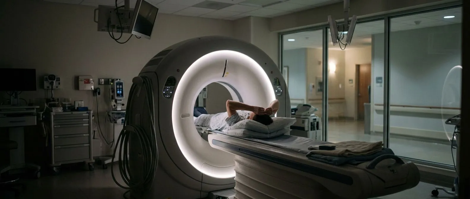
Heart imaging scans are tests that let a medical team look at your heart without opening your chest. They fall under the umbrella of non-invasive heart imaging, meaning nothing enters your body beyond, in some cases, a small intravenous line for contrast dye. The three workhorses are the echocardiogram (an ultrasound of the heart), the cardiac CT scan, and the cardiac MRI.
Each test answers a different question. An echocardiogram asks: how well is the heart pumping, and how are the valves working? A CT scan asks: are the coronary arteries — the vessels that feed the heart muscle — narrowed or blocked? An MRI asks: what is the heart muscle itself made of, and has it been damaged or scarred? Understanding this division of labour is the first step in any honest cardiac imaging comparison.
Why does this matter to you as a patient? Because no single test does everything, and more tests are not automatically better. When heart imaging scans compared properly are matched to the right question, you get the answer you need with the least expense, the least radiation and the least time. That is the principle behind every recommendation a good cardiac team makes.
Echocardiogram vs CT vs MRI: Types of Heart Scans Explained
Before heart imaging scans compared in detail make sense, it helps to see the whole picture at a glance. The three main tests overlap in some areas but each has a clear strength. Here is a quick summary you can come back to:
- Echocardiogram (echo): an ultrasound scan, done with a probe pressed on the chest. Shows the pumping function, valve movement, chamber sizes and fluid around the heart. No radiation, widely available, results the same day.
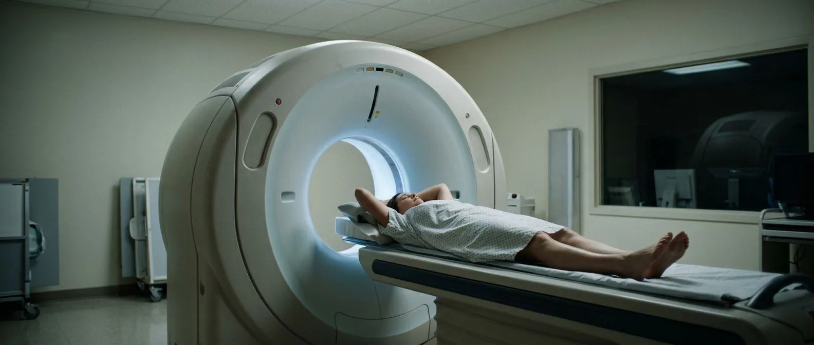
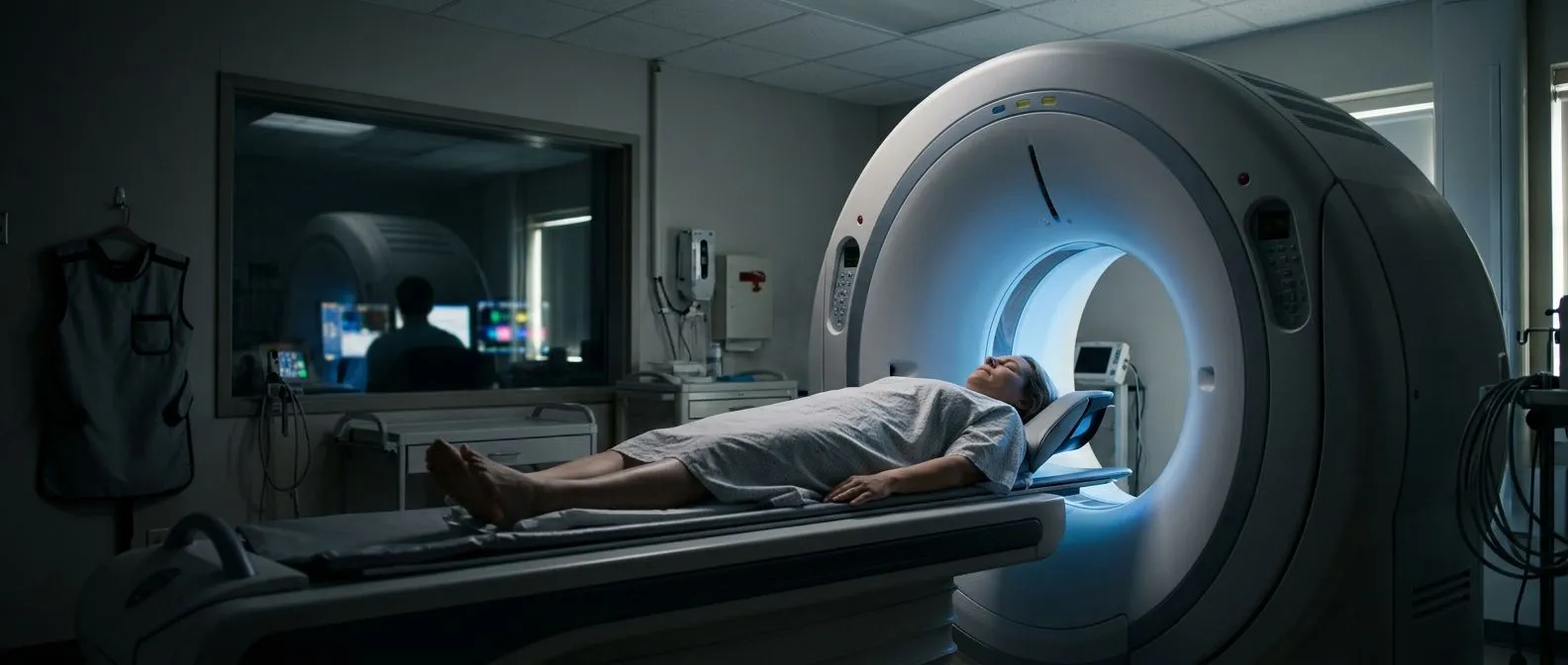
- Cardiac CT: an X-ray scan, sometimes with contrast dye injected into a vein. Shows the coronary arteries in fine detail, plus heart structure, the aorta and — with newer techniques — valve anatomy.
- Cardiac MRI: a magnetic scan, no radiation. Shows the heart muscle in exquisite detail: scars, inflammation, congenital defects and the most accurate measurement of pumping function.
These are the core types of heart scans explained in the simplest possible terms, but each also has variations. An echocardiogram can be a resting scan, a stress scan, or a transoesophageal scan where a slim probe is swallowed to look from inside the oesophagus. A CT can be a simple calcium score or a full coronary angiogram with dye. An MRI can include a stress element or a delayed scan after contrast to reveal scar tissue.
The key point in an echocardiogram vs CT vs MRI decision is that your symptoms and your diagnosis, not preference or convenience, drive the choice. A test chosen because it answers your actual question is worth far more than the most advanced scan chosen at random.
Heart Ultrasound vs MRI and CT: What the Echocardiogram Shows
When people say "heart ultrasound vs MRI", they are usually asking whether the simple scan is enough. For most patients, the echocardiogram is the first and sometimes only scan needed. It is painless, takes 20 to 40 minutes, involves no needles and no radiation, and can be repeated as often as necessary to track changes over time — something doctors call surveillance imaging.
What does the echo actually measure? It shows the four chambers of the heart, the four valves (aortic, mitral, tricuspid and pulmonary), and the pumping strength, expressed as an ejection fraction — a percentage of the blood the left ventricle pushes out with each beat. International standards for these measurements have been set out in the Recommendations for Cardiac Chamber Quantification by Echocardiography in Adults from the American Society of Echocardiography and the European Association of Cardiovascular Imaging, which is why two careful echo labs anywhere in the world should reach similar numbers.
At the first video consultation, the team explains in plain words what the echocardiogram shows. Where the echo raises a question it cannot answer — for example, exactly how a valve has been damaged, or whether the heart muscle is scarred — that is when we move on to CT or MRI. Heart imaging scans compared in this way become a sequence, not a competition.
Coronary CT Angiography vs Echo: Which Test Looks at the Arteries?
The classic debate of coronary CT angiography vs echo comes down to a simple anatomical fact: the coronary arteries are very small — two to four millimetres across — and they move constantly with every heartbeat. An echocardiogram can see the consequences of a blocked artery (weakened heart wall motion) but cannot reliably see the blockage itself. A CT scan can.
A coronary CT angiogram uses an intravenous contrast dye and a scanner timed to the heartbeat to produce three-dimensional pictures of the artery lumen, the channel blood flows through. It can show whether a narrowing is caused by soft plaque or calcified plaque, which influences treatment. A simpler CT, the coronary calcium score, takes just a few minutes with no dye and quantifies the amount of calcium in the artery walls — a useful marker in intermediate-risk patients.
When we plan a case, the coronary question and the valve question are answered with separate, purpose-chosen studies; we never order an echo and hope it will reveal the arteries. That discipline is part of reviewing every case against international practice guidelines before recommending anything, and it means fewer scans for you, not more.
Cardiac MRI vs Echo: When Your Doctor Needs the Finest Detail
The debate of cardiac MRI vs echo arises when the echo result and the clinical picture do not match. MRI uses a powerful magnet and radio waves, with no radiation, and it measures things the other tests cannot: scar tissue inside the heart muscle (using a technique called late gadolinium enhancement), inflammation of the heart muscle (myocarditis), infiltrative diseases, and subtle congenital defects.
MRI is also the gold standard for measuring pumping function precisely, especially in the right ventricle, which is hard to see well with ultrasound. It is the test of choice for cardiomyopathy — disease of the heart muscle itself — and for clarifying whether a patient who had chest symptoms years ago has permanent scar tissue.
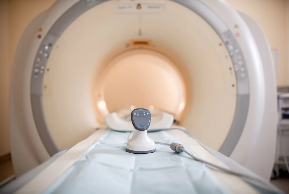
The trade-offs are real. An MRI takes 45 to 90 minutes inside a narrow, noisy tube, some people with claustrophobia need preparation or a mild sedative, and patients with certain implants (some pacemakers, older metal clips) need careful screening first. Recent research published in Frontiers in Cardiovascular Medicine continues to expand what cardiac MRI can detect, and its role keeps growing — but it complements the echo rather than replacing it.
Heart Imaging for Chest Pain: Which Scan Comes First?
Heart imaging for chest pain follows a logic, not a menu. If the pain occurs with exertion — climbing stairs, walking uphill — and stops with rest, the question is whether the coronary arteries are narrowed, and a CT coronary angiogram or a functional test is usually the right starting point. If the pain is unrelated to effort, other causes are considered first, and heart imaging may not be needed at all.
In an emergency with ongoing chest pain, hospitals use echocardiography first because it is immediate: a large heart attack shows up as a wall of the heart that suddenly stops moving. Later, if the diagnosis is still unclear, CT or MRI is added. The sequence matters because each test narrows the possibilities.
One sentence of practical advice from our consulting rooms: in the free first video consultation, we talk through the symptom story with the patient and say exactly which studies are still needed, so that no unnecessary scan is ordered — and we see many patients arrive with imaging that never needed to be done. Bring any previous results; they often answer questions a new scan would otherwise raise.
Heart Imaging for Valve Problems and TAVR Planning
Heart imaging for valve problems deserves its own section, because valves drive many treatment decisions. The echocardiogram is the cornerstone: it measures how narrow a stenotic valve is (in millimetres of mercury, as a pressure gradient), how much a leaky valve regurgitates, the size of the heart chambers, and the pumping function. These numbers determine when valve disease crosses the threshold for intervention.
When a treatment is being planned — especially a transcatheter aortic valve replacement (TAVR), where a new valve is delivered through a tube in the groin artery rather than by open surgery — much more detailed imaging is required. A CT scan is used to measure the aortic annulus (the ring where the valve sits), the size and calcification of the arteries used for access, and the exact dimensions of the valve to be chosen. The European Society of Cardiovascular Radiology has published a consensus document on CT and MR imaging prior to transcatheter aortic valve implantation that standardises exactly which measurements must be taken and reported, and careful imaging is one of the strongest predictors of a smooth TAVR procedure.
The quality of the pre-procedure CT scan shapes the quality of the procedure itself, which is why the multidisciplinary team reviews every case against international guidelines before offering a patient TAVR, minimally invasive surgery or conventional surgery. The final decision always rests with the patient.
Are Heart Scans Safe? Heart CT Radiation Concerns and MRI Safety
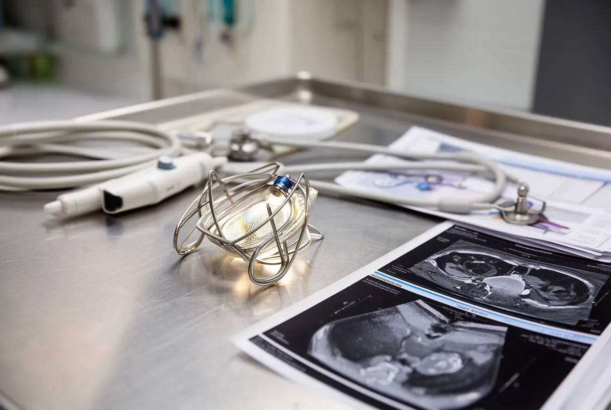
Heart CT radiation concerns are legitimate and worth discussing openly, without alarm. A modern cardiac CT uses far less radiation than scans of a decade ago — techniques such as dose modulation, lower tube voltage and prospective triggering have reduced typical doses dramatically — but it is still an X-ray test, and reasonable practice is to order it only when the answer will change your management.
MRI safety: magnetic resonance involves no radiation at all, but the strong magnet demands screening for pacemakers, defibrillators, metal heart valves from certain eras and other implants. Most modern devices are now MRI-compatible, but the check is always done before the scan is booked.
Contrast dye: CT uses iodine-based contrast and MRI uses gadolinium. Both are generally well tolerated; serious allergic reactions are rare, and kidney function is checked beforehand when it is relevant. In our practice, every patient's imaging plan is reviewed by the multidisciplinary team against international guidelines precisely so that the smallest number of safe, necessary tests is done — and we explain any risk in plain words before you agree to anything.
Heart Scan Cost in Mexico: Prices, Insurance and Practicalities
Heart scan cost in Mexico is a practical concern for local and international patients alike, and honest teams talk about it plainly. Costs vary by city, hospital and the sophistication of the scanner, and a full coronary CT angiogram with contrast costs more than a simple calcium score; a cardiac MRI costs more than an echo. The echo, being quick and equipment-light, is by far the most affordable of the three.
For patients travelling from abroad, the sequence matters as much as the price. A remote review of your existing echocardiogram and previous reports often reveals that much of the needed work-up is already done, so new scans are only ordered where a genuine gap exists. Insurance coverage differs between policies, so ask your insurer specifically whether "cardiac imaging" or "diagnostic studies" are covered and whether pre-authorisation is needed.
Our team works with patients from Guadalajara, Querétaro and Hermosillo and from further afield, and the first contact is a free video consultation in which we get to know the patient, discuss the condition, and specify only the studies that are genuinely still needed. We encourage second opinions as a normal part of deciding about heart care — a good team is never threatened by one.
The Best Heart Imaging Test: What Heart Scan Do I Need?
Patients often ask what the best heart imaging test is, and the honest answer is: the one that answers your question. If the question is "how are my valves and my pumping?" the echo is best. If it is "are my arteries blocked?" the CT is best. If it is "is my heart muscle scarred or inflamed?" the MRI is best. Asking "what heart scan do I need?" is a question for a team that has examined you and read your history, not a question a website can settle.
A useful way to think about heart imaging scans compared in your own case is as a staircase: start with the test that answers the biggest question, and only climb higher if the answer remains unclear. Most patients never need all three. Those who do, need them because each adds a piece to the diagnosis that no other scan can supply.
If you have been told something about your heart — a murmur, a valve problem, chest pain, an abnormal test — the next step is a conversation with a qualified cardiac team, not a self-diagnosis. You can discuss your situation with our multidisciplinary team led by Dr Alejandro Molina and book a consultation; we will explain what your studies actually show, in words you understand, and give you a clear proposal with all the alternatives before you decide anything.