Cardiac CT Scan: What It Shows and How to Prepare
Cardiac CT scan preparation made simple: what this heart imaging test shows, how to get ready step by step, and what to expect.
Good cardiac ct scan preparation is the single biggest factor in whether your heart images come out crisp or blurry, and blurry images often mean repeating the test. This guide walks you through what a cardiac CT scan actually is, what it shows, and exactly how to get ready, step by step. It is written for patients and families, not for doctors, so every term is explained in plain words as we go.
What Is a Cardiac CT Scan?
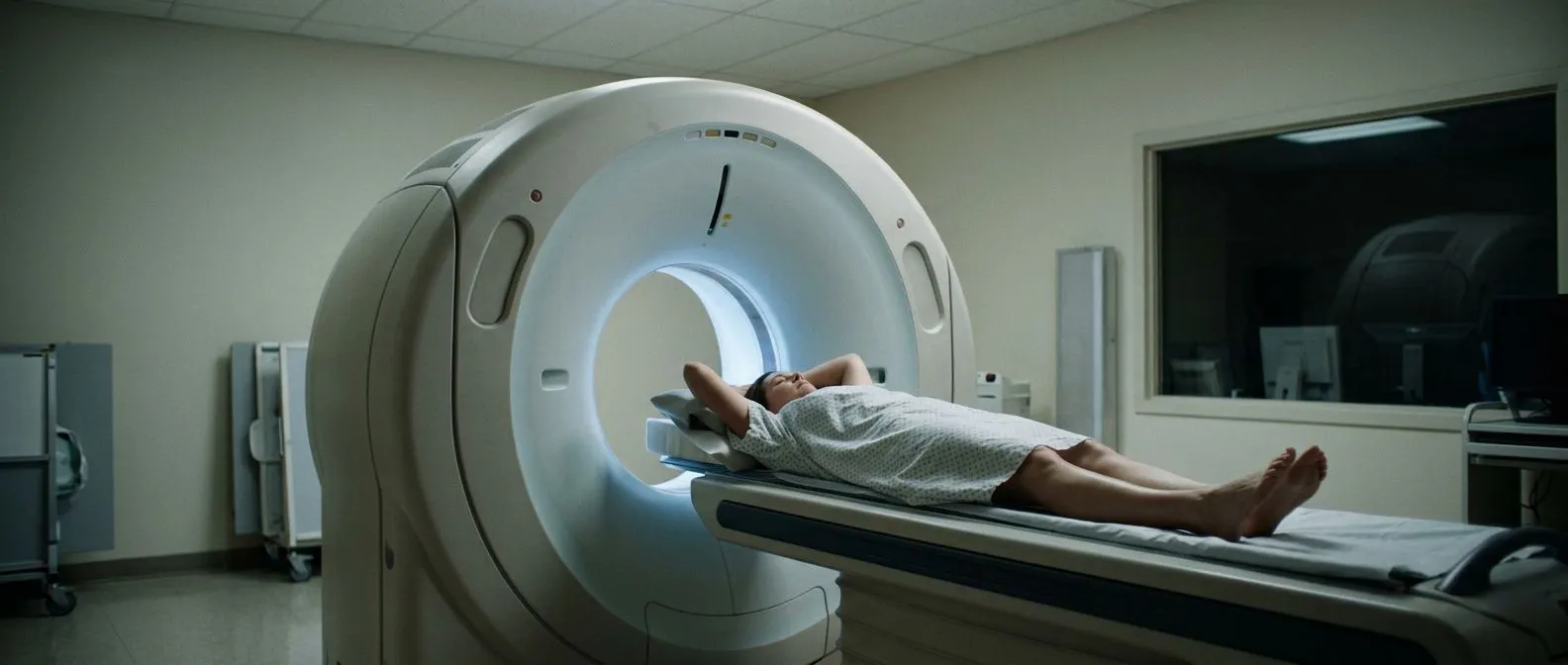
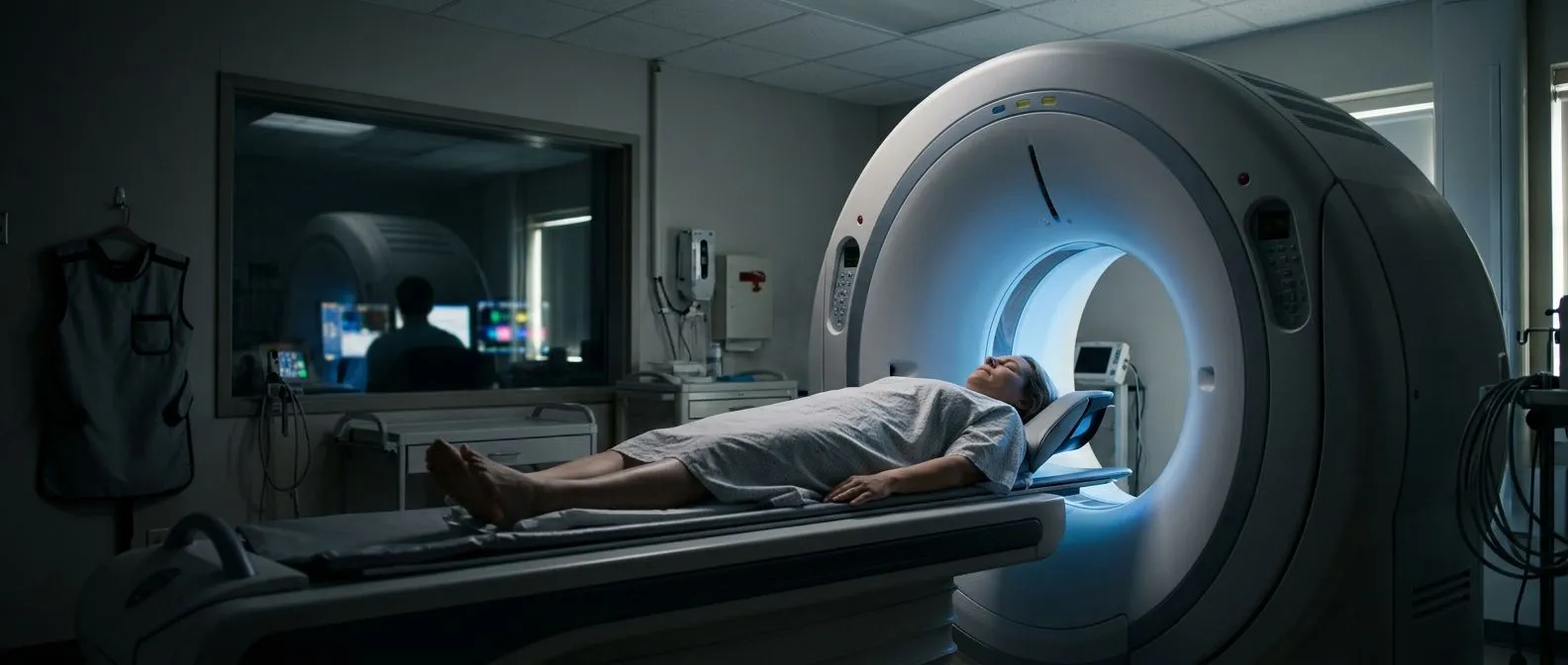
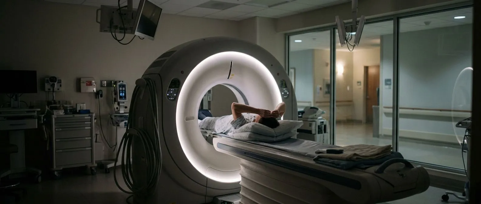
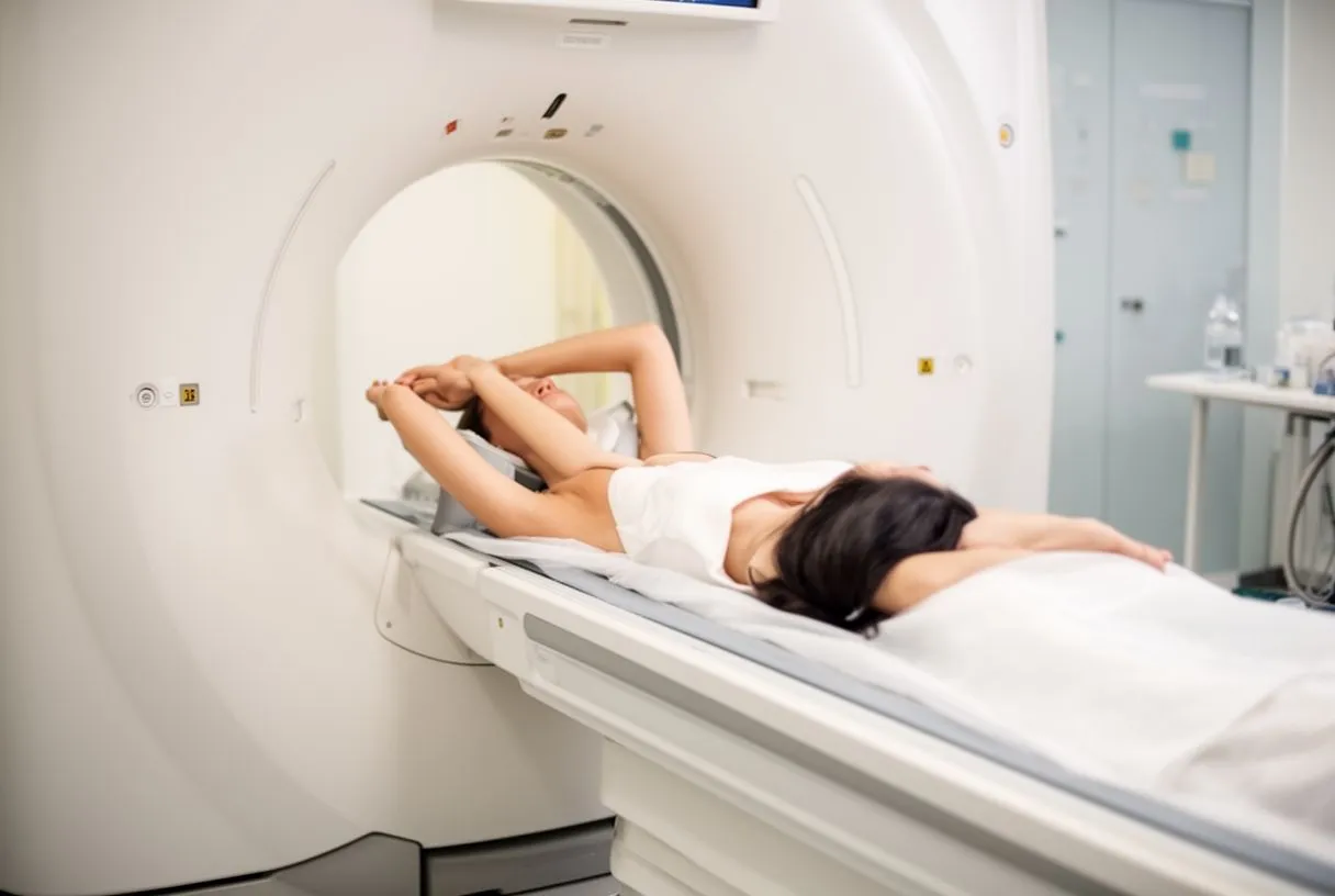
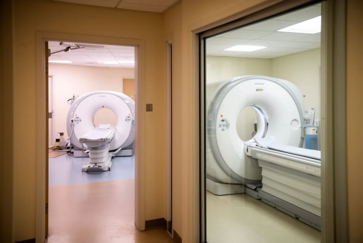
A cardiac CT scan is an imaging test that uses X-rays and a computer to build detailed pictures of your heart while it is beating. The machine is a large ring with a table that slides through it. You lie still on the table, and the scanner rotates around your chest, capturing hundreds of thin cross-sectional images in seconds.
Computed tomography explained simply. "Computed tomography" (CT) means the computer combines many X-ray snapshots into slices, like slices of bread, that a doctor can view one by one. Modern scanners take an entire sweep of the heart in a single heartbeat, which is why the test is fast despite the complexity of the pictures it produces.
Why contrast dye is usually used. Most cardiac CT scans involve an iodine-based contrast agent, a dye injected through a small line in a vein in your arm. The dye travels through your bloodstream and lights up the coronary arteries, the small vessels that feed the heart muscle, so radiologists can see narrowings or blockages inside them. The injection often causes a warm flush through the body for a few seconds; this is normal and passes quickly.
What Does a Cardiac CT Scan Show?
A cardiac CT scan can reveal a remarkable amount about your heart in one session. It shows the coronary arteries, the heart muscle itself, the four heart valves, the outer sac around the heart (the pericardium), and the aorta, the main artery carrying blood away from the heart.
Coronary artery disease and plaque. The most common reason for the test is coronary artery disease, where cholesterol deposits called plaque build up inside the artery walls. The scan shows both hard, calcified plaque and softer plaque, which matters because soft plaque can rupture and cause a heart attack even when it is not severely narrowing the vessel. Research published in the JAMA Network has shown that identifying soft plaque on CT can change how a patient is managed.
Calcium scoring. A related, simpler test called a calcium score scan takes no contrast dye. It measures the amount of calcium in your coronary arteries and produces a number that estimates your future risk of heart attack. Many centers perform this first to decide whether the full contrast scan is needed.
Valves, the aorta, and beyond. The scan can also evaluate the aortic and mitral valves, look for aneurysms (bulges) in the aorta, and check whether a structure called the left atrial appendage contains clot in patients with atrial fibrillation. When we plan surgery for a patient, we routinely review the CT images ourselves rather than relying only on the written report, because the pictures answer questions a summary cannot.
What Is the Difference Between Cardiac CT and Coronary CT Angiography?
People often ask whether "cardiac CT" and "coronary CT angiography" are different tests. In practice, they overlap heavily, and the names are used loosely.
Cardiac CT as the umbrella term. Cardiac CT covers any CT imaging of the heart: calcium scoring, coronary CT angiography (also called CCTA), valve imaging, and aortic imaging. Coronary CT angiography, or CT angiography as it is often called, is the specific study where contrast dye is injected to visualize the coronary arteries from the inside.
What CT angiography adds. CT angiography does not just show plaque on the artery wall. With modern software, it can also simulate blood flow through a narrowing and tell you whether a blockage is actually limiting blood supply to the heart muscle. This is called FFR-CT, and it helps doctors decide whether a blockage needs treatment or can simply be watched with medication.
Which one will you get? That depends on the question your doctor is asking. For chest pain in a patient with intermediate risk, coronary CT angiography is usually the first-line test. For a general risk check, a calcium score may be enough. At our practice in Guadalajara, Querétaro and Hermosillo, every case is reviewed by the multidisciplinary team against international practice guidelines before any test or treatment is recommended, so you receive a clear proposal with the alternatives.
How to Prepare for a Cardiac CT Scan: Step-by-Step
Preparing for a coronary CT scan is not difficult, but it is specific. Follow these heart ct scan preparation steps and your scan is far more likely to be diagnostic on the first attempt.
- Confirm your booking details: arrival time, the imaging center address, and whether you need any lab work beforehand, such as a kidney function test if you have diabetes or reduced kidney function.
- Review your medications with the clinic: tell the team everything you take, including over-the-counter drugs and supplements, and ask about any cardiac ct scan medication instructions specific to you.
- Avoid caffeine for 12–24 hours: coffee, tea, energy drinks, cola, and chocolate all speed the heart rate, and a fast heart makes motion blur on the images.
- Do not smoke that day: nicotine raises the heart rate the same way caffeine does.
- Wear comfortable, metal-free clothing: you will change into a gown anyway, but leave necklaces and jewelry at home.
- Bring prior imaging and reports: old ECGs, stress tests, or previous scans help the team interpret the new study in context.
Arriving early. Plan to arrive 30 to 45 minutes before your appointment. This gives staff time to check your blood pressure, attach monitors, place the IV line, and give any heart-rate medication you may need. Rushing in late often means a rushed scan.
Cardiac CT Scan Fasting Requirements: Can I Drink Water Before?
Fasting is one of the most common sources of confusion in cardiac ct scan preparation, and the rules are simpler than most patients expect.
Light fasting, not a full fast. Most centers ask you to avoid solid food for 4 to 6 hours before a contrast scan. The reason is not the heart itself but safety: if contrast dye makes you feel briefly unwell, an empty stomach reduces the risk of nausea or reflux during the scan.
Water is not only allowed, it is encouraged. Drinking water before the scan helps the veins stay plump for easy IV placement and supports your kidneys as they clear the contrast afterward. Unless your clinic gives you different instructions, drink plain water freely up to your arrival time. Patients taking metformin or with reduced kidney function may be given specific hydration instructions, so always confirm with the imaging center.
Cardiac CT Scan Caffeine Restrictions and Medication Instructions
Caffeine and certain heart medications can affect image quality, which is why cardiac ct scan caffeine restrictions and beta blocker prep deserve their own attention.
Why caffeine is restricted. Caffeine increases your resting heart rate and can also blunt the effect of the beta blocker given to slow your heart during the scan. A heart rate above roughly 60–70 beats per minute can create motion artifacts, blurred sections where the heart was moving too fast to image cleanly. In our consultations we always explain in plain words why each instruction matters, because patients who understand the reason actually follow it.
Beta blocker preparation. A beta blocker is a medication that slows the heart. Many centers give one as a tablet an hour before the scan, or intravenously just before imaging, to bring your rate into the ideal range. If you already take a beta blocker at home, you will typically be told to take your usual dose, but confirm this rather than assuming, since the answer varies by center and by study type.
Nitrates and other medications. Some protocols also give a small dose of nitroglycerin under the tongue just before imaging to widen the coronary arteries so their inner walls are easier to see. Blood thinners, diabetes tablets such as metformin, and Viagra-type medications (which must be stopped 24–48 hours before, because combining them with nitrates drops blood pressure) each have their own rules. Give the imaging center a complete medication list and let them tell you what to hold and what to take.
Cardiac CT Scan: What to Expect During the Test and How Long It Takes
Patients are often surprised at how quick the scan itself is. A common question is how long it takes to do a CT of the heart.
The timeline in real terms. Plan for about 30 to 60 minutes at the imaging center from arrival to departure. The actual scanning takes only seconds to a few minutes; the rest is preparation, IV placement, heart-rate medication, and the breathing instructions. You will be asked to hold your breath for 5 to 10 seconds at a time while images are captured.
What it feels like. The scan is painless. You lie on the table, the ring moves around your chest, the contrast injection brings a warm or metallic-tasting flush for several seconds, and then it is over. There is no tunneling and no loud hammering like an MRI. If you feel anxious about enclosed spaces or the IV line, tell the staff; they deal with this every day and can help you through it.
After the scan. You can usually leave right away and resume normal eating, drinking, and activity unless told otherwise. The main aftercare is simple: drink plenty of water over the next 24 hours to help your kidneys flush the contrast dye. We tell every patient this at the end of the appointment, because it genuinely reduces the small risk of contrast-related kidney strain.
Cardiac CT Scan vs Stress Test: Which Do You Need?
Another frequent comparison is the cardiac CT scan vs stress test, and the two answer different questions.
What a stress test measures. A stress test, whether on a treadmill or with a nuclear perfusion scan, evaluates how your heart functions under exertion. It looks at the consequences of blockages, that is, whether parts of the heart muscle are starved of blood when you exercise, rather than looking at the arteries directly.
What CT looks at instead. A coronary CT angiogram looks directly at the arteries and the plaque inside them. It can find disease before it causes symptoms and can rule out coronary artery disease with high confidence when the arteries are clean. A nuclear perfusion scan, by contrast, shows whether blood flow is already impaired.
How doctors choose. Guidelines generally favor CT angiography as the first test for chest pain in patients with low to intermediate risk, because a normal CT reliably excludes significant disease. A stress test or nuclear perfusion scan may be preferred when there are known blockages already, abnormal heart rhythms that interfere with CT imaging, or specific questions about blood flow under load. The right answer depends on your individual history, which is exactly why the choice should be made with a cardiac team rather than guessed at.
Which Is Better, Cardiac CT or MRI?
The comparison of cardiac CT versus cardiac MRI (magnetic resonance imaging) comes up constantly, and the honest answer is that neither is universally better, because they answer different questions.
Where CT wins. Cardiac CT is faster, more comfortable, and unmatched for seeing coronary arteries, calcified plaque, and the aorta. A scan that takes minutes with CT can take an hour or more with MRI. For suspected coronary artery disease and for pre-surgical planning, CT is usually the tool of choice.
Where MRI wins. Cardiac MRI gives superior pictures of the heart muscle itself. It can identify scar tissue from a previous heart attack, measure how well the heart pumps, and diagnose conditions such as myocarditis (inflammation of the heart muscle) or cardiomyopathies (diseases of the heart muscle) that CT cannot characterize well. It also uses no radiation, which matters for younger patients needing repeated imaging.
Radiation and safety in context. Modern CT scanners use dose-reduction techniques, and a single cardiac CT delivers a radiation dose comparable to everyday environmental exposure over a period of years, an acceptable trade-off when the information is genuinely needed. Consensus documents such as the one from the European Society of Cardiovascular Radiology on imaging before transcatheter aortic valve implantation (Springer) show how standardized CT protocols are used to plan complex structural procedures safely. When a CT is medically indicated, the benefit of the information outweighs the small dose; the same reasoning applies to other studies such as a cardiac CT or a ventilation-perfusion scan, where each test is chosen for the question it answers best.
Cardiac CT Scan Results: When They Arrive and What Comes Next
Patients frequently ask about cardiac ct scan results time, and the honest answer depends on the center.
How quickly you get answers. Many imaging centers can flag a critical finding the same day, but a full formal radiology report typically takes 2 to 7 days. If you had the scan urgently because of chest pain, ask specifically when and how you will be contacted, and make sure the center has a working phone number for you.
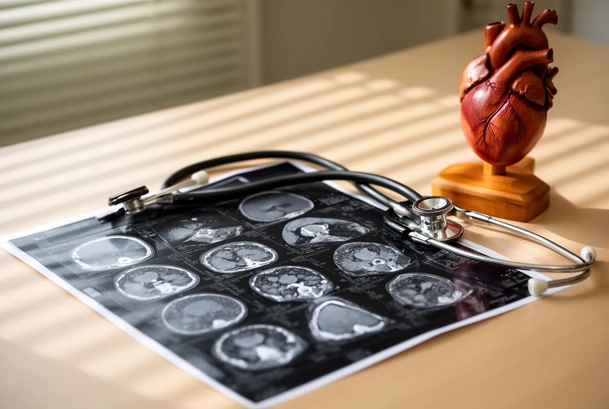
Understanding what the report says. A radiology report uses technical language: terms like "no significant stenosis" (no meaningful narrowing), "calcified plaque burden," or "soft plaque in the LAD" (the left anterior descending artery, one of the three main coronary arteries). Rather than trying to decode it alone, ask for a review appointment. In our own consultations we take the time to explain what the studies actually show, in plain words, and to answer every question before any decision is made, and we encourage patients to seek a second opinion and treat it as a normal part of deciding about heart care.
What happens after an abnormal scan. A positive CT does not automatically mean surgery. Many patients with moderate plaque are managed with medication, cholesterol control, and repeat imaging over time. Others may need an invasive coronary angiogram, a catheter test where dye is injected directly into the arteries through a wrist or groin catheter, or treatment such as a stent or bypass surgery. If a blocked valve or severe coronary disease is found, the CT images often serve as the roadmap for the procedure itself, including minimally invasive and transcatheter options like TAVR (transcatheter aortic valve replacement).
Thorough cardiac ct scan preparation is genuinely simple: know what to stop, what to drink, and when to arrive. Avoid caffeine, follow the fasting and medication instructions your clinic gives you, drink water beforehand, and arrive early with your questions ready. A well-prepared scan usually means clear images, one appointment instead of two, and faster answers about your heart. If chest pain or other heart symptoms have prompted this test, bring the results to a qualified cardiac team and book a consultation rather than trying to interpret them on your own. Dr Molina and our multidisciplinary team review every case together and will explain your options, including less invasive ones where the evidence supports them, and the final decision always remains yours.