

Cardiac CT
Learn how cardiac CT works, from calcium scoring to angiograms, plus preparation, safety, costs in Guadalajara, and what your results mean.
What is a Cardiac CT Scan?
A cardiac CT scan is a non-invasive imaging test that uses specialized X-ray equipment to take detailed, cross-sectional pictures of the heart and its blood vessels. If you or a loved one are preparing for heart surgery, your surgical team may use this advanced technology to gain a precise, three-dimensional view of your cardiac anatomy. This safe, outpatient procedure provides invaluable information without requiring a single incision, allowing physicians to thoroughly evaluate your heart health before any intervention is considered.
By combining multiple X-ray images taken from different angles around the body, a computer generates a highly detailed, 3D view of the heart, the coronary arteries, and surrounding structures. The resulting images are far more detailed than a standard chest X-ray, revealing not just the heart's overall shape but also the intricate pathways of the blood vessels that supply the heart muscle.
For a patient preparing for cardiac surgery, understanding the condition of the heart prior to an operation is paramount. Cardiac computed tomography provides the surgical team with a comprehensive roadmap of your unique anatomy. This advanced visualization allows the surgeon to meticulously plan the procedure, anticipate any potential anatomical challenges, and ultimately ensure the safest and most effective surgical outcome for you or your loved one.
How Does Cardiac Computed Tomography Work?
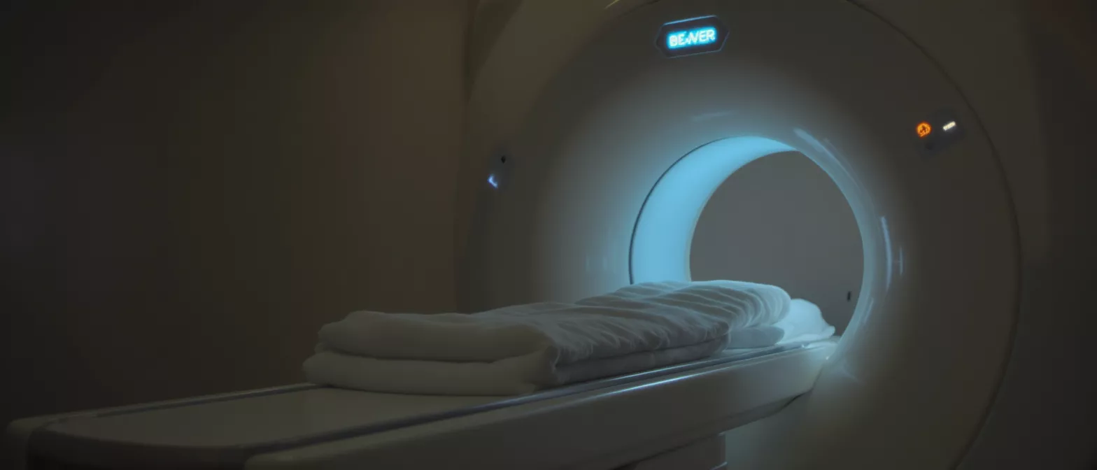
During cardiac computed tomography, the patient lies flat on a comfortable, motorized examination table that slowly slides into a large, donut-shaped machine known as a gantry. Inside this ring, an X-ray tube rapidly rotates around the patient's body, emitting precisely controlled, narrow beams of radiation. Detectors positioned directly opposite the X-ray tube meticulously capture the radiation that passes completely through the body, recording thousands of distinct measurements in a fraction of a second.
Because the human heart is constantly contracting and relaxing, capturing a sharp, clear image requires precise synchronization with the cardiac cycle. The CT scanner is directly connected to an electrocardiogram (ECG) monitor that tracks your exact heart rate. The machine uses this ECG data to take its X-ray snapshots during the brief, split-second pauses between heartbeats—known as diastole—ensuring the final images remain perfectly crisp and free of motion blur.
A powerful computer then processes all the collected data, stacking the individual cross-sectional slices together to construct a fully interactive, three-dimensional model of your heart. This detailed digital model allows your cardiologist and surgeon to rotate the image, view it from any conceivable angle, and essentially take a virtual tour of the heart's chambers and coronary vessels before any physical operation begins.
Why Would a Doctor Order a CT Heart Scan?
Your cardiologist may recommend a CT heart scan to investigate a variety of cardiovascular symptoms or to monitor known heart conditions before proceeding to surgery. If you experience unexplained chest pain, shortness of breath, or irregular heartbeats, this scan provides vital clarity. It helps doctors determine if there are physical blockages in the coronary arteries, structural defects in the heart muscle itself, or complications within the heart valves.
Cardiac ct for plaque detection: One of the most frequent uses of this imaging tool is to detect and evaluate the buildup of plaque within the coronary arteries. Plaque, which is composed of fat, cholesterol, and calcium, can gradually accumulate and severely narrow the arteries supplying blood to the heart. Identifying these blockages non-invasively helps your medical team determine if a surgical intervention, such as a coronary bypass, is strictly necessary.
Pre-surgical structural evaluation: When a patient is being evaluated for a complex procedure like valve replacement or congenital defect repair, surgeons need to know the exact size and shape of the heart's internal structures. The three-dimensional images provided by this scan allow for highly precise surgical planning, reducing the risk of unexpected complications once you are in the operating room.
Cardiac CT for Calcium Scoring
One specific, highly valuable application of this technology is the calcium score test. Over time, microscopic amounts of calcium can deposit within the walls of the coronary arteries as atherosclerotic plaque builds up. Using a quick, non-invasive scan specifically tailored for this purpose, physicians can accurately measure the exact volume of this calcium and assign a numerical score that reflects the burden of disease.
This score is a powerful and reliable indicator of your overall risk for experiencing a future cardiac event. A higher calcium score indicates more significant, hardened plaque buildup, which may prompt your medical team to be more aggressive in their medical management or push for surgical intervention. By accurately quantifying your risk early, calcium scoring helps guide life-saving treatment decisions before a severe heart attack occurs.
Because calcium scoring does not typically require the injection of IV contrast dye, it is a remarkably simple and fast screening tool. It gives the cardiac surgical team a rapid, reliable snapshot of long-term disease progression, helping them confidently decide if more advanced imaging or a direct surgical approach is needed. For many patients, coronary artery calcium scoring serves as the initial stepping stone toward a comprehensive surgical plan.
CT Coronary Angiogram vs Invasive Angiography
For decades, the only definitive way to directly visualize the inside of the coronary arteries was through a traditional, invasive coronary angiogram. That highly effective but invasive procedure requires inserting a long, thin catheter directly into a major artery in the groin or wrist, carefully threading it up to the heart, and injecting a special contrast dye while taking live X-ray videos. While still the ultimate gold standard, it requires actual physical instrumentation inside the body.
Today, non-invasive imaging provides a highly accurate, outpatient alternative to diagnostic catheterization. Instead of threading physical wires through your arteries, a simple IV is placed in the arm, and a specialized contrast dye is safely injected into the bloodstream. The CT scanner then captures incredibly detailed images of the dye as it flows through the heart's vessels, clearly highlighting any dangerous narrowing or blockages without ever entering the body.
For many cardiac surgery candidates, opting for the non-invasive route first is highly preferred to eliminate entirely unnecessary procedures. If the scan shows completely clear arteries, the patient avoids the inherent risks and prolonged recovery time associated with an invasive diagnostic procedure. Non-invasive CT coronary angiogram testing is often the safest first step before committing to surgical catheterization.
Cardiac CT vs MRI: Which is Better?
Both computed tomography and magnetic resonance imaging (MRI) are highly advanced, non-invasive ways to look inside the chest, but they operate on completely different physical principles. CT uses rapidly rotating X-rays to generate its images, making it exceptionally fast and highly sensitive to dense structures like bone and calcified plaque. MRI, by contrast, uses powerful magnetic fields and radio waves, making it excellent at differentiating between soft tissues without using any radiation at all.
When evaluating coronary arteries for dangerous blockages, CT is generally the preferred modality due to its incredible speed and unmatched spatial resolution. It can easily capture the tiny, fast-moving coronary vessels during a single held breath, reliably providing a clear picture of any obstructive plaque. MRI is often reserved more for evaluating the heart muscle tissue itself, looking for inflammation, scarring, or complex structural issues that require detailed soft-tissue contrast.
Your cardiologist and cardiac surgeon will choose the specific imaging modality based entirely on the clinical question they need answered. If you have severe coronary disease and the surgical team needs to evaluate arterial blockages, CT is almost always the logical choice. In some complex cases, patients may safely require a cardiac MRI with contrast to rule out myocardial scarring before a final surgical plan is finalized.
Cardiac CT Preparation: What to Expect
Preparing for your heart ct imaging is surprisingly straightforward, but it requires a few important steps to ensure the resulting images are as clear and accurate as possible. You will typically be asked to avoid consuming any food or drinks for a few hours before your scheduled appointment time. This fasting is especially critical if your specific scan requires the use of an iodine-based contrast dye to properly highlight the blood vessels.
Medication and heart rate control: Because the scanner captures images in sync with your heartbeat, a slower, steady heart rate produces the absolute best results. If your resting heart rate is naturally high, your doctor may prescribe a short-acting beta-blocker to take shortly before the test to gently and temporarily slow your pulse. This ensures the scanner captures crisp, blur-free images of the rapidly moving heart muscle.
During the procedure: Once you are on the table, the technologist will place small, painless sticky pads on your chest to monitor your ECG. You will be asked to hold your breath for short intervals—usually just 5 to 15 seconds at a time—while the table moves through the scanner. The entire process, from the time you walk into the imaging center to the time you leave, typically takes less than half an hour, and you can resume normal activities immediately.
Cardiac CT Radiation Dose and Safety
A very common concern for patients facing multiple pre-surgical tests is the amount of radiation exposure involved. It is perfectly reasonable to be cautious about radiation, but modern CT scanners are engineered with highly sophisticated software and hardware specifically designed to minimize exposure. Techniques like prospective triggering and dose modulation ensure that the machine only emits X-rays during the exact fraction of a second when the heart is at rest, drastically reducing the total dose delivered.
While you are exposed to some ionizing radiation during the scan, the absolute dosage is relatively low and carefully calibrated by a medical physicist. For the vast majority of surgical candidates, the immediate, life-saving diagnostic benefits of clearly identifying coronary blockages or dangerous heart defects far outweigh the minimal, theoretical long-term risks associated with this low level of radiation. Your medical team will never order an unnecessary scan; the information gained is strictly essential for your surgical safety.
If you have any existing kidney concerns, the medical team will also carefully evaluate your kidney function before the test, as the contrast dye is filtered out of the blood by the kidneys. In most instances, a minor IV hydration protocol is utilized to help safely and efficiently flush the contrast out of your system.
Understanding Cardiac CT Interpretation Results
Once the raw scan is complete, the immense amount of data is sent directly to a specialized radiologist or cardiologist for detailed, careful interpretation. These highly trained physicians spend hours meticulously reviewing hundreds of individual cross-sectional slices and complex 3D reconstructions of your heart. They are looking for dangerous narrowing of the vessels, plaque buildup, congenital anomalies, and any subtle signs of structural disease that could impact your upcoming surgery.
The resulting medical report is sent directly to your referring surgeon and cardiologist, who will use these highly detailed findings to finalize your ultimate surgical approach. It is important to remember that you will not typically receive a diagnosis directly from the imaging technologist operating the machine. The data requires complex, expert analysis, and your surgical team will sit down with you to explain exactly what the images show and how it influences your specific treatment plan.
The clear images produced by 3d cardiac ct allow your surgical team to confidently visualize the exact geography of your internal anatomy, ensuring that every surgical cut and graft is precisely planned before the operation begins.
Cardiac CT Cost and Accessibility in Guadalajara
Access to state-of-the-art cardiac imaging is highly critical for any patient facing heart surgery, and this technology is widely available across major medical centers in Mexico. The cardiac ct cost can vary depending on the exact type of scan performed, whether contrast dye is utilized, and the technical complexity required to accurately capture the specific anatomical structures in question. It is generally considered an affordable, highly accessible diagnostic tool compared to more invasive surgical diagnostic alternatives.
When considering cardiac ct guadalajara facilities, patients have access to modern, internationally accredited hospitals equipped with the absolute latest generation of CT scanners. These centers provide the exact same level of diagnostic accuracy and safety that you would expect to find in the United States or Europe, making Guadalajara a premier destination for comprehensive cardiac care, safe diagnosis, and complex heart surgery.
Ultimately, the goal of any imaging test is to provide peace of mind and a clear path forward for the patient. By utilizing non-invasive imaging, Dr Alejandro Molina and the dedicated surgical team can approach your specific heart condition with the highest possible level of confidence. Knowing exactly what to expect inside the chest before making a single incision leads to safer operations, significantly faster recoveries, and ultimately, far better long-term outcomes for your heart health.






