

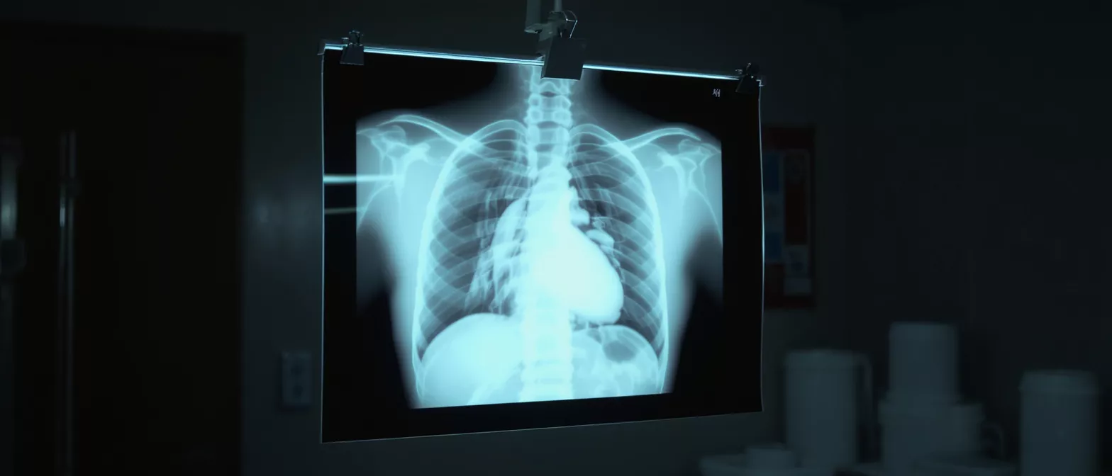
Chest X-ray
Learn how a chest x-ray helps heart surgeons evaluate your heart and lungs before and after cardiac surgery.
What is a Chest X-ray?
A chest x-ray is a fast, painless imaging test that creates pictures of the structures inside your chest, including your heart, lungs, blood vessels, and bones. It uses a small amount of electromagnetic radiation to capture these internal images on a digital detector or special film. For patients preparing for or recovering from heart surgery, this scan provides your medical team with vital information about your cardiovascular and respiratory health.
Why a chest x-ray matters for cardiac patients: Before any surgical intervention, your surgeon needs a clear baseline picture of your chest anatomy. This baseline helps them identify pre-existing conditions—such as an enlarged heart or fluid in the lungs—that could affect your surgical plan or anesthesia management. Without this imaging, your surgical team would be navigating blind to underlying structural issues.
Think of this imaging study as a foundational diagnostic tool. It is often the first step in a comprehensive cardiac evaluation because it is widely available, quick to perform, and yields immediate visual feedback that guides more advanced testing like echocardiograms or CT scans.
How Does a Chest X-ray Work?
The technology behind a chest x-ray relies on the principle that different tissues absorb radiation at varying rates. Dense materials, like your ribs and spine, absorb a significant amount of radiation, appearing white on the final image. Softer tissues, like your heart and blood vessels, appear in shades of gray. Because air absorbs the least radiation, your lungs look dark, which provides excellent contrast for spotting abnormalities.
The physics of radiographic contrast: A machine emits a controlled beam of radiation that passes through your body for a fraction of a second. The radiation that makes it through strikes a digital sensor on the other side, creating a shadow-like image. This precise contrast is what allows a cardiac specialist to distinguish the borders of your heart from the surrounding lung tissue.
You will typically stand against a specialized flat plate for the image. If you are unable to stand safely, the equipment can be adjusted to take the picture while you sit in a chair or lie in a hospital bed, ensuring every patient can undergo the scan comfortably.
What Does a Chest X-ray Show?
When looking at a chest x-ray, a trained medical professional evaluates several critical structures simultaneously. The scan reveals the size and shape of the heart, the condition of the lungs, the positioning of major blood vessels, and the integrity of the rib cage. It also clearly shows any medical devices, such as pacemakers or artificial valves, ensuring they remain in the correct position.
Specific cardiovascular indicators visible on film: Your surgeon looks for an enlarged cardiac silhouette, which can suggest heart failure or valve disease. They also examine the pulmonary vasculature—the blood vessels within the lungs—to see if blood is backing up, which is a common sign of cardiac distress. This comprehensive view makes the scan indispensable for cardiovascular diagnosis.
For a patient facing heart surgery, the scan provides a roadmap. It also rules out simultaneous lung issues, like pneumonia, that could complicate your recovery.
Why Heart Surgeons Order a Chest X-ray
Your surgical team relies on a chest x-ray before any major cardiac procedure to confirm you are physically ready for the operating room. The scan checks for underlying respiratory infections, evaluates the overall cardiac silhouette, and ensures the heart is not carrying an excessive fluid load. Identifying these issues early allows your medical team to optimize your health before surgery begins.
Pre-surgical risk assessment and planning: The surgical team uses the images to map out their approach, noting the size of your heart relative to your chest cavity. Knowing exactly how much space the heart occupies helps the surgeon anticipate technical challenges they might face once the chest is opened.
This simple imaging study is a critical safety net. It ensures that no unexpected anatomical surprises will arise during the operation, ultimately making your surgical journey safer and more predictable.
What to Expect During a Chest X-ray Procedure
Undergoing a chest x-ray is remarkably straightforward and entirely painless. A radiologic technologist will guide you to the imaging room and ask you to remove any metal objects, such as jewelry, glasses, or button-up shirts, as metal can obscure the final images. You will then be positioned against the imaging plate, usually with your hands on your hips and your shoulders rolled forward to push your shoulder blades out of the way of the heart.
The patient experience and positioning: The technologist will ask you to take a deep breath and hold it perfectly still for just one or two seconds while the machine hums softly. Holding your breath inflates your lungs fully, pushing the heart against the ribs and providing the clearest possible view. The entire appointment usually takes about fifteen minutes from start to finish.
If you are in the intensive care unit following heart surgery, a portable x-ray machine can be brought directly to your bedside. The technologist will carefully slide a flat plate behind your back and adjust the machine arm over your chest to capture the necessary images without requiring you to leave your bed.
Understanding Your Chest X-ray Results
Once the images are captured, a radiologist—a doctor specializing in medical imaging—analyzes them and sends a report to your cardiologist or surgeon. The results are usually available quickly, often within an hour in a hospital setting. Your doctor will review the findings with you, explaining what the shadows and shapes on the film mean for your specific cardiac condition.
Interpreting clinical findings: A normal result means your heart is a normal size, your lungs are clear of fluid, and your skeletal structure is intact. However, abnormal findings do not immediately mean disaster; they simply guide your doctor. Finding an enlarged heart or mild pulmonary congestion helps your medical team tailor your treatment plan and medications accordingly.
It is completely normal to feel anxious while waiting for these results. When looking at your films, utilizing patient education materials for radiology can help you feel more grounded. Understanding what your doctors are looking for eases the tension of the unknown, empowering you to engage actively in your care.
How Chest X-rays Help Assess Heart Size
One of the most common reasons a cardiologist orders a chest x-ray is to measure the size of your heart. On the image, the heart sits prominently in the center of the chest, and its overall size is evaluated by comparing it to the width of your rib cage—a measurement known as the cardiothoracic ratio. If the heart takes up more than half the width of the chest cavity, it is considered enlarged.
Cardiac silhouette and heart enlargement: An enlarged heart, or cardiomegaly, is not a disease itself but a symptom of an underlying issue, such as high blood pressure, valve disease, or heart failure. The x-ray provides a quick, objective way to track heart size over time, allowing your doctor to see if a condition is worsening or if treatments are working.
For a patient preparing for valve repair, seeing the exact degree of heart enlargement helps the surgeon understand the severity of the condition. This information directly influences surgical timing and the specific technique chosen for your repair.
- Cardiothoracic Ratio: Compares heart width to chest width to check for cardiomegaly.
- Cardiac Contours: Examines the specific bumps and outlines of the heart to spot enlarged chambers.
- Vascular Pedicle Width: Measures the width of the central blood vessels to evaluate fluid status.
Evaluating Lungs and Heart After Cardiac Surgery
After your heart surgery is complete, chest x-rays become a routine part of your recovery monitoring. In the intensive care unit, your surgical team will order frequent scans—sometimes daily—to ensure everything inside your chest is healing as expected. These postoperative images verify that there is no excess air, fluid, or blood accumulating around your heart or lungs.
Postoperative monitoring and complications: It is very common to have some collapsed lung tissue or mild pleural effusion (fluid around the lungs) immediately after cardiac surgery. The x-ray helps your team track these expected changes, ensuring they resolve naturally or intervening quickly if a chest tube is needed to drain excess fluid. It also confirms that central lines, breathing tubes, and pacing wires are perfectly positioned.
This continuous visual monitoring is crucial for catching potential complications before you ever feel them. Knowing that your medical team is looking inside every day builds confidence in the recovery process.
Are Chest X-rays Safe for Cardiac Patients?
A very natural concern for patients and families is whether radiation from a chest x-ray is safe, especially when recovering from major heart surgery. Fortunately, the radiation dose from a standard chest x-ray is extremely low. You are exposed to a similar amount of naturally occurring background radiation simply by living your normal life for a few days. The diagnostic benefits of this scan vastly outweigh any negligible risk.
Radiation exposure and safety protocols: Medical facilities follow strict safety protocols, known as ALARA (As Low As Reasonably Achievable), to minimize radiation exposure. The technologist will provide a lead apron to protect parts of your body that do not need imaging. If you require multiple scans throughout your recovery, your care team carefully tracks your cumulative radiation exposure.
For pregnant patients or those who might be pregnant, it is critical to inform the medical team beforehand. While a chest x-ray is generally considered safe, special precautions or alternative imaging methods may be used to protect the developing baby from any unnecessary radiation.
- Low Dose: Equivalent to a few days of natural background radiation.
- High Value: Provides essential diagnostic data that far outweighs the minimal risk.
- Strict Protocols: ALARA principles ensure exposure is kept to the absolute minimum necessary.
Key Takeaways About Chest Imaging
The chest x-ray remains one of the most valuable and foundational tools in cardiac care. From the initial evaluation of your heart's size and shape to the vigilant monitoring of your lungs after surgery, this quick and painless scan provides your medical team with critical, real-time insights. It allows them to see the hidden impacts of your heart disease and catch postoperative complications early.
Your role in the imaging process: As a patient, understanding why these images are taken helps alleviate anxiety and builds trust in your care team. Remember to hold still, follow breathing instructions, and communicate any mobility issues to the technologist so they can capture the best possible images. Your cooperation directly impacts the quality of the scan.
At its core, chest imaging is about safety and precision. It gives your surgeon the visual clarity needed to guide your treatment plan and ensures that your journey through heart surgery is as safe and successful as possible. Trust in the process, knowing that every image taken is a step toward a healthier heart.






