

Cardiac MRI
Cardiac MRI uses magnets and radio waves to create detailed heart images.
What is a Cardiac MRI?
A cardiac MRI is a non-invasive imaging test that uses powerful magnets and radio waves to create detailed pictures of your heart. It does not use ionizing radiation, making it a safe option for evaluating complex heart conditions. The scan provides high-resolution images of the heart muscle, valves, and blood vessels.
Why does this matter for heart surgery patients? Before any surgical intervention, your cardiac surgeon needs a precise roadmap of your heart anatomy. This imaging test reveals the exact size of your heart chambers, how thick the muscle walls are, and the severity of any tissue damage.
Understanding this test helps alleviate anxiety. When you know what to expect and why it is ordered, you become an active participant in your pre-operative care, which often leads to better outcomes and reduced stress before your procedure.
How Does Cardiac MRI Imaging Work?
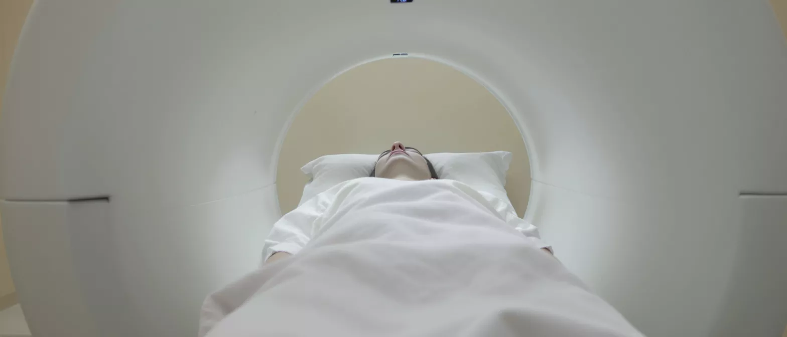
Cardiac MRI imaging relies on a large, tube-like machine containing a powerful magnet. When you lie inside the scanner, the magnetic field temporarily realigns hydrogen atoms in your body. Radio waves then cause these atoms to emit faint signals, which a computer translates into highly detailed, three-dimensional images of your heart.
How does the scanner capture a moving heart? The machine uses a technique called electrocardiogram (ECG) gating. Small sticky patches placed on your chest track your heartbeat, allowing the scanner to take pictures at the exact same point in each cardiac cycle, resulting in crisp, motion-free images.
Because it captures both structure and function simultaneously, this scan is unmatched in its ability to assess blood flow and tissue characteristics. It can distinguish between healthy heart muscle and scarred tissue from a previous heart attack.
Why is a Heart MRI Scan Recommended Before Surgery?
Detailed tissue characterization: A heart MRI scan is unique in its ability to distinguish between different types of heart tissue. For patients awaiting surgery, it shows the exact location and extent of scar tissue, which is crucial for planning complex repairs.
Assessing heart muscle viability: Surgeons need to know if heart muscle is temporarily stunned or permanently dead. This imaging test helps determine if restoring blood flow through a bypass will actually revive the muscle and improve heart function.
By providing comprehensive data on anatomy and blood flow, the test ensures surgical teams are fully prepared. It eliminates guesswork, allowing specialists to tailor the surgical approach to the specific needs of the patient.
Cardiac MRI vs Echo: Which is Better?
When comparing a cardiac MRI vs echo, it is important to understand that both are valuable but serve different purposes. An echocardiogram uses sound waves and is often the first-line test because it is quick, portable, and inexpensive. It provides excellent real-time views of how the heart pumps.
Differences in image clarity and scope: While an echo is excellent for routine evaluations, an MRI provides far superior image resolution. It is not hindered by air in the lungs or the shape of the patient's ribcage, which can sometimes block ultrasound waves during an echo.
- Detail level: MRI provides unmatched clarity of the heart muscle tissue, allowing for precise scar detection.
- Radiation exposure: Neither test uses ionizing radiation, making both extremely safe for repeated use.
- Time and accessibility: Echo exams take about 20 minutes and are widely available; MRI scans take longer and require specialized equipment.
How to Prepare for a Cardiac MRI Test?
Preparation for a cardiac MRI test is straightforward. You will be asked to wear loose, comfortable clothing without metal zippers, buttons, or fasteners. Because the machine uses a powerful magnet, you must remove all jewelry, watches, and any metal objects before entering the scanning room.
Dietary and medication guidelines: You can usually eat, drink, and take your prescribed medications as normal unless your doctor specifically instructs you otherwise. If contrast dye will be used, you may be asked to refrain from eating for a few hours prior to the test.
It is vital to inform the technologist if you have any implanted medical devices. While many modern pacemakers and defibrillators are MRI-safe, older models may require special protocols or alternative imaging. Providing a complete medical history ensures your safety.
What to Expect During the Cardiac MRI Procedure?
Entering the scanner environment: The cardiac MRI procedure takes place in a specialized room. You will lie down on a movable table that slides into the center of the cylindrical machine. The technologist will leave the room to operate the scanner from behind a glass window, but they can see, hear, and speak with you at all times.
Using contrast agents safely: To enhance the images, a contrast dye called gadolinium is often injected through an IV line in your arm. This dye helps highlight areas of inflammation or scarring, though it is processed by the kidneys and typically leaves your body within 24 hours.
During the scan, the machine will produce loud thumping and humming noises. You will be given earplugs or headphones to block the noise and help you stay relaxed. You must hold very still, as movement can blur the images and require the technologist to repeat certain sequences.
Are There Any Cardiac MRI Risks?
For the vast majority of patients, there are no significant cardiac MRI risks. Because the test does not use radiation, it does not carry the risks associated with X-rays or CT scans. The magnetic field itself is harmless to human tissue.
Rare complications with contrast: The primary consideration involves the use of gadolinium contrast dye. While allergic reactions are exceedingly rare, the dye is filtered through the kidneys. Patients with severe kidney disease may require a blood test beforehand to ensure their kidneys can safely clear the contrast agent.
It is completely normal to feel claustrophobic inside the scanner. If you have a history of anxiety in tight spaces, your doctor may prescribe a mild sedative to help you remain calm and still during the imaging process.
How Long Does a Cardiac MRI Scan Take?
Patients often ask how long a cardiac MRI scan takes. The entire appointment usually lasts between 60 and 90 minutes, though the actual time spent inside the machine is typically 45 minutes. The duration depends on the complexity of the images required.
Factors affecting scan duration: If the test is specifically looking for detailed tissue scarring or complex congenital defects, the radiologist may need to acquire more image sequences. Additionally, the technologist may need to pause and coach you on your breathing to ensure optimal image quality.
You should plan to spend a couple of hours at the imaging center to account for check-in, preparation, and post-scan observation. If you received a sedative to relax, you will need someone to drive you home afterward.
Understanding Your Cardiac MRI Diagnosis Results?
A specialized cardiologist or radiologist will analyze your images and send a detailed report to your surgeon. The results provide a definitive cardiac MRI diagnosis, pinpointing structural defects, valve abnormalities, and the exact percentage of scarred muscle tissue.
Reviewing findings with your surgeon: Dr Alejandro Molina will review these images with you, pointing out exactly where the problem lies and how surgery will correct it. Seeing the visual evidence of the condition helps patients understand the necessity of the operation.
It serves as a baseline for tracking your recovery after surgery, allowing your medical team to compare future imaging against the pre-surgical state.
Cardiac MRI Centers in Jalisco and Next Steps?
For patients in Guadalajara, accessing high-quality imaging is straightforward. Several cardiac MRI centers in Jalisco offer state-of-the-art equipment capable of performing these specialized scans. Choosing a center recommended by your cardiac surgeon ensures the images meet specific surgical planning requirements.
Coordinating care with your surgical team: Share the imaging results with your surgical team so your pre-operative pathway is not delayed. Once the scan is complete, the results are sent to your doctor for review.
Scheduling your evaluation is the critical first step, and our team is ready to guide you through every phase of the process.






