CT Angiography of the Heart and Aorta: Safety and Results
CT angiography heart scan reveals blocked arteries safely without surgery.
What Is CT Angiography of the Heart?
CT angiography heart imaging is a scan that takes detailed pictures of your coronary arteries — the small vessels that feed the heart muscle — without inserting any tube into your body. A CT (computed tomography) scanner rotates around your chest, and contrast dye injected into a vein makes the blood vessels glow clearly on the images. In one painless session, it can show calcium, soft plaque, and narrowed segments inside the arteries that a plain CT scan or a stress test cannot see directly.
Why patients hear about it so often. For years, the only way to look inside the coronary arteries was coronary angiography, a catheter procedure where a thin tube is threaded from the wrist or groin up to the heart. CT angiography changed that. It is a non invasive heart artery test, so it is often the first study ordered when a doctor suspects blockages but wants to avoid an invasive procedure unless it is truly needed.
More than just the coronary arteries. The same scanner can also image the aorta — the large main artery that carries blood from the heart to the body. CT angiography of the thoracic aorta and abdominal aorta is how teams measure aneurysms, dissections, and other aortic disease. The technique is the same; only the area of focus changes. Your doctor selects which vessels the scan must cover before it begins.
How a CT Angiography Heart Scan Works
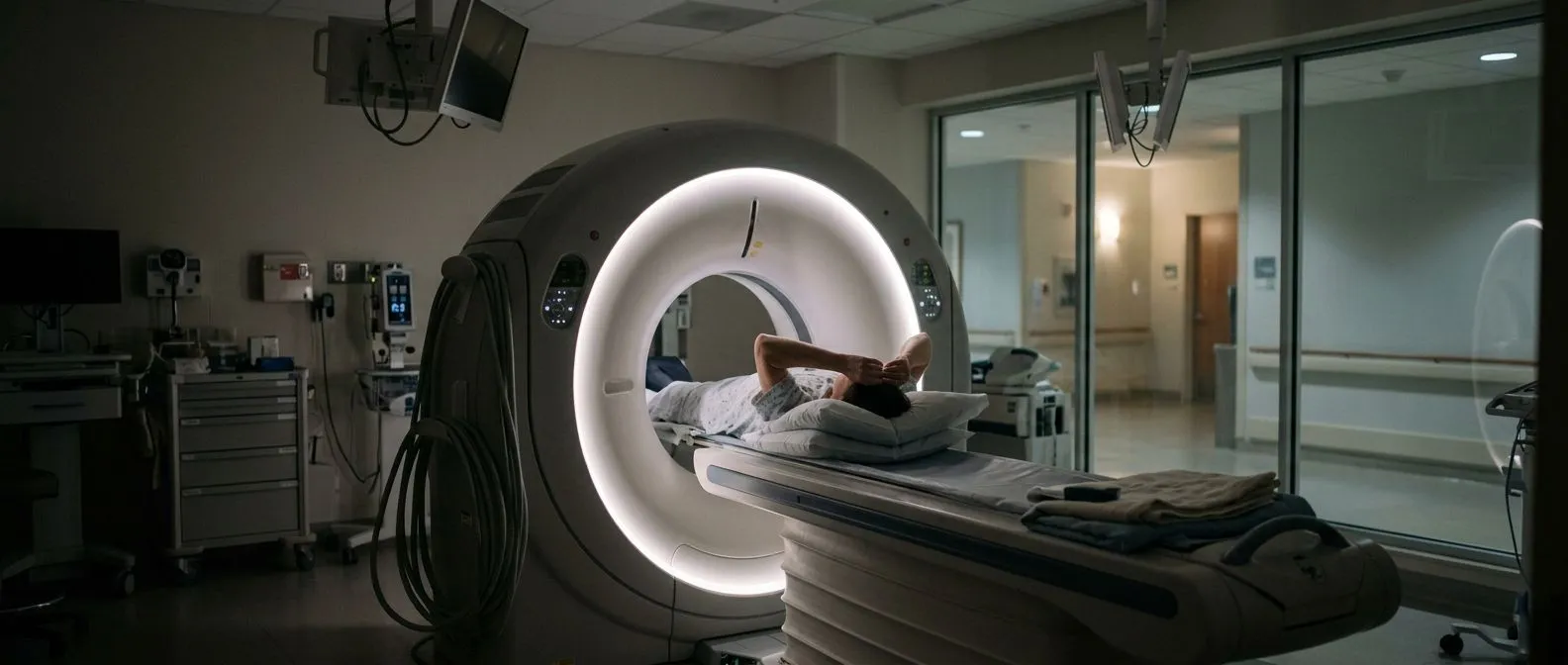
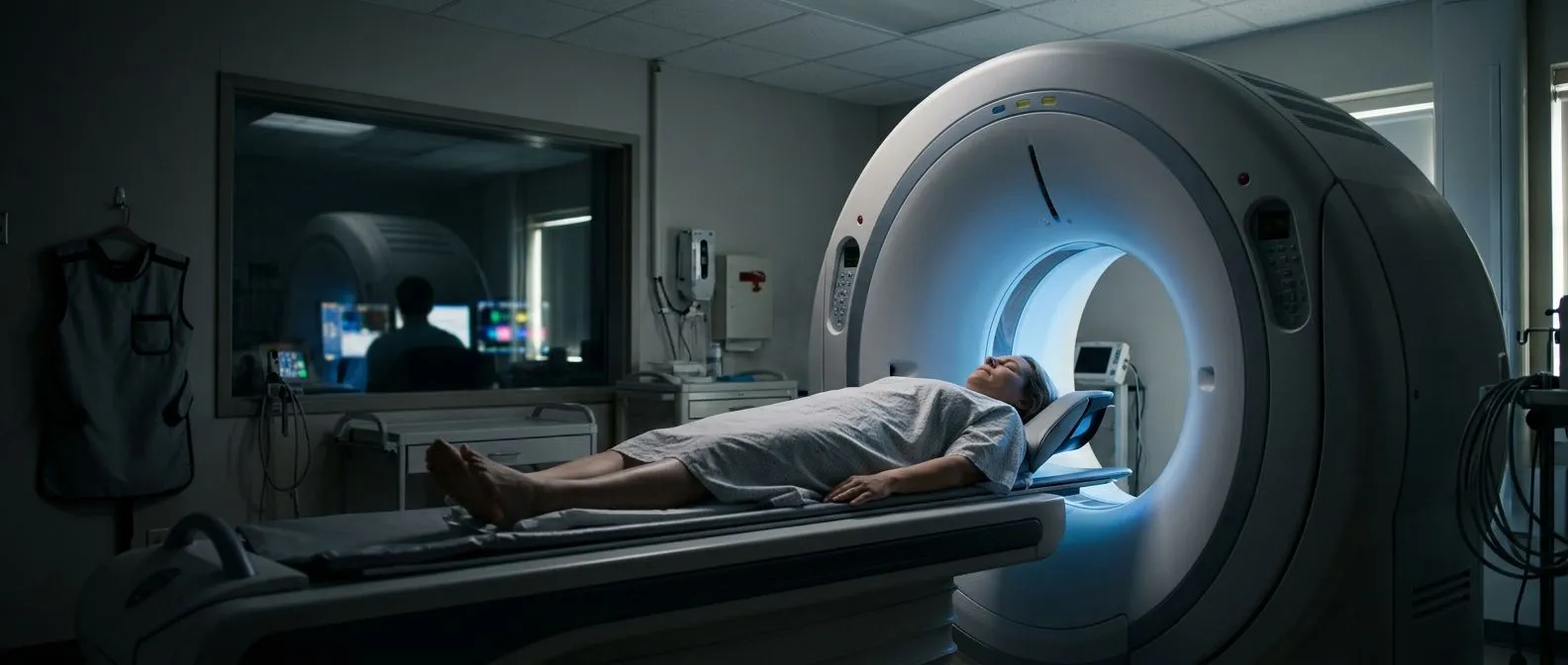
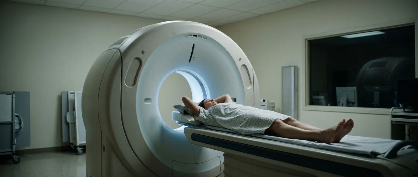
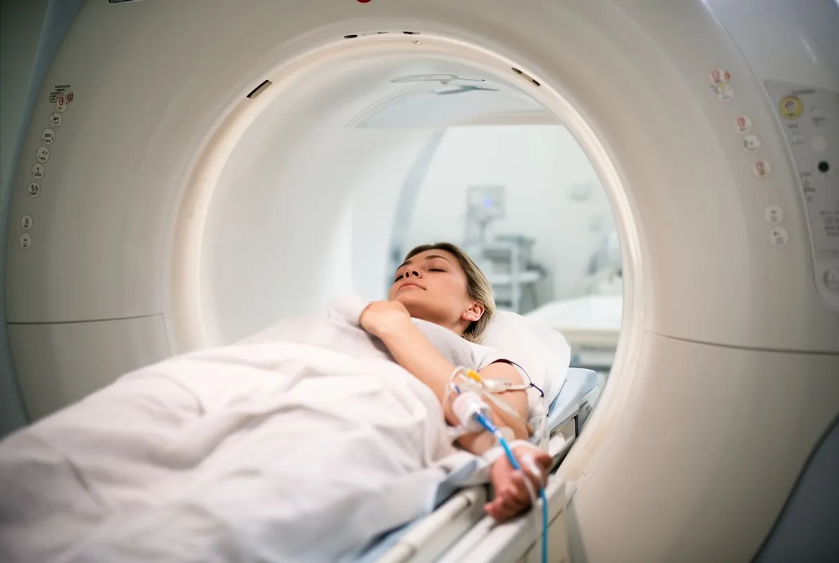
Step by step. You lie on a table that slides into a ring-shaped scanner. A radiology nurse places a small plastic tube (a cannula) in a vein in your arm. Contrast dye — an iodine-based fluid — is injected, and the scanner captures hundreds of thin image slices in seconds. Modern machines take pictures between heartbeats, which is why you may be asked to hold your breath for a few seconds and why your heart rate is checked beforehand.
The role of the contrast. A heart CT scan with contrast works because the dye fills the arteries and makes their outline visible. Without it, the scanner sees mostly calcium and anatomy, not the inside of the vessel. Some scans also use a beta-blocker medication, given orally or intravenously, to slow the heart temporarily so the images stay sharp. The team checks your blood pressure and pulse before giving anything.
What happens to the images. The computer rebuilds the slices into three-dimensional pictures that show each coronary artery from every angle. A radiologist and your cardiologist review them together: where the plaque is, how much it narrows the channel, and whether the narrowing looks stable or risky. That review is where the real value of the test lies — the scan itself is just the beginning.
Why Would a Doctor Order a CT Angiogram?
Common reasons. Doctors order a CT coronary angiogram heart scan when a patient has chest pain, breathlessness, or abnormal results on an exercise or stress test, and the cause needs to be seen directly. It is also used to investigate possible aortic problems — an enlarged or torn aorta — and to check whether previous stents or bypass grafts remain open. In each case, the question is the same: which vessel, if any, is blocked, and how severely?
Ruling things out safely. One of the most useful roles of the test is what it excludes. When a coronary CT angiography procedure shows normal, smooth arteries, many patients can avoid a catheter procedure entirely, and their chest pain can be investigated for other causes — muscle, reflux, or anxiety. That "negative" result is genuinely good news, and it is one of the test's main contributions to everyday practice.
Planning treatment. When the scan does show disease, it shapes the conversation. A blockage found on a heart blockage CT scan with no catheter tells the team where the problem is and how tight it is, which then guides whether medication, a stent, or surgery makes sense. In our practice, every such case is reviewed by the multidisciplinary team against international practice guidelines before a recommendation is given, and the patient receives a clear proposal with the alternatives.
What Is the Difference Between a CT Scan and a CT Angiogram?
The plain CT scan. A standard CT scan of the chest takes pictures of the lungs, ribs, and general anatomy. It is quick and useful for many questions, but it was not designed to see inside moving coronary arteries. On a plain scan, the heart's motion blurs the vessels, and the blood inside them looks much the same as the tissue around them. You cannot reliably see blockages that way.
The angiogram adds contrast, timing, and heart synchronization. CT angiography is the same machine, but with three additions: iodine contrast injected at the precise moment the scanner captures the heart, images timed to the quietest part of the heartbeat, and software that corrects for motion. Those three differences turn a general picture of the chest into a roadmap of the arteries. It is the contrast and the timing — not the scanner itself — that make the difference.
Why the distinction matters to you. If your doctor says "CT scan," you will get anatomy pictures; if they say "CT angiogram," you will get vessel pictures with an injection. Knowing which one is being ordered helps you prepare and helps you ask the right question: is this study going to answer the specific concern about my heart? If not, it is fair to ask why it is being done at all.
Is CT Angiography Painful? What the Scan Actually Feels Like
The honest answer. CT angiography is not a painful test. The only uncomfortable parts are the venous cannula — the same brief pinprick as any blood draw — and, for some patients, a warm or flushing feeling as the contrast enters the arm. That warmth lasts ten to twenty seconds and can feel like it spreads through the chest, which surprises people who were not expecting it. It is normal and passes quickly.
What the rest feels like. You lie still on a padded table, sometimes with your arms above your head. The machine is quiet apart from a soft whirring, and the table moves a few centimetres with each image set. You will be asked to hold your breath for five to ten seconds a few times. Total scan time on the table is usually under fifteen minutes; for most people the wait before and after takes longer than the scan itself.
If you are anxious or claustrophobic. Modern scanners are wide and well-lit — very different from older machines — and you can see out and talk to the technologist the whole time. Tell the staff if you feel nervous; they deal with this every day and can adjust the pace. Before any of our patients has a scan, we take the time in consultation to explain exactly what the study will involve, in plain words, and to answer every question before the day arrives.
Cardiac CT Angiography Preparation: How Long Does It Take?
Before the day. Good cardiac CT angiography preparation mostly involves timing and caffeine. Most centres ask you to avoid coffee, tea, energy drinks, and smoking for at least 12 hours, because caffeine raises the heart rate and blurs the images. You can usually eat a light meal and take your regular medicines, but confirm this with the imaging centre, especially if you take diabetes medication or blood thinners. Drink water normally — being well hydrated helps your kidneys clear the contrast.
On the day. Arrive early enough for registration and a pre-scan check of your pulse and blood pressure. Wear comfortable clothing without metal, and leave jewellery at home. If your heart rate is high, the team may give a medication to slow it and wait for it to take effect. From arrival to leaving the department, most people spend about one to two hours; the actual scanning takes only minutes.
After the scan. There is no recovery period — this is a heart imaging test with no downtime. You can eat, drink, drive, and return to work immediately. Drinking extra fluids over the next day helps flush the contrast through your kidneys. If a catheter was not involved (and in CT angiography it never is), there is no wound and no leg or wrist restriction afterwards.
- Drink water well before and after the scan to protect your kidneys.
- Skip caffeine and tobacco for at least 12 hours beforehand.
- Bring your medication list — some drugs need adjusting before contrast.
CT Angiography Heart vs Cardiac Catheterization
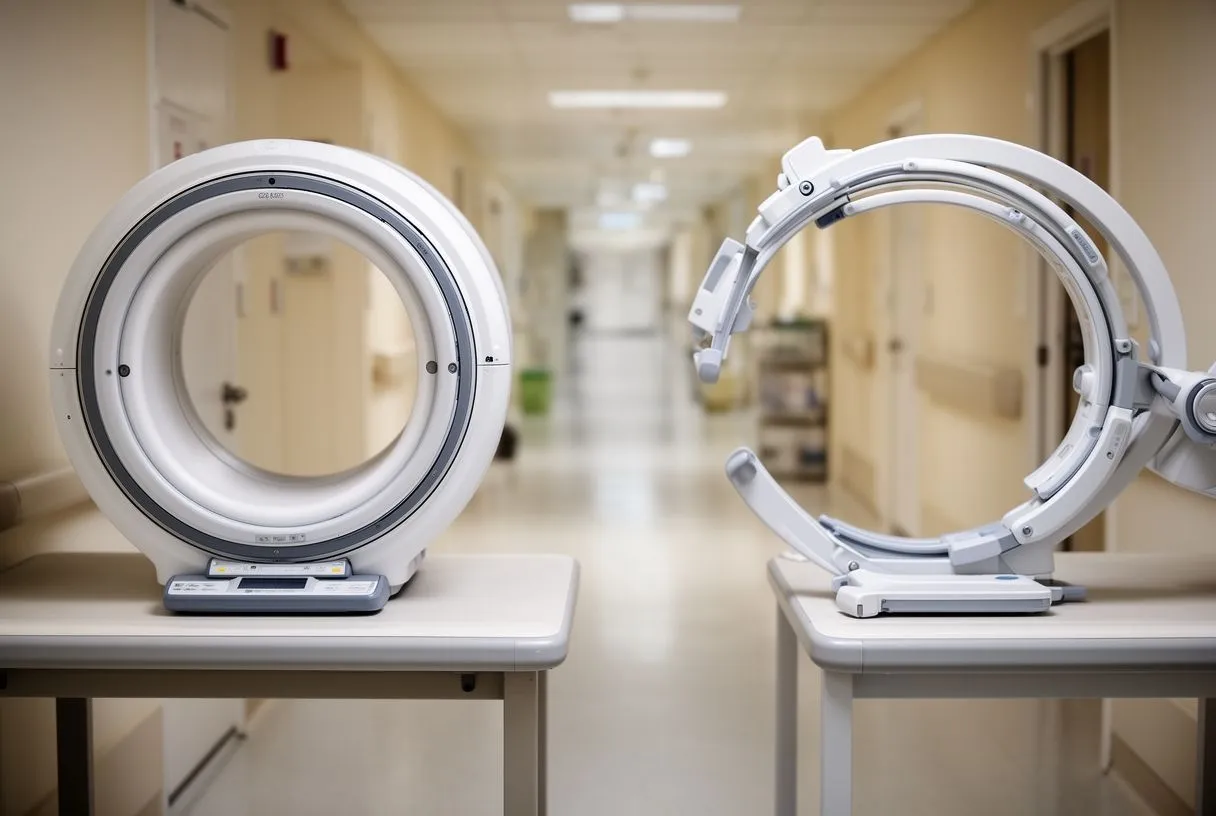
Two different jobs. CT angiography heart scanning and catheter-based coronary angiography answer overlapping questions, but they are not interchangeable. Catheter angiography is invasive: a tube enters an artery at the wrist or groin and dye is injected directly into the coronary arteries, giving real-time moving pictures. It is more precise inside a single vessel, and — crucially — a stent or balloon can be treated in the same session if a blockage is found.
What CT does better. CT shows the artery wall, not just the channel. It can identify soft plaque, calcified plaque, and plaque that looks "positive" — the type associated with future risk — even when the channel is not severely narrowed. For patients with stable chest pain, guidelines generally support CT angiography as a first-line test, reserving the catheter for those whose scan shows significant disease or whose case needs direct measurement.
Incidental findings worth knowing about. Invasive angiography occasionally reveals unexpected things, and CT has a role there too. A published case report described an incidental large mobile aortic arch mass found during conventional angiography — a finding that changed the entire treatment plan for that patient (see the report in the Journal of Tehran Heart Center). Scans that image the whole chest and aorta can also surface such findings before any catheter is inserted.
How we decide which one you need. Our team, led by Dr Alejandro Molina, performs conventional, minimally invasive, robotic and transcatheter procedures, and we recommend the less invasive route only where the evidence supports it for that specific patient. For many patients, a good CT scan answers the question without any catheter at all. For others — complex stent decisions, valve work, or unclear images — the catheter remains the right tool. The choice is made patient by patient, not by habit.
CT Heart Scan Safety and Radiation: What You Should Know
Radiation in context. Any CT scan uses ionizing radiation, and it is fair to ask how much. Modern CT angiography protocols use dose-reduction technology — including prospective triggering and lowering tube output during parts of the heartbeat — which has cut typical doses dramatically compared with scanners of a decade ago. The dose of a single heart scan is generally comparable to a range of common imaging studies, and it varies with the machine and the protocol used for you.
Contrast risks. The iodine contrast is generally safe, but two issues deserve honesty. A small number of people have an allergic-type reaction, usually mild (itching or hives), which the imaging team is prepared to treat immediately. People with significant kidney disease need a blood test beforehand to confirm the contrast is safe for them. If you have kidney problems, asthma, or a previous contrast reaction, say so before the scan — it changes the preparation, not usually the plan.
What the evidence says. Large outcome trials have shown that using CT angiography as a first test in patients with suspected coronary disease is a sound strategy; the SCOT-HEART investigators reported that adding CT angiography to standard assessment improved diagnostic certainty and guiding of treatment, published in JAMA. No test is risk-free, but the question is always whether the information gained outweighs the small and manageable risks — and in properly selected patients, it does.
Is it safe enough for you? For most adults being investigated for chest pain, the answer is yes. Pregnant patients, very young people, and those needing repeated scans deserve a specific conversation about cumulative radiation, and alternatives such as ultrasound or MRI may be discussed. Every case we review is checked against international practice guidelines before a recommendation is given, and radiation and contrast exposure are part of that review.
What Happens if a CT Angiogram Shows Blockage?
First, the scan is read in degrees. Not every blockage is the same. The radiologist reports the percentage of narrowing in each artery: mild (under about 30%), moderate, or severe (typically 70% or more in a main vessel). The report also describes whether the plaque is calcified or soft and whether it involves the left main artery, the descending aorta territory, or smaller branches. Those details determine what happens next far more than the word "blockage" alone.
Then, the treatment conversation. Mild blockage is usually managed with medication and lifestyle changes — statins, blood pressure control, exercise — with follow-up as advised. Severe blockage in an important artery raises the question of a stent or bypass surgery. Neither decision is made from the CT alone; the team weighs your symptoms, other test results, and your overall health. In our consultation we explain what the studies actually show, what each option would involve, and what the alternatives are — and the final decision is always the patient's.
False positives — the honest caveat. CT angiography is excellent at ruling out disease, but it slightly overestimates how tight a narrowing is. A heavily calcified artery can look worse on CT than it truly is, which is why every reported ct angiography heart false positive possibility is resolved by looking at the whole picture — and, when needed, by invasive measurement before anyone commits to a stent. We encourage second opinions and treat seeking one as a normal part of deciding about heart surgery, especially when a scan suggests a major intervention.
CT Angiography Heart Results Time and Cost
When you get your results. Typical ct angiography heart results time is one to three working days, though it varies by centre. The images are ready immediately, but they need careful reading and reporting by a radiologist or cardiologist. Many centres can share a preliminary impression on the same day. Ask your centre's timeline when you book, and make sure a follow-up appointment is arranged so the results are explained to you rather than just handed over.
What the cost involves. Heart CT angiography cost varies with the centre, the scanner used, and whether medications to control your heart rate are included. Ask whether the quoted price covers the radiologist's report, the rate-control medications, and a consultation to explain the results.
How we make it simple for patients. Our first contact is a free video consultation: we get to know the patient, talk through the condition, and say which studies are still needed so no unnecessary ones are ordered. That matters with CT angiography, because the test is only worth doing when it answers a specific question. Our consulting rooms are in Guadalajara, Querétaro and Hermosillo, and a face-to-face visit follows when a patient decides to go ahead.
CT angiography heart scanning has earned its place in modern cardiac care: it is quick, painless, non-invasive, and accurate enough to rule out blockages in most patients — and to guide treatment when blockages are real. It does not replace the judgement of an experienced team, and it is not needed by everyone with chest pain. The right sequence is a careful history, the simplest test that answers the question, and a clear conversation about what the results mean for you. If you have symptoms or a scan result you do not fully understand, discuss it with a qualified cardiac team — and book a consultation rather than trying to interpret it on your own.