

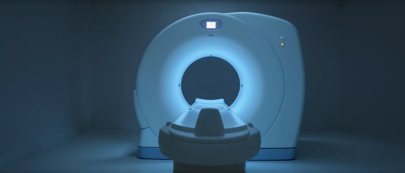
CT Angiography
CT angiography creates 3D heart images to diagnose coronary artery disease.
What is CT Angiography?
CT angiography is an advanced, noninvasive heart imaging test that allows your medical team to see the blood vessels in and around your heart without making any surgical incisions. If you are facing heart surgery or experiencing chest pain, your cardiology team needs a clear map of your coronary arteries to plan the safest route for your care. This scan provides exceptionally detailed, 3D images of your vascular system, helping your surgeon understand exactly what they are working with before you ever enter the operating room.
By using a specialized X-ray machine that rotates around your body, this technology captures hundreds of thin slices of information in just a few seconds. A contrast dye is injected into a vein in your arm to illuminate your blood vessels, highlighting any areas where plaque or cholesterol might be narrowing the arteries. The entire outpatient process is fast, painless, and requires no recovery time.
For patients and families navigating the anxiety of heart disease, the value of a clear coronary artery disease diagnosis cannot be overstated. Knowing the exact location and severity of a blockage empowers you and your surgical team to make confident, informed decisions about the best path forward, whether that means lifestyle changes, stenting, or a more complex surgical intervention.
How does a cardiac CT scan work?
The technology behind a cardiac CT scan relies on a sophisticated doughnut-shaped machine called a CT scanner. You will lie comfortably on a narrow table that slowly slides into the center of the machine. To ensure the clearest pictures possible, the technician will place small sticky pads on your chest to monitor your heartbeat, ensuring the camera only takes pictures in the brief milliseconds between heartbeats when the heart muscle is naturally still.
How the contrast dye illuminates your arteries: The iodine-based contrast material is introduced through a small intravenous line, usually in your arm. You might experience a brief, entirely normal wave of warmth spreading through your body and a metallic taste in your mouth. This dye temporarily makes your blood vessels glow bright white on the X-ray images, providing a stark contrast against the surrounding heart muscle.
As the scanner rotates around you, it gathers data to build a high-resolution, three-dimensional model of your beating heart. A computer then removes the tissue and bone from the image, leaving behind only a pristine, glowing map of your arteries. This final rendering gives your cardiologist a clear view of the entire coronary tree, allowing them to rotate and zoom into specific vessels on a computer screen.
CT angiography vs angiogram key differences
Understanding the difference between a CT scan and a traditional invasive angiogram is crucial for easing pre-surgical anxiety. A traditional angiogram is a more invasive procedure where a catheter is carefully threaded from your groin or wrist directly into your heart's arteries. While it remains the definitive gold standard for certain complex cases, it carries slightly higher risks because it involves entering the body and physically manipulating instruments inside your blood vessels.
Conversely, ct angiography is strictly noninvasive. There are no catheters threaded into your heart, and the entire process is essentially an advanced X-ray. If your surgical team needs to screen for issues before a major operation, they will typically start here because it provides an excellent diagnostic overview without the need for surgical recovery or an overnight hospital stay.
Doctors often use these two tools in tandem rather than in competition. If your scan reveals a complex issue that requires a stent or a deeper look, your specialist may follow up with a traditional angiogram. In many cases, however, the noninvasive scan provides enough definitive information to confidently proceed with surgical planning, sparing the patient from an unnecessary invasive procedure.
Why do doctors order coronary CT angiography?
Your heart specialist will order a cardiac imaging test for several critical reasons, primarily to investigate the root cause of symptoms like unexplained chest pain or shortness of breath. If an exercise stress test yields ambiguous results, or if you have a strong family history of heart disease, this scan offers the most direct way to physically see if plaque is restricting blood flow to your heart muscle. It is an invaluable tool for ruling out dangerous blockages.
Identifying high-risk plaque before surgery: Not all plaque is created equal. This imaging technique allows specialists to analyze the actual composition of the buildup in your arteries. Spotting soft, vulnerable plaque that is prone to rupturing can be life-saving information, guiding your surgical team to prioritize specific arteries for bypass or intervention before a heart attack occurs.
Additionally, the scan helps surgeons evaluate the overall anatomy of your heart to determine if you are a suitable candidate for specific surgical approaches. By understanding the precise size, shape, and path of your coronary vessels beforehand, your surgical team can anticipate challenges and tailor their operative strategy to your unique anatomy, ultimately leading to better outcomes and a safer recovery.
CT angiography preparation what to expect
Proper preparation ensures the imaging center can capture the highest quality images of your heart. You will generally be asked to avoid eating or drinking anything for a few hours before your scheduled appointment, and you should strictly avoid caffeine for at least 12 hours prior. Caffeine can elevate your heart rate, which makes it more difficult for the scanner to capture those perfectly still, crisp images between your heartbeats.
When you arrive at the clinic, the nursing staff will review your medical history and establish your IV line. Because the contrast dye is filtered out through your kidneys, they will need to confirm that your kidney function is healthy enough to process the material safely. You will also be asked to change into a hospital gown to ensure no metal buttons or zippers interfere with the X-ray imaging process.
- Avoid heavy meals: Fasting for 4-6 hours prevents nausea from the contrast dye.
- Discuss your medications: Inform your team about all prescriptions, especially beta-blockers or diabetes drugs.
- Plan your time: The entire visit takes about an hour, though the actual scan is under 15 seconds.
Understanding ct angiography risks and safety
For the vast majority of patients, noninvasive heart imaging is remarkably safe and well-tolerated. The test does involve a small dose of ionizing radiation, but modern cardiac scanners are heavily optimized to keep this exposure to an absolute minimum. Your medical team will only recommend the scan if the diagnostic benefits significantly outweigh any theoretical risk associated with radiation exposure.
Allergies to contrast dye: A small percentage of patients have a mild allergic reaction to the iodine-based contrast material, experiencing hives or itching. Be sure to tell your technologist about any prior allergies to seafood or contrast agents, as they can administer preventive medication beforehand. Patients with severe asthma or compromised kidney function will be closely monitored or offered alternative imaging approaches.
You might notice some minor bruising or tenderness around the IV site in your arm for a day or two, which is entirely normal. Once the scan is complete, you will be encouraged to drink plenty of water to help your body naturally flush the contrast dye from your system. Aside from that, you can immediately return to your normal daily activities without restriction.
What do ct angiography results mean?
Interpreting the final images requires the trained eye of a specialized cardiologist who will analyze the 3D renderings of your arteries. They are looking for areas where the vessel walls have narrowed, indicating the presence of atherosclerotic plaque. The results are typically categorized to show whether the narrowing is mild, moderate, or severe, which directly informs the urgency of your surgical or medical intervention.
If the results show minimal or no significant plaque, your doctor can confidently rule out coronary artery disease as the source of your symptoms. This provides immense emotional relief for patients worried about impending heart surgery, allowing the medical team to pivot and investigate other potential causes. It is a powerful tool for definitively clearing patients of major arterial disease.
When the scan reveals clinically significant blockages, your surgical team will use the detailed images to plan their exact approach, whether that involves minimally invasive cardiac bypass tools or stenting. The 3D map shows them exactly where the healthy tissue ends and the blockage begins, allowing for precise measurements that are critical for a successful operation. Your surgeon will sit down with you to explain these findings and outline the recommended steps.
How noninvasive heart imaging guides surgery
For a heart surgeon, having a preoperative ct angiography scan is like having a detailed GPS before embarking on a complex journey. While the surgeon still relies on their direct vision in the operating room, the pre-surgical scan allows them to assess the overall calcium load in your arteries. Heavy calcification can change how they handle the vessels during bypass surgery, and knowing this in advance prevents unexpected surprises during the operation.
The scan also provides vital information about the structure of the heart itself, allowing the surgeon to evaluate the size of the heart chambers and the function of the heart valves. If a patient is undergoing a combined procedure, this comprehensive anatomical overview helps the surgical team anticipate the need for specialized heart valve repair devices or additional grafting materials before they even make the first incision.
Ultimately, this level of preoperative visualization significantly reduces the time a patient spends on the cardiopulmonary bypass machine. By entering the operating room with a complete map of the patient's unique vascular anatomy, the surgical team can work more efficiently and decisively. This careful planning directly translates to shorter surgeries, less trauma to the body, and a smoother, faster recovery for the patient.
Finding a ct angiography near me
When you or a loved one requires urgent cardiac evaluation, locating a facility equipped with advanced imaging technology is a top priority. Not every local clinic has the specialized, high-speed CT scanners required for detailed cardiac work, so your primary cardiologist will refer you to a dedicated imaging center or hospital that specializes in comprehensive heart diagnostics. They will ensure the facility has the exact equipment needed to capture high-resolution images of your beating heart.
If you are experiencing symptoms or have been told you need advanced screening, reaching out to a specialized cardiac diagnostic center in Guadalajara ensures you have access to the latest noninvasive technology.
When choosing a center, it is important to ask if they have a dedicated cardiac radiologist on staff to interpret the results. The subtle nuances of a coronary scan require an expert eye to correctly identify soft plaque and subtle vessel narrowing. Working with an experienced cardiovascular team ensures that your images are not just taken correctly, but interpreted with the surgical perspective in mind.
Moving forward after your cardiac imaging test
Completing your cardiac imaging test is a major milestone in your journey toward heart health. Whether the results provided the reassuring news of clear arteries or the critical information needed to plan a surgical intervention, you now have a clear roadmap for your next steps. The anxiety of the unknown is often the heaviest burden for heart patients, and this diagnostic clarity provides a profound sense of relief and direction.
Your cardiology team will review the comprehensive results with you in a detailed consultation, explaining exactly what the images show and how they impact your specific treatment plan. If surgery is recommended, the surgical coordinator will guide you through the scheduling process, ensuring all the necessary preoperative clearances are obtained. You will never be left to navigate these complex decisions alone; your medical team is with you at every step.
Remember that undergoing this scan is a proactive, powerful step toward protecting your long-term health. By choosing to undergo advanced imaging, you have given your doctors the vital information they need to provide the safest, most effective care possible. Take a deep breath, trust your specialized medical team, and know that you are taking the right steps to ensure a stronger, healthier heart for the future.






