Nuclear Perfusion Scan: Testing Blood Flow to the Heart
A nuclear perfusion scan heart test reveals blood flow problems before surgery.
When a cardiologist suspects that part of your heart muscle is not getting enough blood, one of the most common tests ordered is a nuclear perfusion scan heart study. It uses a tiny amount of radioactive tracer and a special camera to show where blood is flowing — and where it is not. This guide walks you through what the test involves, how to prepare, what the results mean, and what happens next if a blockage is found.
What Is a Nuclear Perfusion Scan Heart Test?
The basic idea. A nuclear perfusion scan — doctors often call it myocardial perfusion imaging — is a picture of blood flow through the heart muscle itself. A safe tracer is injected into a vein, travels with your blood, and the parts of the heart receiving good flow absorb more of it than areas with poor flow. A gamma camera reads that signal and builds detailed images of the heart at work.
Why flow matters more than anatomy alone. Some narrowed arteries never cause trouble; some narrowings that look mild on a picture actually limit blood flow badly when the heart is working hard. Perfusion imaging answers the question that matters most to your cardiologist: is any part of the heart muscle actually being starved? That answer shapes whether medication, a stent, or surgery is the right path.
Stress and rest images. Almost always the test is done twice: once with the heart under stress (exercise or medication) and once at rest. Comparing the two sets of images shows whether an area is permanently damaged or only struggling under load. The technique itself is well established in nuclear cardiology and standardised across imaging centres worldwide — see the American Society of Nuclear Cardiology's overview at ASNC.
How Does a Nuclear Perfusion Scan Heart Work?
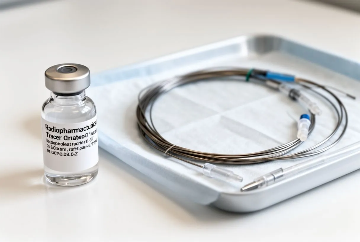
The tracer. The tracer — usually a substance such as technetium-based sestamibi — behaves like a red blood cell. It flows wherever blood flows, but it releases a faint signal the camera can detect. The dose is very small, similar in effective radiation to other routine diagnostic imaging, and it leaves the body naturally over a day or two.
Creating stress safely. If you cannot walk on a treadmill, or the doctor wants images under precise, controlled conditions, a medication is given instead. Drugs such as adenosine or regadenoson dilate the healthy arteries, mimicking the blood demand of exercise. Imaging protocols for these studies follow consensus guidelines on scanner settings and image reporting — an example is the European Society of Cardiovascular Radiology's consensus document on cardiac imaging protocols, published in European Radiology.
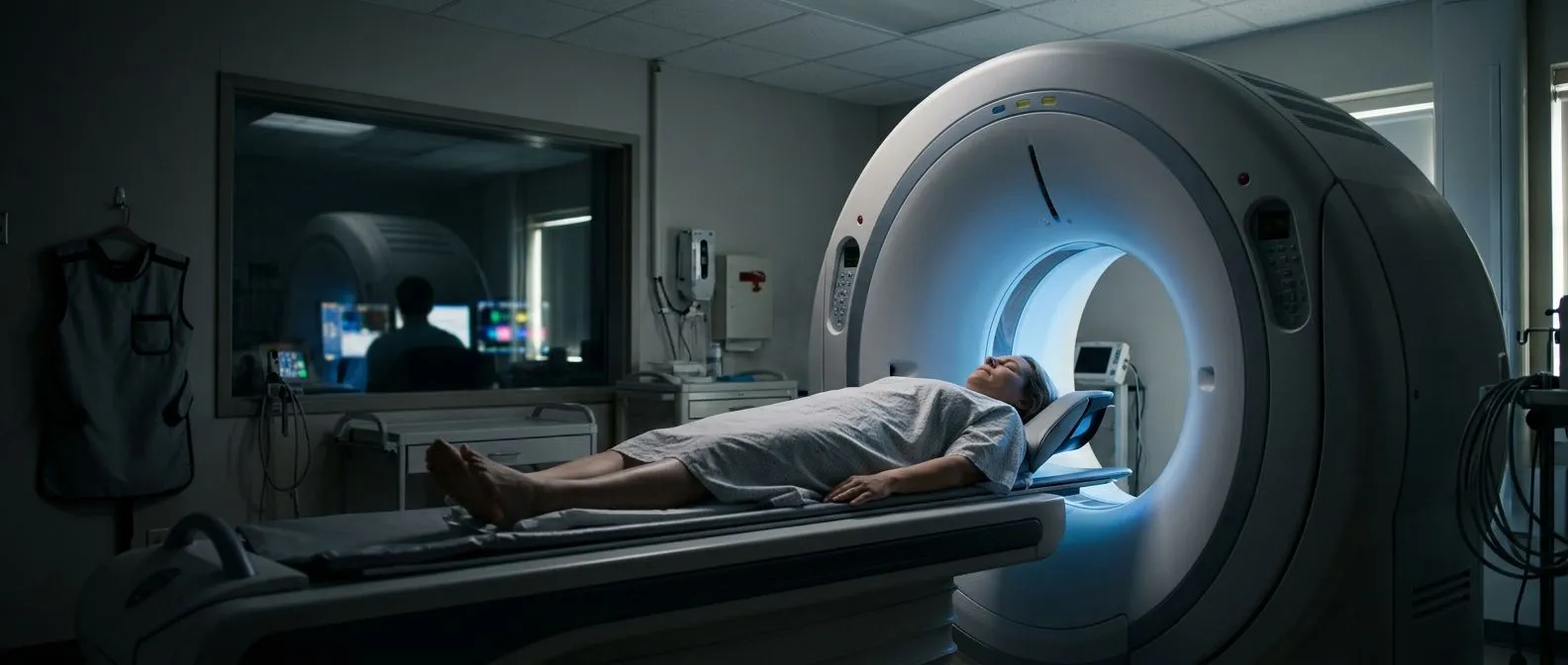
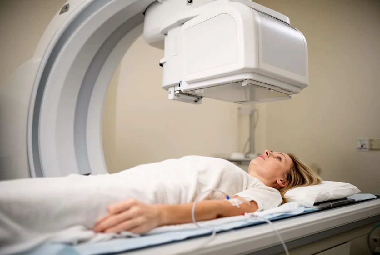
The camera and computers. You lie flat under a gamma camera (SPECT) — in some centres, a PET scanner is used instead, which takes faster, sharper images. Computers reconstruct cross-sections of the heart so the reading doctor can compare every wall of the muscle between the stress and rest studies.
What Happens During a Myocardial Perfusion Scan?
Before the first images. A small intravenous line is placed, and the first tracer injection is given. Then you wait — the tracer needs time to reach the heart. If you are exercising, you walk on a treadmill while the heart rate climbs to the target, and the tracer is injected near peak effort.
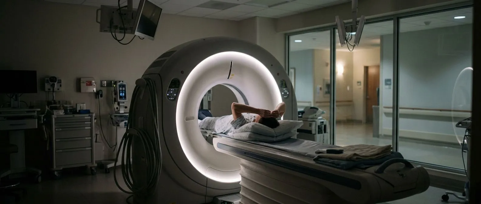
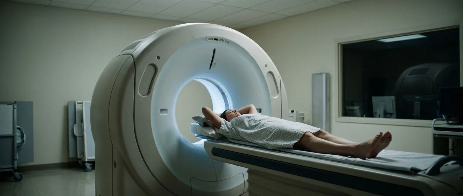
Under the camera. Imaging itself is painless. You lie still on a table for roughly 15 to 25 minutes while the camera moves slowly around your chest. The main request is simple: stay still. Movement blurs the images, and blurred images sometimes need repeating.
The second half. After the first images, you rest for a few hours — sometimes the rest images are taken on a separate day. Then the second injection and second scan are done. Most people go home the same morning and feel back to normal within hours. Ask for every step to be explained before the appointment, so you do not walk into the scanning room unsure of what comes next.
Sometimes a subtle question remains — for example, whether soft tissue near the breast or diaphragm is masking part of the heart. In those cases the team may add a specialised attenuation-correction acquisition (sometimes called a SIP sequence in modern scanners) to sharpen the images before the report is written.
Nuclear Perfusion Scan Preparation: What Should You Avoid?
Caffeine is the big one. Caffeine blocks the very receptors the stress medications use, so if your scan uses a pharmacological stress agent, you will be asked to avoid coffee, tea, cola, energy drinks and chocolate for roughly 24 hours beforehand. Skipping this can make the test unusable — it is the single most common reason a scan has to be repeated.
Medications. Some heart medicines, and certain asthma drugs, may need pausing the night before or the morning of the test — but only ever on your doctor's explicit instruction. Never stop a medication on your own; the team will give you a precise list.
Eating and other practicalities.
- Food: a light meal is usually fine a few hours before the test; nothing heavy right before stress.
- Hydration: drinking water helps your body clear the tracer afterwards.
- Jewellery and lotions: leave metal at home; the camera works better without it.
Tell the team about pregnancy, breastfeeding, allergies and recent other nuclear scans before the appointment. These do not usually prevent the test, but they change how it is planned.
Nuclear Perfusion Scan vs Angiogram: Which Test Do You Need?
What each one shows. A nuclear perfusion scan shows how blood is behaving in the heart muscle — function under stress. An angiogram (invasive cardiac catheterisation) shows the anatomy: the exact location and severity of blockages inside the coronary arteries, in fine detail.
How they work together. Perfusion imaging is often the first gate: if it is normal, an invasive angiogram may be avoidable altogether. If it shows significant areas of poor flow, the angiogram (or a CT angiogram) confirms where the blockages are so a treatment plan can be built. Neither test replaces the other; they answer different questions.
Because radiation, cost and comfort all matter, most cardiologists use the least invasive test that will answer the question — and reach for catheterisation when the perfusion result demands it.
What Are the Downsides of a Nuclear Stress Test?
Radiation exposure. Every nuclear scan involves a small dose of ionising radiation — roughly comparable to other diagnostic imaging such as a CT scan. It is not a reason to avoid a medically necessary test, but it is a fair reason to ask whether a repeat scan is truly needed.
False positives and false negatives. No test is perfect. Body habitus, breast tissue, or a perfectly healthy heart under-exercised on the day can produce images that over- or underestimate disease. This is why results are always interpreted alongside your symptoms, your history, and sometimes other tests.
Short-lived side effects. Stress medications can cause flushing, chest pressure, headache or breathlessness for a few minutes — they wear off quickly, and an antidote exists for adenosine if needed. Patients who arrive anxious usually do better when they know these sensations are expected and temporary.
Nuclear Perfusion Scan Results: What Do They Mean?
The four possible patterns. Reading a perfusion study comes down to comparing stress and rest images, and there are four broad outcomes:
- Normal perfusion: good flow under stress and at rest — reassuring result.
- Reversible defect: poor flow under stress that returns to normal at rest — this suggests a significant narrowing that is still treatable.
- Fixed defect: poor flow on both scans — usually a scar from a previous heart attack.
- Mixed pattern: areas of both scar and threatened muscle — common and important for surgical planning.
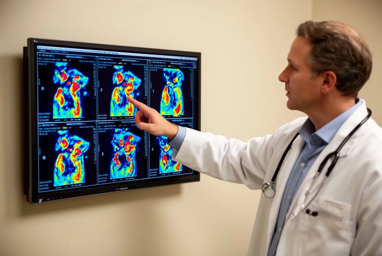
Additional measurements. The report also often includes the heart's pumping function (ejection fraction), the size of the heart chambers, and signs of strain. Together these numbers tell your cardiologist not only whether there is a problem, but how serious it is and how urgently it needs addressing.
How long until results. A report is typically ready within a few days. We take the time to go through the images with our patients in plain words — what the study shows, what it does not show, and what the realistic options are — before any decision is made.
What Is a Nuclear Medicine Ventilation Perfusion Scan?
A different question entirely. Not every nuclear perfusion scan concerns the heart. A ventilation-perfusion scan — often shortened to V/Q scan — looks at the lungs. "Ventilation" means how well air moves in and out of each part of the lung; "perfusion" means how well blood reaches those same areas.
Why it is done. The classic reason is to look for a pulmonary embolism — a blood clot lodged in the lung's arteries. A normal V/Q match (air and blood reaching the same regions) makes a significant embolism unlikely; a mismatch points to one. It is a valuable alternative for people who cannot have a contrast CT scan, such as those with severe kidney problems or contrast allergy.
What to expect. You breathe a mildly radioactive gas or aerosol for the ventilation images, then receive a tracer injection for the perfusion images. Like the cardiac version, it is painless and the radiation is low. Your doctor will tell you which type of scan — cardiac or lung — has been ordered, because the two are often confused by name.
What Happens If They Find a Blockage During a Nuclear Stress Test?
The result is a starting point, not a verdict. An abnormal perfusion scan rarely leads to emergency treatment by itself. It tells your cardiologist where to look next — usually an angiogram to map the blockage precisely, and sometimes additional tests of heart muscle viability.
Treatment options with trade-offs. Depending on what the follow-up tests show, the realistic options usually fall into three groups:
- Medication: for stable, mild disease — drugs to lower heart workload, prevent clots and manage cholesterol, together with lifestyle changes.
- Stent (angioplasty): a balloon and metal scaffold opened inside the narrowed artery, suited to one or a few significant blockages.
- Bypass surgery: grafts that carry blood around severe or multiple blockages — the choice when the anatomy, or other factors like diabetes or valve disease, favour surgery.
How the decision is made. The right choice depends on which arteries are involved, how much muscle is at risk, and your overall health. Every case we see is reviewed by our multidisciplinary team of cardiac surgeons against international guidelines before a recommendation is made, and the patient always receives the alternatives with the reasoning behind each one. We perform conventional, minimally invasive, robotic and transcatheter procedures, and we recommend the less invasive route only where the evidence genuinely supports it for that patient.
Nuclear Perfusion Scan Cost and Choosing a Cardiac Team
What drives the price. The cost of a nuclear perfusion scan varies widely by country, imaging technology (SPECT versus PET), and whether the study is bundled into a broader work-up. Ask any centre for a written quote that states which scanner, which protocol, and whether the cardiologist's interpretation is included.
Before the test, ask about the rest of the work-up. A good centre orders only the studies that will change the plan — a Full Blood Count, kidney function, and a review of existing imaging are typical starting points, and your doctor will explain exactly which of them your case needs before anything is ordered. When a patient first contacts us, the video consultation is free: we get to know them, talk through the condition, and say which studies are still needed — so no unnecessary ones are ordered and no money is spent on tests that will not change the plan.
Second opinions are normal. If you have been told you need surgery based on a perfusion result, a second opinion is not a betrayal of your doctor — it is a standard part of making a well-informed decision about a major operation. We actively encourage patients to seek second opinions, and we treat that as the normal, sensible step it is. If you would like your scan results and options reviewed by Dr Alejandro Molina's team, book a consultation at our consulting rooms in Guadalajara, Querétaro or Hermosillo, or start with the free video consultation.
In summary, a nuclear perfusion scan heart study is a safe, well-proven way to show whether your heart muscle is actually receiving the blood it needs — information that decides between medication, a stent, or surgery. Know what the test involves, prepare properly, and make sure someone explains the images to you in plain language. Bring your questions, ask for a second opinion if a major decision hangs on the result, and never act on an abnormal scan alone — discuss it with a qualified cardiac team before deciding on treatment.