

Ventilation-perfusion Scan
Learn how a ventilation-perfusion scan evaluates lung health before cardiac surgery.
What is a ventilation-perfusion scan
A ventilation-perfusion scan is a specialized nuclear medicine imaging test that evaluates how well air and blood are flowing through your lungs. Often referred to simply as a V/Q scan, this diagnostic tool creates detailed images showing the distribution of air as you breathe and blood as it circulates through the pulmonary arteries. For patients preparing for cardiac surgery or managing complex heart conditions, understanding lung function is a critical part of the preoperative assessment process.
What is a V/Q scan used for in cardiac patients: In the context of cardiology and cardiac surgery, a ventilation-perfusion scan helps your medical team identify potential risks before you enter the operating room. If your heart has been struggling to pump efficiently, it can affect how well blood reaches your lungs, and a V/Q scan provides precise data on this dynamic. This test allows your surgeon, such as Dr Alejandro Molina, to accurately assess your respiratory health and optimize your surgical plan.
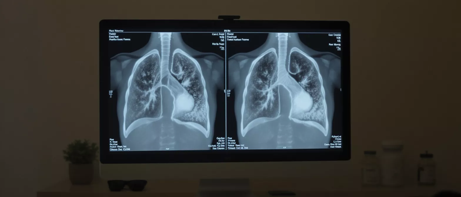
By comparing the airflow (ventilation) and blood flow (perfusion) images side by side, doctors can detect even subtle abnormalities that might complicate anesthesia or recovery. It is a painless, highly detailed way to ensure your lungs are healthy enough to withstand the physical demands of a major procedure. The scan is particularly useful for diagnosing a pulmonary embolism, which is a potentially dangerous blood clot in the lungs.
How does a V/Q scan work
A ventilation-perfusion scan works by using tiny amounts of safe radioactive materials called radiotracers. The test is split into two distinct phases: one that measures airflow and another that measures blood flow. These tracers emit a small amount of energy that a special gamma camera detects, translating the data into comprehensive images of your lung function.
The ventilation phase: During the first part of the test, you breathe in a slightly radioactive gas through a mask or mouthpiece. The gas mixes with the air in your lungs, and the camera takes pictures that show exactly where the air travels. If air cannot reach a specific area of the lung, the scan will reveal a gap in ventilation.
The perfusion phase: Immediately after the ventilation phase, a slightly radioactive substance is injected into a vein in your arm. This tracer travels directly to your heart and into the blood vessels of your lungs, allowing the gamma camera to capture detailed images of your lung's blood supply. By comparing these two sets of images, your medical team can accurately identify any ventilation-perfusion ratio mismatch.
Why is a lung blood clot test needed before surgery
Before undergoing any major cardiac surgery, your medical team must ensure that your lungs are functioning properly to handle the stress of cardiopulmonary bypass and anesthesia. A lung blood clot test like a V/Q scan is often ordered to rule out a pulmonary embolism. Finding a blood clot before surgery is essential, as operating on a patient with an untreated clot significantly increases the risk of serious complications.
Detecting pulmonary embolism before cardiac procedures: A pulmonary embolism occurs when a blood clot breaks loose from the deep veins of the leg or arm and travels to the lungs, blocking a pulmonary artery. If this happens during or immediately after surgery, it can be life-threatening. A pulmonary embolism scan ensures that no hidden clots are present before the surgical intervention begins.
Beyond clots, the test assesses baseline lung health. Patients with complex heart disease often have concurrent lung issues like chronic obstructive pulmonary disease or heart-failure-related fluid buildup. Understanding the ventilation perfusion ratio mismatch helps your anesthesiologist and surgeon prepare the most tailored, safe approach for your specific anatomy and physiology.
- Risk assessment: Identifies blood clots that could complicate anesthesia or post-operative recovery.
- Blood flow mapping: Shows how well blood circulates through the lungs, highlighting any vascular blockages.
- Surgical planning: Allows your surgical team to tailor their approach based on your actual lung capacity and reserve.
Ventilation-perfusion scan procedure step by step
Understanding the ventilation-perfusion scan procedure can help alleviate any anxiety you or your family might feel before the test. The scan is completely painless, requires no incisions, and is performed entirely on an outpatient basis in the nuclear medicine department. A trained nuclear medicine technologist will guide you through the entire process.
Step-by-step experience: You will be asked to change into a hospital gown and remove any metal objects. During the ventilation phase, you sit or lie down and breathe in the radioactive gas through a specialized breathing apparatus. Once the ventilation images are captured, you will receive an injection for the perfusion phase, and the gamma camera will take additional images.
The entire procedure typically takes between 30 and 45 minutes to complete. You will be asked to remain perfectly still while the camera is taking pictures, but you can breathe normally unless instructed otherwise. The camera itself does not emit radiation; it simply detects the tracer inside your body, making the experience very safe and comfortable.
V/Q scan results and what they mean
Reading V/Q scan results involves comparing the airflow images directly with the blood flow images. In a healthy lung, air and blood should distribute evenly to the exact same areas. When the patterns match perfectly, it means your lungs are functioning optimally and no dangerous blockages are present.
Interpreting a ventilation perfusion mismatch: If the scan reveals that areas of the lung have good airflow but poor blood flow, it strongly suggests a pulmonary embolism diagnosis. This specific finding is known as a ventilation perfusion mismatch, because the blood vessels are blocked while the airways remain open.
In some cases, the scan might show matched defects, where both air and blood are absent from a specific area. This usually indicates a different type of lung disease, such as severe pneumonia or lung tissue damage from old infections. Your medical team will interpret these results alongside your cardiac history to make the most accurate diagnosis possible.
V/Q scan vs CT angiography which is better
When a pulmonary embolism is suspected, doctors must choose between a ventilation-perfusion scan and a CT pulmonary angiography. Both are excellent diagnostic tools, but they have distinct advantages depending on the patient's condition. The choice often depends on kidney function, allergies to contrast dye, and whether the patient has underlying lung disease.
V/Q scan versus CT pulmonary angiogram: A V/Q scan is a nuclear medicine lung scan that is particularly beneficial for patients who have poor kidney function or severe allergies to the iodinated contrast used in CT scans. It specifically highlights how well the lungs are oxygenating blood without putting extra strain on the renal system.
CT angiography is faster and provides highly detailed images of the actual blood vessels, showing the exact size and location of the clot. However, for patients who already have compromised kidney function due to heart disease, the contrast dye required for a CT scan can be dangerous. In these cases, the ventilation-perfusion lung scan is the preferred diagnostic method.
Is a pulmonary embolism scan safe
Safety is a primary concern for any patient facing heart surgery, and you may wonder about the risks associated with a pulmonary embolism scan. The radiation exposure during a ventilation-perfusion scan is very low. The radioactive tracers used are quickly eliminated from the body through natural biological processes, minimizing any long-term side effects.
Understanding radiotracer safety: The amount of radiation you receive during a V/Q scan is comparable to standard diagnostic X-rays and is well below the threshold that would cause harm. The benefits of accurately diagnosing a life-threatening blood clot far outweigh the minimal risks associated with this nuclear medicine test. Radiotracers are carefully formulated to ensure they do not cause allergic reactions in the vast majority of patients.
If you are pregnant or breastfeeding, it is crucial to inform your physician before scheduling the test. While the radiation dose is low, special precautions or alternative imaging methods may be considered to protect the baby. Otherwise, you can resume your normal daily activities immediately after the scan is complete, as no recovery time is needed.
- Low radiation exposure: The dose is minimal and comparable to natural background radiation over a few months.
- No contrast dye: Eliminates the risk of kidney damage or iodine-allergy reactions common with CT scans.
- Quick elimination: The radiotracer leaves the body naturally within a few hours to days.
How to prepare for a nuclear medicine lung scan
Preparing for a nuclear medicine lung scan is remarkably straightforward and requires very little effort on your part. Unlike other medical procedures, a ventilation-perfusion scan usually does not require fasting, dietary restrictions, or bowel preparation. You can eat, drink, and take your prescribed medications unless your doctor specifically advises otherwise.
Pre-scan checklist for patients: On the day of your appointment, wear comfortable clothing that is easy to remove, as you will need to change into a hospital gown. Leave all metal jewelry at home, as metal can interfere with the gamma camera's imaging capabilities. If you have recently had a chest X-ray, bring the images with you, as comparing them to the V/Q scan helps your radiologist make a more accurate diagnosis.
Be sure to inform the technologist if you use supplemental oxygen at home. They may ask you to bring your portable oxygen with you so they can monitor your breathing safely during the ventilation phase of the test. It is also helpful to bring a list of your current medications and a brief summary of your cardiac history.
Understanding ventilation perfusion ratio mismatch
A ventilation perfusion ratio mismatch is a medical term used to describe a situation where the amount of air reaching the air sacs does not match the amount of blood reaching the capillaries surrounding them. In a perfectly functioning respiratory system, this ratio is tightly balanced to ensure maximum oxygen exchange. A V/Q scan is the gold standard for detecting this specific imbalance.
What does a ventilation perfusion mismatch mean for you: When there is a mismatch, it typically points to an issue like a pulmonary embolism, where blood is blocked but air flows freely. Understanding the ventilation perfusion ratio mismatch is critical for your surgical team.
Not all mismatches are caused by blood clots. Conditions like asthma, chronic bronchitis, or localized lung damage can also alter the ratio. By identifying the exact type and location of the mismatch, your cardiologist and surgeon can develop a targeted treatment plan that addresses both your heart and lung health simultaneously, ensuring a safer surgical outcome.
What to expect after your lung scan
Immediately following your ventilation-perfusion scan, you can return to your normal daily routine without any restrictions. There is no recovery time required, and you will not feel any lingering effects from the radiotracer. The small amount of radioactive material will naturally clear from your body through your urine and exhaled breath over the next 24 hours.
Following up on your scan results: The images from your scan will be carefully reviewed by a radiologist specializing in nuclear medicine. Within a day or two, the results will be sent to your cardiologist or surgeon. They will discuss the findings with you during a follow-up appointment or incorporate the data directly into your pre-surgical planning conference.
If the scan shows a pulmonary embolism, your doctor will likely prescribe blood thinners to dissolve the clot before any elective cardiac surgery proceeds. If the scan is clear and shows healthy, matched airflow and blood flow, it provides immense peace of mind. Knowing your lungs are healthy gives you and your surgical team the confidence to proceed with the necessary cardiac intervention safely.






