Cardiac MRI: What to Expect During the Scan
Cardiac MRI what to expect during your scan: preparation steps, scan duration, noise levels, contrast dye, and breathing instructions.
If your cardiologist has ordered a cardiac MRI, you may be wondering exactly what to expect during a cardiac MRI — how long it takes, whether it hurts, and what all the knocking sounds mean. A cardiac MRI (magnetic resonance imaging of the heart) uses a strong magnetic field and radio waves, not radiation, to build detailed pictures of your heart muscle, valves, and major vessels. It is one of the most information-rich tests in cardiology, and knowing what the appointment involves can take most of the anxiety out of it. This guide walks you through the whole experience, from preparation to receiving your results.
What Is a Cardiac MRI and What Is It Looking For?
A cardiac MRI is a scanning test that creates moving and still images of your heart using magnets and radio waves. Because it involves no ionizing radiation, it can be repeated safely when your team needs to follow a condition over time.
What shows up on a cardiac MRI? The scan can reveal the size and pumping strength of the heart's lower chambers (the ventricles), inflammation or scarring of the heart muscle, congenital defects present from birth, tumors or fluid around the heart, and disease of the heart valves such as the aortic or mitral valve. A related technique, magnetic resonance angiography (MRA), uses the same machine to image the blood vessels themselves — an MRA scan is used to look at the aorta, the body's main artery, and other vessels for aneurysms, narrowing, or tears.
At our practice, we use Nature-published evidence on cardiovascular imaging alongside standard guidelines when deciding whether a cardiac MRI will genuinely change a patient's treatment plan. In the consultation we take the time to explain what the studies actually show, in plain words, before any decision is made.
Why Your Cardiologist Might Order a Cardiac MRI
Cardiologists order this scan when an echocardiogram (an ultrasound of the heart) or other tests leave important questions unanswered. It is especially valuable for assessing heart valve disease, measuring the aorta before surgery, quantifying how much heart muscle is damaged after a heart attack, and diagnosing conditions like myocarditis, which is inflammation of the heart muscle often triggered by a virus.
Chest pain diagnosis without clear answers. If you have cardiac MRI chest pain workups in mind, know that the scan can show whether the small vessels of the heart are restricted even when the main arteries look normal. For patients being considered for valve surgery or TAVR (transcatheter aortic valve replacement, a minimally invasive way of replacing the aortic valve through a catheter), imaging is central to planning. Consensus documents from the European Society of Cardiovascular Radiology define standardized scanning protocols and measurements used before valve procedures, which is the standard we follow when preparing patients for intervention (Insights into Imaging).
Every case at our clinic is reviewed by the multidisciplinary team against international practice guidelines before a recommendation is given, and the patient always receives a clear proposal with the alternatives.
What Should You Not Do Before a Cardiac MRI?
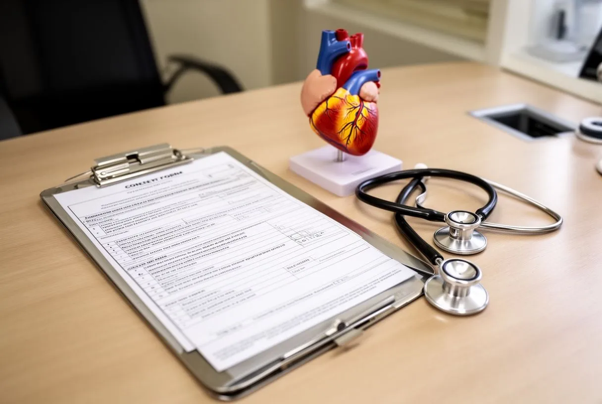
Preparation for a cardiac MRI is straightforward, but a few rules matter for safety and image quality.
- Avoid heavy meals and caffeine beforehand: caffeine can raise your heart rate and interfere with the breathing-gated images; most centers ask you to skip food for 4–6 hours if contrast is planned.
- Bring your full medication list: most medicines are taken as usual, but your team will confirm; do not stop anything without instructions.
- Metal Items to remove: jewelry, watches, hearing aids, and hairpins must come off; tell the team about pacemakers, defibrillators, stents, cochlear implants, or metal fragments from prior injuries — many modern devices are MRI-safe, but each must be verified first.
- Pregnancy: MRI without contrast is generally considered safe in pregnancy, but always tell the staff so they can adjust the protocol.
What to wear. Choose clothing without metal zippers or clasps, or change into a gown at the facility. Arrive 30–45 minutes early for paperwork and an IV line placement if contrast is planned.
What Happens During a Cardiac MRI, Step by Step
Knowing the sequence of a cardiac MRI procedure step by step helps many patients relax once they are on the table.
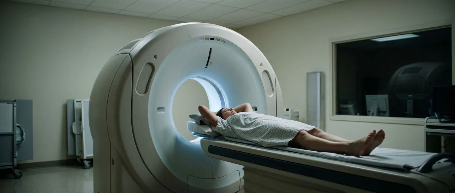
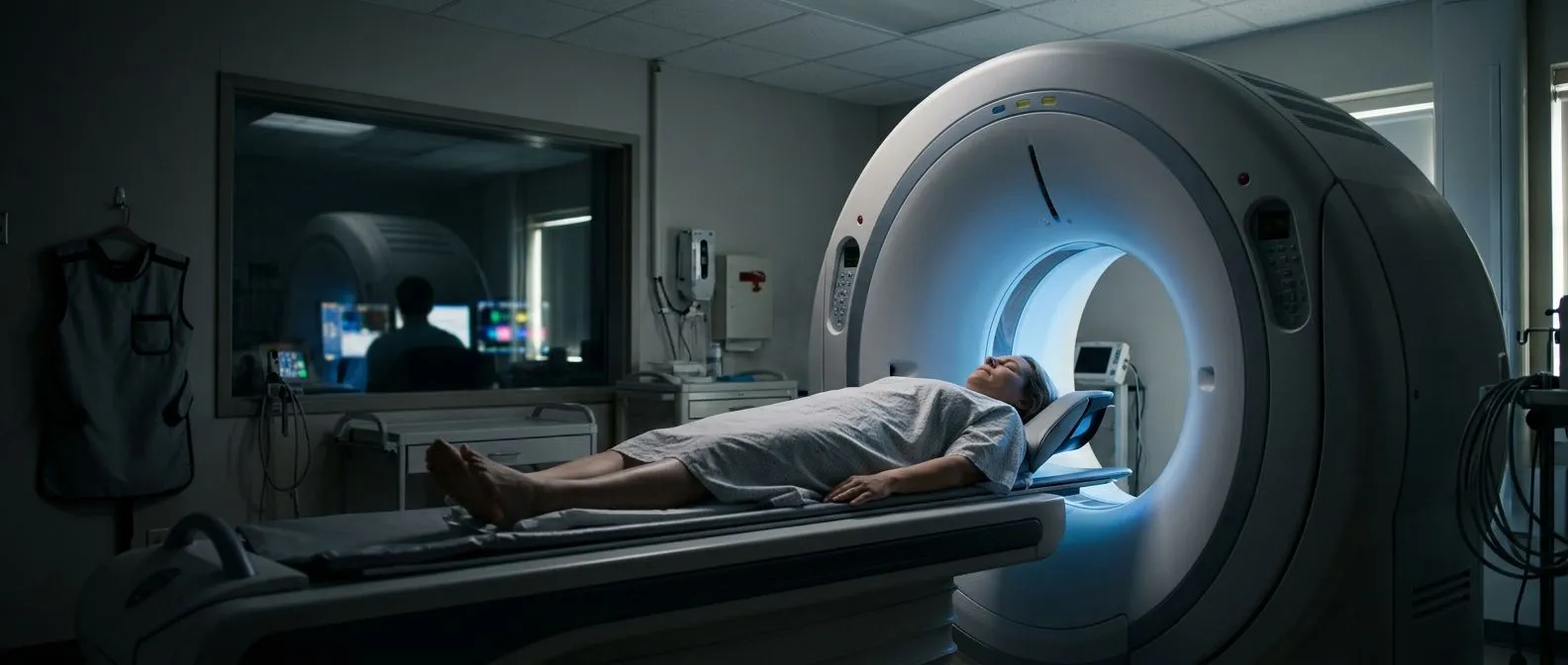
Before the scan. You will be asked about metal implants and allergies, given ECG electrodes (small sticky patches on your chest) so the machine can time images to your heartbeat, and positioned on a sliding table. A coil — a flat receiver pad that looks like a heavy blanket or plastic panel — is placed over your chest.
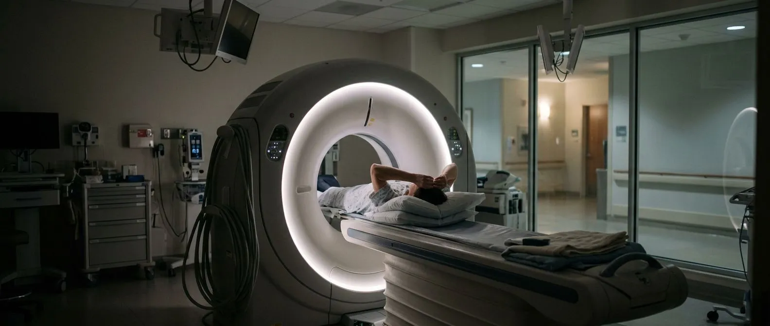
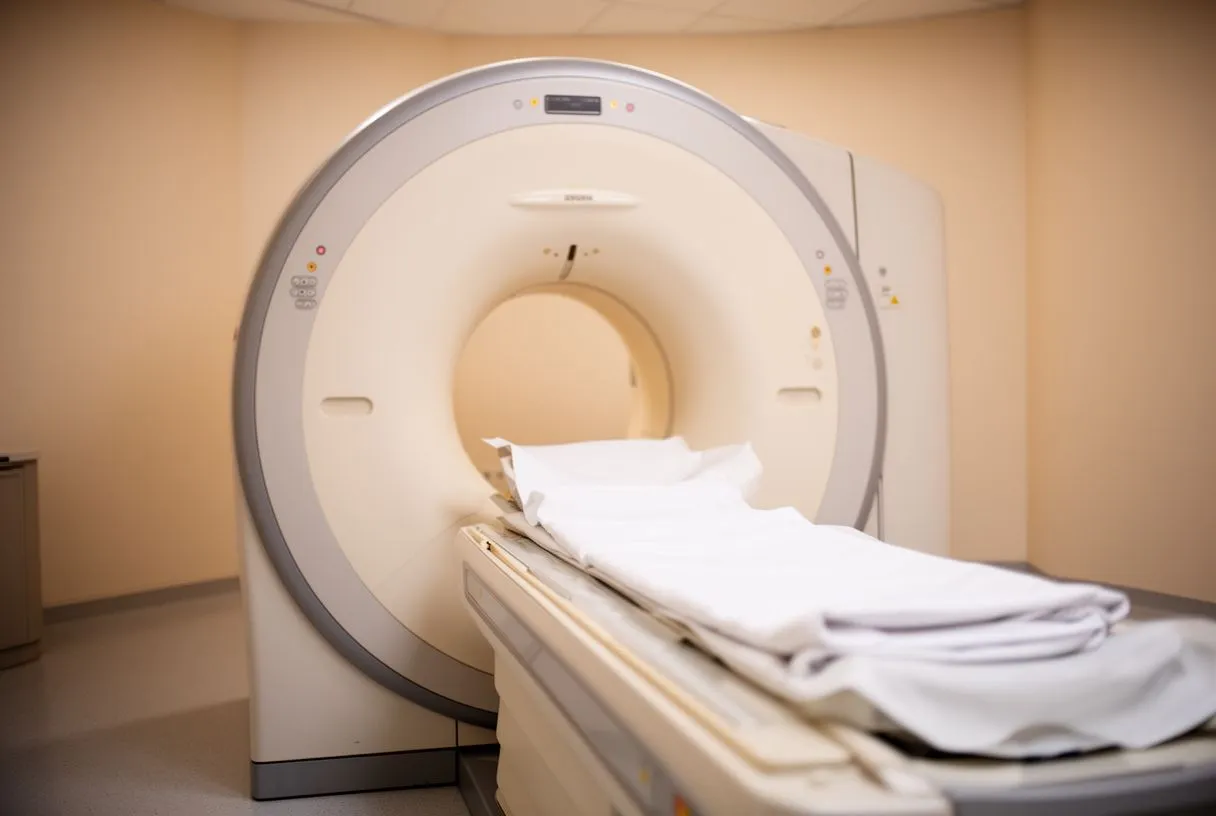
During the scan. The table slides into a tunnel that is open at both ends. The scanner makes loud tapping and thumping sounds while it works; you will be given earplugs or headphones. You will hear instructions through an intercom: "Breathe in… hold… breathe normally." Holding your breath for 8–15 seconds at a time keeps the pictures sharp, because the machine captures images between breaths. The rest of the time you simply lie still and relax.
These breathing instructions are usually explained before the scan begins, and a short rehearsal makes the whole appointment calmer.
How Long Are You in the Machine for a Cardiac MRI?
This is the question patients ask most. A typical cardiac MRI takes between 45 and 90 minutes, with most studies finishing in about an hour. The time varies because the scan captures many different image sequences — some look at pumping function, others at scarring, others at the vessels — and each sequence runs for a few minutes.
If the scan runs longer. If your heart rate is fast or irregular, or if a breath-hold is difficult, the technologist may repeat sequences to get clear images. This is routine and not a sign that anything is wrong. Complex protocols, such as those done before valve intervention, can run toward the upper end of the range.
There is no recovery period as such: cardiac MRI recovery is same-day, and you can drive yourself home unless you received a sedative.
Is a Cardiac MRI Claustrophobic and Noisy?
Many people ask whether the tunnel will trigger claustrophobia. The honest answer: the space is narrower than most people imagine, but most patients manage well with simple measures.
Cardiac MRI sedation options if you need them. If you know that enclosed spaces trouble you, tell the team when booking. Options range from keeping your eyes closed with a mirror or music, to a family member in the room, to mild oral sedation given before the scan. Only rarely does a patient need deeper sedation, and that can be arranged in advance. Sedated patients will need someone to drive them home.
The noise is real — the scanner can reach the volume of a jackhammer in short bursts — but earplugs plus headphones with music reduce it to a background rhythm. Many patients describe the experience as lying still inside a drum while someone plays an uneven drumbeat. If you go in knowing that the noise is expected and harmless, it loses most of its power to unsettle you.
Cardiac MRI Contrast Dye: What to Expect
Many cardiac MRI scans use a contrast agent called gadolinium, a fluid injected through a small IV line in your arm. It highlights blood flow and makes scar tissue or inflammation visible in ways plain images cannot.
How it feels. The injection itself feels like a cool sensation moving up your arm; some people notice a brief metallic taste. It is not the iodine-based dye used in CT scans, so iodine allergies are not a concern, and gadolinium reactions are uncommon and usually mild. Kidney function is checked beforehand in most adults, because gadolinium is cleared by the kidneys.
After the injection. The scan continues with new sequences lasting another 10–20 minutes. The contrast is later passed naturally by the body. If your scan is scheduled without contrast, the appointment is usually shorter.
Does a Cardiac MRI Show Blockages?
This is one of the most common questions, and the answer needs nuance. A standard cardiac MRI is excellent at showing heart muscle function, scarring, valve problems, and the anatomy of the aorta — but it is not the primary test for narrowing of the coronary arteries, the small vessels that feed the heart muscle.
What it can and cannot do. Some cardiac MRI techniques, such as perfusion imaging (watching contrast flow through the heart muscle) — sometimes described as a nuclear perfusion scan when done with radioactive tracers instead — can detect areas of reduced blood supply that suggest blockage. However, the definitive test for coronary artery blockages remains coronary angiography, a catheter procedure that directly outlines the arteries. Your cardiologist chooses the test based on your risk profile and symptoms.
When our team reviews a case, we ask which question each test is actually answering before any study is ordered, so no unnecessary ones are done. In a free video consultation we get to know the patient, talk through the condition, and say which studies are still needed.
Cardiac MRI vs Stress Test: Which One Do You Need?
Patients often compare a cardiac MRI vs stress test and wonder why one was chosen over the other. Both assess the heart, but they answer different questions.
- Stress test: shows how your heart performs under physical or medication-induced exertion; useful for symptoms that appear with activity and for tracking known coronary disease.
- Cardiac MRI: shows structure and tissue in exceptional detail — muscle scarring, valve function, heart chamber sizes, congenital defects, and the aorta — without radiation.
- They often work together: a stress test may flag a problem, and a cardiac MRI may then clarify its cause; or a scan may reveal valve disease that a stress test alone cannot explain.
If you have already had one of these tests and are unsure why the other was suggested, it is a fair question to raise with your cardiology team — a good explanation is part of good care.
Cardiac MRI Results, Cost, and Next Steps
Cardiac MRI results waiting time varies by center: a radiologist or cardiologist who specializes in heart imaging reviews hundreds of images, and a formal report is usually ready within a few days, with urgent findings communicated sooner.
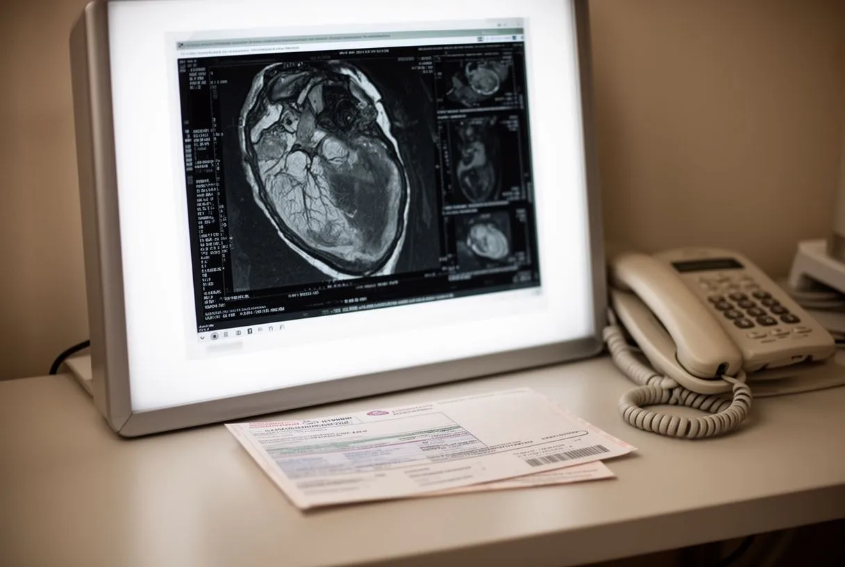
Understanding your report. A cardiac MRI report typically describes the pumping strength of the heart (called ejection fraction), the presence or absence of scarring, valve function, and any structural findings. When we review results with patients at our consulting rooms in Guadalajara, Querétaro, or Hermosillo, we explain what the numbers mean for their specific situation and what the realistic options are — including, where appropriate, conventional surgery, minimally invasive approaches, robotic surgery, or transcatheter procedures such as TAVR.
Cost considerations. The cost of a heart MRI varies widely by country, facility, and whether contrast is used, so ask the imaging center for a written estimate in advance. For international patients considering treatment in Mexico, the team can review existing studies and explain which studies are still needed. Dr Molina trained in cardiothoracic surgery at the Universidad de Guadalajara, in cardiac and minimally invasive surgery at the University of Montreal, and in structural cardiology and TAVR at CHU Bordeaux — and we encourage every patient to seek a second opinion as a normal part of deciding about heart surgery.
Is a cardiac MRI worth it? For most patients who are told they need one, yes — it answers questions that other tests cannot, involves no radiation, and often determines whether surgery or a catheter-based treatment is the right path. Heart disease remains one of the leading causes of lost healthy years worldwide according to the Global Burden of Disease Study (The Lancet), and getting an accurate diagnosis is the first step toward protecting your future.
Knowing what to expect during a cardiac MRI — the preparation, the noise, the breathing instructions, the contrast — turns an intimidating appointment into a manageable one. If you have been told you need a cardiac MRI, or if you have valve disease, chest pain, or an aortic condition being evaluated, we invite you to book a free video consultation with our team. We will review your case, explain which studies are truly needed, and help you decide with confidence. Do not self-diagnose: talk with a qualified cardiac team about your own situation.