

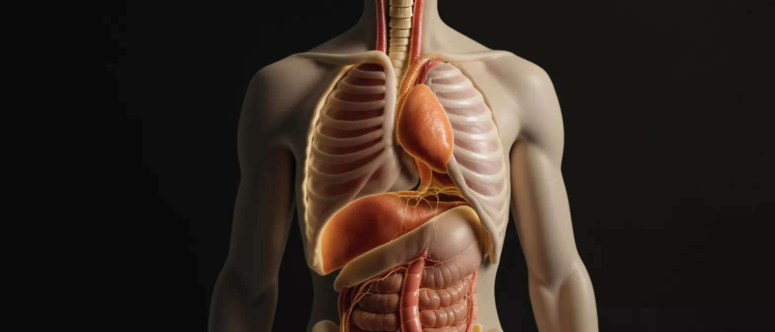
Descending Aorta
Learn about the descending aorta, its anatomy, branches, and conditions like aneurysms and dissection.
What is the Descending Aorta?
The descending aorta is the largest artery in the chest and abdomen, responsible for carrying oxygen-rich blood from the heart down to the rest of the body. After the aortic arch curves over the heart, the vessel transitions downward, forming the descending section. Understanding this structure helps patients and families make sense of complex cardiac diagnoses. For anyone facing heart surgery, knowing the basic layout of these vessels can ease anxiety and clarify why a specific procedure is necessary.
This blood vessel is divided into two main portions: the thoracic section in the chest and the abdominal section below the diaphragm. Because it supplies blood to vital organs like the kidneys, liver, and intestines, its health is critical to overall well-being. When surgeons discuss operating near or on this vessel, they are dealing with the body's main high-pressure blood highway.
Why the descending aorta matters before surgery: If an aneurysm or tear occurs here, the risk of life-threatening internal bleeding increases significantly. Surgeons must carefully manage blood pressure and vessel integrity when operating in this area. Recognizing its role helps patients appreciate the precision required in cardiovascular procedures.
Descending Aorta Anatomy and Branches
Descending thoracic aorta path: Beginning just distal to the left subclavian artery, this segment travels downward through the chest cavity. It supplies oxygenated blood to the ribs, esophagus, and portions of the lungs via small branching vessels. As it passes behind the heart, it remains a deeply protected but crucial structure.
Abdominal aorta transition: Once the vessel passes through the aortic hiatus in the diaphragm, it becomes the abdominal section. Here, it branches out to supply the digestive tract and paired organs. Major branches include the renal arteries, which are vital for kidney function and blood filtration.
Understanding the vascular anatomy of the chest and abdomen is essential for diagnosing and treating conditions like aneurysms or blockages. If you are preparing for a surgical consultation, you might want to heart anatomy reference models for patient education to help visualize the structural layout of the heart and its major vessels.
How the Descending Aorta Function Supports Blood Flow
The primary descending aorta function is to act as the main pipeline for distributing freshly oxygenated blood from the heart to the lower body. Unlike the ascending section, which deals with the immediate high-pressure output from the left ventricle, the descending segment absorbs and smooths out this pressure. The elastic walls expand and contract with each heartbeat, maintaining a steady, continuous flow rather than a series of sharp pulses.
Pressure regulation and flow dynamics: By absorbing the mechanical force of the heartbeat, the descending aorta protects the smaller, delicate arteries further down in the legs and organs from pressure damage. This buffering effect is essential for preventing vascular disease and organ stress. When this elasticity degrades due to age or hypertension, the risk of arterial dilation or tearing increases.
If the vessel walls weaken, patients might require an aortic stent graft for endovascular repair to reinforce the artery and restore normal blood flow without open surgery.
What Conditions Affect the Descending Aorta?
Several disorders can compromise the descending aorta, with aortic aneurysms and aortic dissections being the most prevalent. An aneurysm occurs when the vessel wall weakens and balloons outward, while a dissection happens when the inner layer of the artery tears, allowing blood to split the wall apart. Both descending aorta conditions require immediate medical evaluation and careful monitoring.
Other potential issues include atherosclerosis, which narrows the arterial passage with plaque, and rare inflammatory diseases like Takayasu arteritis. Patients with genetic conditions such as Marfan syndrome or vascular Ehlers-Danlos syndrome are at a significantly higher risk for structural weakness in this artery, necessitating regular cardiovascular screening.
- Aneurysm: Abnormal widening or bulging of the vessel wall due to tissue degradation.
- Dissection: A life-threatening tear in the inner lining that forces blood between the artery layers.
- Atherosclerosis: Plaque buildup that restricts blood flow and increases the risk of blockages.
Descending Aorta Symptoms and Warning Signs
Recognizing descending aorta symptoms can be challenging because the signs often mimic other conditions. In the chest, a thoracic aneurysm might cause deep back pain, jaw ache, or chest tightness. If a dissection occurs in the descending thoracic aorta, patients frequently describe sudden, severe, and tearing pain radiating straight through to the back between the shoulder blades.
When an abdominal aneurysm grows large enough to press against surrounding structures, it can lead to a pulsing sensation near the navel, deep back pain, or groin discomfort. Because these warning signs can escalate rapidly, sudden severe pain in the chest, back, or abdomen warrants an immediate emergency room visit.
For postoperative patients or those managing chronic vascular disease, home blood pressure monitors for cardiovascular health are highly recommended to catch silent hypertension before it causes dangerous symptoms.
How is Aortic Disease Diagnosed?
Cardiologists rely heavily on advanced medical imaging to visualize the descending aorta and identify any structural abnormalities. A CT angiogram is the gold standard for viewing the entire vessel, offering detailed cross-sectional images that reveal the exact size, shape, and location of an aneurysm or tear. This scan is critical for pre-surgical planning.
Echocardiograms and magnetic resonance imaging (MRI) are also utilized to assess blood flow dynamics and vessel wall integrity. A transthoracic echocardiogram can sometimes capture the upper portion of the descending thoracic aorta, while a transesophageal echocardiogram (TEE) provides closer, more detailed images from inside the esophagus.
Early detection saves lives: Many aneurysms are discovered incidentally during routine X-rays or ultrasounds performed for unrelated medical issues. Once an aneurysm is identified, doctors establish a baseline measurement and schedule regular surveillance to track its growth rate over time.
Aortic Aneurysm Treatment Options
Active surveillance for stable vessels: If a descending aneurysm is small and not causing descending aorta symptoms, surgical intervention may be delayed. Cardiologists typically recommend regular imaging tests every six to twelve months alongside strict blood pressure medication management to minimize vessel stress.
Endovascular stent grafting: For larger aneurysms that risk rupturing, a minimally invasive approach is often preferred. Surgeons insert a fabric-covered metal tube through a small incision in the groin, threading it up into the aorta to reinforce the weak wall and prevent leakage.
The choice between open surgical repair and endovascular stent grafting depends on the patient's overall health, the specific anatomy of the aortic arch, and the location of the aneurysm. The goal is always to prevent a catastrophic rupture while minimizing recovery time for the patient.
Aortic Dissection Surgery and Recovery
When a dissection occurs in the descending aorta, the immediate medical goal shifts from surgery to medical stabilization, provided blood flow to vital organs remains intact. Doctors focus on aggressively lowering blood pressure using intravenous medications to halt the progression of the tear and prevent complete rupture.
Emergency open surgery becomes necessary if the dissection blocks blood supply to the kidneys, intestines, or legs. During this procedure, the damaged segment of the aorta is removed and replaced with a durable synthetic graft. Recovery from open aortic surgery requires close monitoring in an intensive care unit for several days.
Post-surgical rehabilitation focuses on allowing the sternum or abdominal incision to heal while gradually restoring physical strength. Patients must commit to lifelong cardiovascular management to protect the repaired vessel and the remaining healthy aorta.
Living with Descending Aorta Disorders
Managing life with a descending aorta condition requires a proactive approach to cardiovascular health and physical safety. The primary objective is maintaining strict blood pressure control to reduce mechanical stress on the arterial walls. Beta-blockers and ACE inhibitors are commonly prescribed to ensure smooth, controlled blood flow through the repaired or weakened vessel.
Lifestyle adjustments are equally critical. Patients are advised to completely avoid tobacco use, as smoking severely degrades vascular tissue and accelerates aneurysm growth. It is also important to avoid heavy lifting and extreme physical exertion that could cause sudden, dangerous spikes in blood pressure.
- Routine Imaging: Attend all scheduled CT scans or ultrasounds to monitor the graft and surrounding vessel.
- Cardio-Safe Exercise: Focus on low-impact, moderate aerobic activities like walking to maintain overall heart health.
- Smoking Cessation: Quit smoking immediately to protect the structural integrity of all blood vessels.
Key Takeaways for Heart Surgery Patients
Understanding the descending aorta transforms a frightening diagnosis into a manageable medical journey. This massive artery is essential for delivering oxygenated blood to the lower body, and its elastic walls naturally buffer the intense pressure generated by each heartbeat. When conditions like aneurysms or dissections strike this specific region, the clinical approach balances careful monitoring with timely, precise surgical intervention.
For patients preparing for or recovering from aortic surgery, trusting your surgical team is paramount. At Cirugía de Corazón, our goal is to provide clear, compassionate education that empowers you to make informed decisions. By understanding your vascular anatomy, adhering to prescribed medications, and attending follow-up appointments, you can successfully navigate your recovery and protect your long-term cardiovascular health.
Always remember that symptoms like sudden, severe chest or back pain require emergency care. With modern medical imaging, safe surgical techniques, and dedicated postoperative management, patients with descending aorta conditions can lead full, active lives.






