

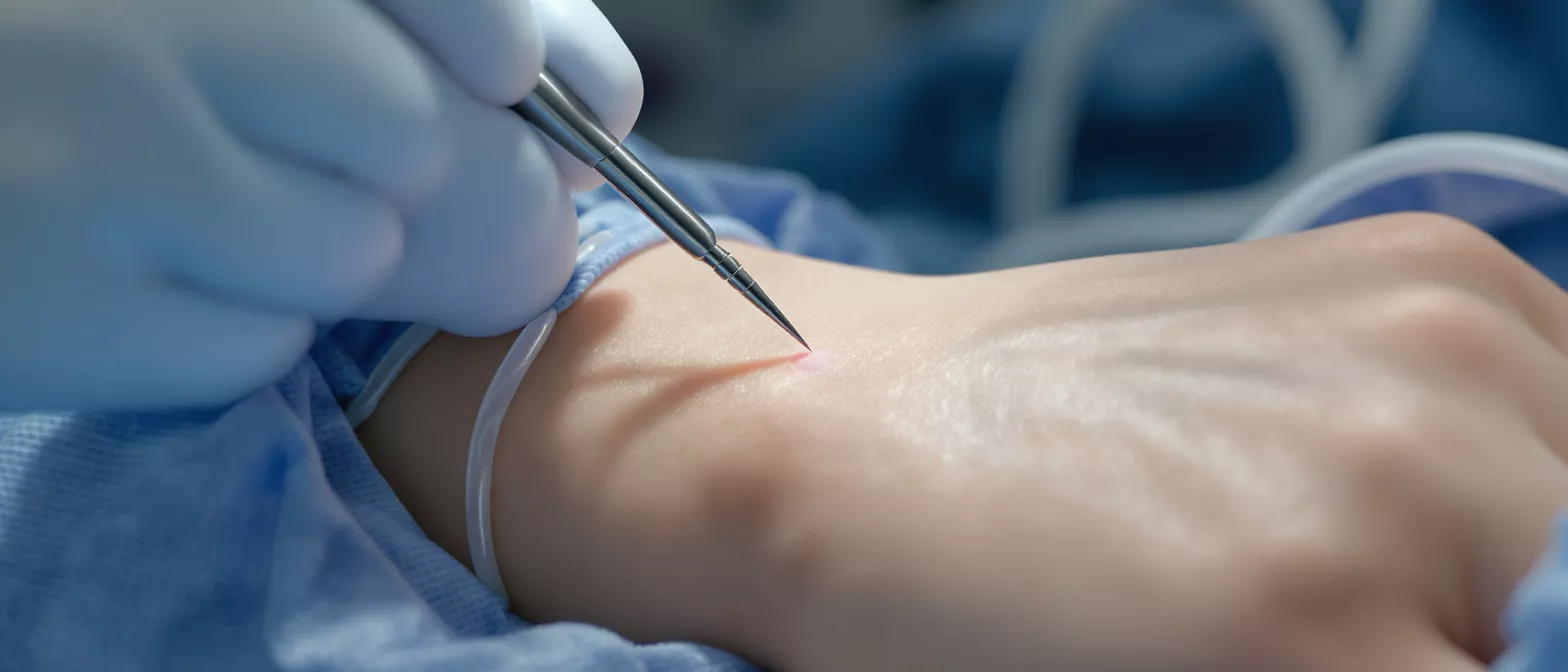
Endomyocardial Biopsy
Discover how an endomyocardial biopsy diagnoses heart tissue health.
What is an endomyocardial biopsy
An endomyocardial biopsy is a specialized diagnostic procedure where a doctor removes a tiny sample of heart tissue for laboratory testing. If you or a loved one are preparing for cardiac surgery, hearing the word "biopsy" can naturally cause anxiety. However, this procedure is a standard and highly important tool that provides your surgical team with critical information about the muscle tissue itself. It allows them to see the exact cellular health of your heart before any surgical intervention is planned or after a specific treatment is completed.
How does the term break down? The word "endomyocardial" refers to the inner lining of the heart, known as the endocardium, and the muscular layer of the heart wall, known as the myocardium. During a heart muscle biopsy, the physician specifically targets these layers to capture cells that show inflammation, infection, or rejection. This precision ensures the tissue retrieved is exactly what the pathology lab needs to make an accurate diagnosis.
Why does this matter for a cardiac-surgery patient? For a patient facing heart surgery, understanding the exact condition of the heart muscle is vital for surgical planning. The surgeon needs to know if the muscle is healthy enough to withstand an operation, if there is an underlying inflammatory process, or if the heart is struggling due to cellular rejection of a previously transplanted organ.
Why is a heart tissue biopsy performed
A heart tissue biopsy is performed to uncover the exact cause of unexplained heart muscle dysfunction, severe inflammation, or cellular rejection. While imaging tests like echocardiograms show how well the heart pumps blood, they cannot show what is happening at a cellular level. When a patient presents with sudden heart failure or abnormal rhythms, a biopsy helps the medical team pinpoint the root cause rather than just treating the symptoms.
Identifying myocarditis and cardiomyopathies: One of the most common reasons for taking a tissue sample is to diagnose myocarditis, which is an inflammation of the heart muscle often caused by a viral infection. It is also used to differentiate between various types of cardiomyopathies, which are diseases of the heart muscle that can lead to heart failure. By examining the tissue, doctors can identify specific structural abnormalities in the heart cells that dictate the correct long-term treatment plan.
In post-surgical or post-transplant patients, the procedure acts as an early warning system. Even if a patient feels completely fine, microscopic changes in the heart tissue can signal that the body is trying to reject a new organ. Catching these cellular signs early allows the medical team to adjust immunosuppressive medications immediately and effectively.
How the cardiac biopsy procedure works
The cardiac biopsy procedure is a minimally invasive technique that is typically performed in a specialized hospital setting, often in a cardiac catheterization laboratory. The doctor uses a flexible tube called a catheter, which is carefully guided through the blood vessels directly into the heart. This approach means no large surgical incisions are required, which significantly reduces physical trauma and speeds up recovery time.
The bioptome and tissue retrieval: At the tip of the catheter is a small, specialized instrument called a bioptome, which functions like tiny forceps. Once the catheter reaches the right side of the heart, the physician opens the bioptome to gently pinch off a microscopic piece of the heart muscle. Patients generally do not feel the actual cutting of the tissue, as the inner heart wall lacks pain receptors, though they may feel some pressure.
Throughout the entire procedure, the medical team uses continuous X-ray imaging to ensure the catheter is positioned safely against the heart wall. The tissue samples are immediately preserved and sent to the pathology laboratory for detailed microscopic analysis.
Endomyocardial biopsy after heart transplant
An endomyocardial biopsy after heart transplant is the most reliable method doctors have for monitoring a patient's immune system. When a new heart is transplanted, the body's natural defense mechanism may recognize the new organ as foreign and attempt to destroy it. This dangerous process is known as graft rejection, and catching it early is absolutely critical for the long-term survival of both the patient and the new organ.
Scheduled surveillance biopsies: Because rejection can happen silently without causing any immediate symptoms, patients undergo a post-transplant biopsy on a regular schedule. These are most frequent in the first few months following the surgery when the risk of rejection is highest. Over time, as the patient and the new heart stabilize, the time between biopsies gradually lengthens.
The procedure itself uses the exact same cardiac graft rejection biopsy technique described above. By routinely sampling the heart muscle, doctors can detect cellular changes before they affect the heart's ability to pump blood. If signs of rejection are found, medications are adjusted immediately to suppress the immune response and protect the transplanted heart.
Diagnosing heart disease with myocardial tissue sampling
Diagnosing heart disease with myocardial tissue sampling is essential when non-invasive tests yield inconclusive results. Sometimes, blood tests and imaging scans suggest a problem, but the specific diagnosis remains elusive. In these complex cases, analyzing the actual heart muscle cells under a microscope provides the definitive answer needed to begin targeted treatment.
Detecting infiltrative diseases and drug toxicity: A myocardial tissue sampling procedure can reveal rare infiltrative conditions, such as amyloidosis or sarcoidosis, where unusual proteins or inflammatory cells build up in the heart muscle. It is also highly effective for diagnosing drug-induced toxicity. Certain chemotherapy drugs or heavy metals can damage the heart, and a biopsy is the only way to measure the exact extent of that cellular damage.
- Infiltrative disorders: Identifies abnormal protein deposits like amyloid that stiffen the heart muscle.
- Drug toxicity: Measures cellular damage caused by specific medications or environmental exposures.
- Severe myocarditis: Confirms viral or autoimmune inflammation of the heart muscle.
By providing a definitive histological diagnosis, the biopsy prevents patients from undergoing unnecessary or incorrect treatments.
Endomyocardial biopsy risks and complications
While the procedure is generally considered safe, it is important to be aware of potential endomyocardial biopsy risks and complications. Because the procedure involves passing instruments into the heart and removing tissue, there are inherent risks. However, the procedure is performed by highly trained specialists who take extensive precautions to minimize any danger to the patient.
Common risks and preventative measures: The most frequent complication is bleeding or bruising at the access site in the neck or groin. More serious but rare risks include cardiac arrhythmias, which are irregular heartbeats that usually resolve on their own once the catheter is removed. In very rare cases, the biopsy instrument can perforate the heart wall, leading to fluid buildup around the heart that might require immediate drainage.
To mitigate these risks, the medical team uses continuous ultrasound and X-ray guidance to navigate the blood vessels and heart chambers safely. The entire procedure is closely monitored by a full surgical team ready to respond instantly to any cardiac changes. Patients are closely observed for several hours after the biopsy to ensure there are no immediate complications.
Understanding myocardial biopsy results
Understanding myocardial biopsy results requires the specialized expertise of a cardiac pathologist. Once the heart tissue is extracted, it is immediately sent to the laboratory where it is processed, sliced into extremely thin sections, and stained with special dyes. These dyes highlight the different cellular structures, allowing the pathologist to see exactly how the heart muscle cells are behaving.
Grading rejection and cellular analysis: In the context of heart transplants, the pathologist will assign a grade to the myocardial histology biopsy. This grade indicates the severity of the immune system's attack on the heart muscle. A low grade typically means no significant rejection is occurring, while a higher grade indicates active rejection that requires immediate medical intervention.
For non-transplant patients, the results might show signs of inflamed cells, viral particles, or abnormal protein deposits. The pathology report provides the surgical team with a definitive diagnosis, which is then used to tailor the patient's medication plan or determine if surgical intervention is necessary.
Heart biopsy recovery and what to expect
Heart biopsy recovery is generally quick and straightforward, primarily because the procedure is minimally invasive. Most patients undergo the biopsy on an outpatient basis and can return home the same day. The actual procedure takes only about thirty to sixty minutes, followed by a brief observation period in the recovery room to monitor vital signs.
Immediate post-procedure care: After the biopsy, the patient must lie flat for a few hours to prevent bleeding from the catheter insertion site. The nursing staff will frequently check the access site for any signs of swelling or bleeding. Most patients are advised to avoid strenuous physical activity and heavy lifting for at least 24 to 48 hours to allow the puncture site to heal properly.
Patients might experience mild soreness at the access site, but significant pain is uncommon. The medical team will provide specific instructions on what to watch out for, such as fever or increasing chest pain, which would require immediate medical attention. Otherwise, normal activities can typically be resumed the following day without restriction.
How safe is an endocardial biopsy technique
The endocardial biopsy technique has been refined over decades, making it a highly safe and routine procedure in major cardiac centers. For a frightened patient or family member, knowing that this technique is performed thousands of times a year with excellent success rates can provide immense comfort. The safety of the procedure relies heavily on the strict protocols followed by the surgical and nursing staff.
Technological advancements and safety protocols: Modern biopsies are performed using advanced imaging technology that allows the physician to see the exact position of the catheter in real-time. This continuous visualization drastically reduces the risk of damaging the heart valves or the coronary arteries. The bioptome itself is designed to take tiny, precise bites of tissue that heal quickly without compromising the overall function of the heart muscle.
Furthermore, the clinical staff is rigorously trained in managing any potential complications before they become serious. Emergency equipment is always on standby in the catheterization lab, ensuring that if a rare event like a cardiac perforation occurs, it can be addressed immediately. This comprehensive preparation is why the procedure remains the gold standard for cardiac tissue sampling.
Key takeaways on cardiac tissue sampling
Ultimately, an endomyocardial biopsy is a powerful and necessary tool in the arsenal of modern cardiac surgery and cardiology. It provides a unique microscopic view of the heart muscle that no other diagnostic test can offer. Whether it is being used to monitor a transplanted heart, diagnose a mysterious case of heart failure, or evaluate the effects of a disease, the information it provides is vital for effective treatment.
Empowering patients through definitive diagnosis: While the idea of having a piece of the heart removed sounds frightening, understanding the procedure helps alleviate fear. The risks are low, the recovery is fast, and the potential benefits are immense. It allows doctors to move from guessing to knowing, which is the safest foundation for any heart surgery or treatment plan.
Always discuss any concerns about a cardiac biopsy with the attending surgeon or cardiologist. They can explain exactly why the procedure is being recommended and how the results will directly impact the patient's care plan moving forward.






