Coronary Angiography: The Procedure and Recovery
Coronary angiography procedure at Cirugía de Corazón: learn preparation, catheter placement, recovery time, and what to expect.
When chest pain or an abnormal stress test leaves you with unanswered questions, a coronary angiography procedure is the study that shows the coronary arteries — the small vessels that feed the heart muscle — from the inside. It remains the most precise way to see whether a narrowing is truly limiting blood flow, and it is often the step that turns uncertainty into a clear plan. In this guide we walk through what the test involves, how to prepare, what recovery actually feels like, and what happens if a blockage is found.
What Is a Coronary Angiography Procedure?
The test in plain words. Coronary angiography is an X-ray movie of the Coronary Arteries taken while a thin, flexible tube called a catheter delivers contrast dye into them. The dye makes the vessels visible on screen, so the cardiologist can see the exact location and severity of any narrowing caused by Coronary Artery Disease. It is a diagnostic test — it looks, it does not treat — although treatment can sometimes follow in the same sitting. To compare the options, see catheterisation versus heart surgery.
People are often surprised at how routine the study has become. It is performed thousands of times a day worldwide, usually through the wrist (radial artery) or the groin (femoral artery), and most patients go home the same day. The value of the test is its precision: a CT scan can suggest a blockage, but angiography shows whether the narrowing is real, how tight it is, and which vessel it affects.
In our practice, the multidisciplinary team led by Dr Alejandro Molina reviews every angiography report together with the patient's full history before proposing any next step, so that the decision to treat — or simply to monitor — is never made on one image alone.
How Does a Coronary Angiogram Work?
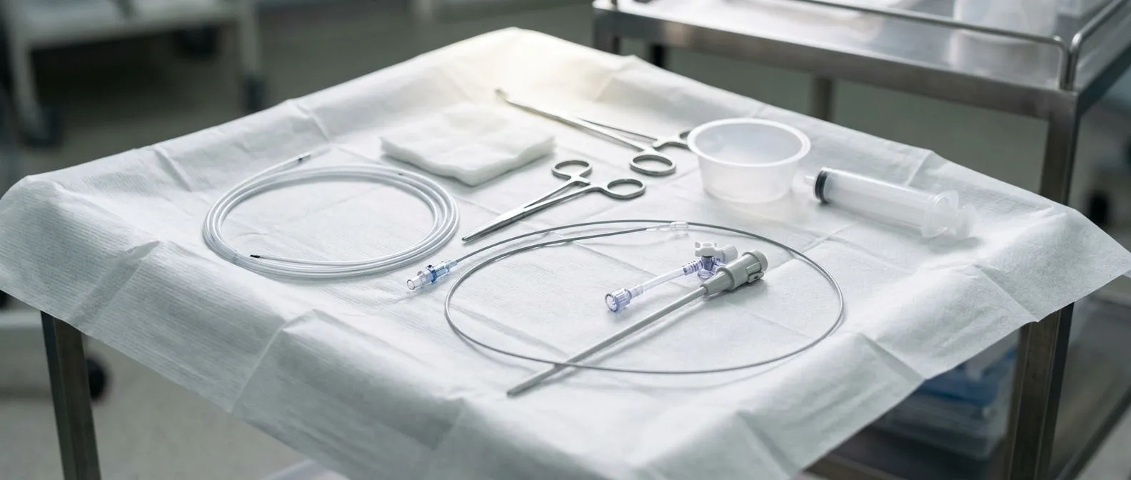
Step by step. The study takes place in a catheterization laboratory, a procedure room with a moving X-ray camera. After local anesthetic and light sedation, the doctor inserts a short sheath (a small plastic tube) into the artery, then threads the diagnostic catheter up through the blood vessels to the heart. Dye is injected selectively into the Left Main Coronary Artery and the Right Coronary Artery, and images are recorded from several angles so no segment of the vessel is hidden.
The contrast agent is iodine-based, similar to the dye used in CT studies. It is injected in small, rapid boluses; you may feel a brief warm flush in your chest when this happens. Each injection lasts a few seconds, and the whole imaging portion usually takes fifteen to twenty minutes. The catheter's position at the origin of each artery — the Coronary Ostia — is confirmed on screen before dye is injected, and any Coronary Buttons or variants are noted in the report.
Because the heart moves, the X-ray camera also moves, sweeping left and right to keep the arteries in focus. This is why the images are far sharper than a still X-ray could ever be, and why radiographic detail remains the reference standard against which other tests are measured.
Coronary Angiography Preparation Checklist
Before the day of the test. Preparation is straightforward, but a few details matter. Your team will give you specific instructions; a typical list looks like this:
- Keep drinking water — good hydration helps your kidneys clear the contrast dye afterwards.
- Bring your full medication list, especially blood thinners and diabetes drugs, which may need pausing or adjusting.
- Usually nothing to eat for six to eight hours beforehand; small sips of water are normally allowed.
- Tell the team about any previous allergic reaction to contrast dye or about kidney problems — a protective plan can be arranged.
- Arrange for someone to drive you home, since sedation makes driving unsafe that day.
Patients on the blood thinner warfarin usually have it stopped several days ahead and an INR check done just before the study; newer anticoagulants follow different rules your cardiologist will explain. If you take metformin for diabetes, the team may hold it for a day or two after the contrast to protect kidney function. A short phone call to the clinic a few days before the test resolves almost every question.
In our first contact we use a free video consultation to review exactly which studies each patient still needs — our team would rather order one well-chosen test than a long list of unnecessary ones — and the face-to-face visit at the consulting rooms in Guadalajara, Querétaro or Hermosillo completes the preparation.
Coronary Angiography: What to Expect During the Test
Are you awake? Yes — you stay lightly sedated but able to respond, because your cooperation helps the team. You will not feel pain during the catheter's passage, only the initial sting of local anesthetic at the puncture site and brief warm sensations when the dye is injected. Many patients describe the experience as far easier than they feared; some even doze through it.
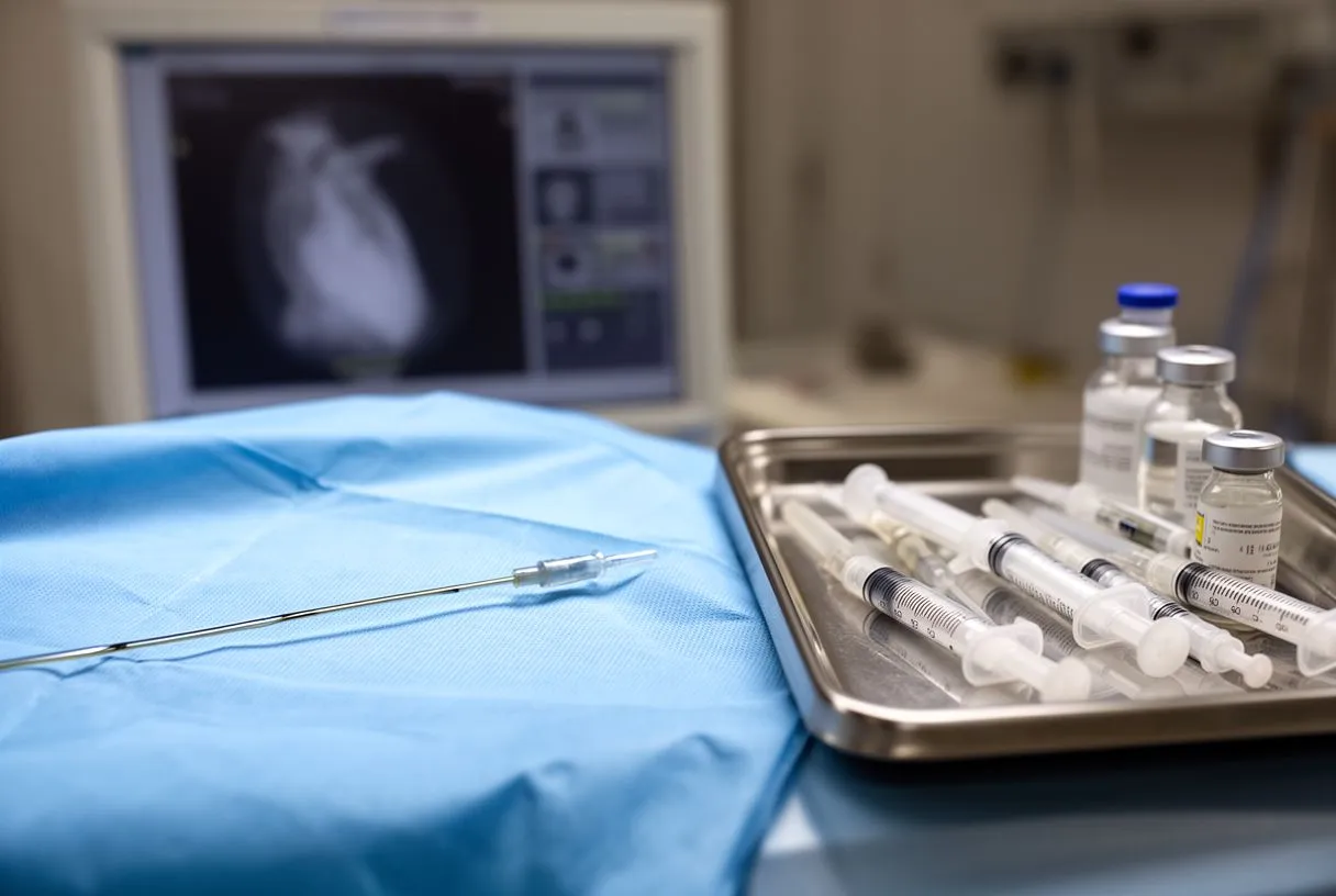
How long does it take? Plan for the whole visit, not just the study. The angiography itself usually lasts twenty to forty minutes, but registration, preparation, sedation and observation afterward stretch the day to roughly half a day. The team monitors your heart rhythm and blood pressure continuously throughout. If the wrist is used, a pressure bracelet is applied at the end; if the groin is used, you will lie flat for a couple of hours while the puncture seals.
Is angiography very painful? For most people, no. The local anesthetic numbs the entry site completely, and the catheter travels inside blood vessels, which do not register pain the way skin does. A mild ache or bruising at the puncture site for a few days is normal. If anything feels sharp or intense during the study, say so — the team can add sedation or pause.
Is a Coronary Angiogram a Serious Procedure?
Where it sits on the risk scale. It is a genuine medical procedure performed inside an artery, so it deserves respect — but it is one of the safest invasive cardiac tests in medicine. Serious complications such as heart attack, stroke or damage to the artery are uncommon, occurring in well under one percent of diagnostic studies in modern registries. The most frequent issues are minor: bruising, a small bleed at the puncture site, or a temporary drop in kidney function from the contrast dye.
Risk is not identical for everyone. Kidney disease, diabetes, advanced age, severe vessel disease and allergy to contrast all raise it somewhat, which is exactly why the team reviews your history beforehand and takes precautions such as hydration protocols and wrist access where appropriate. The radial (wrist) approach, now widely preferred, has reduced access-site bleeding complications markedly compared with the groin approach.
Ask your treating team to walk you through the specific risks of your own case before the date is set; a second opinion is a normal part of deciding — the final decision is always yours.
Coronary Angiography Recovery Time and Aftercare
The first 24 hours. Coronary angiography recovery time is short for a diagnostic study. After a wrist puncture, the pressure bracelet stays on for a few hours and you can usually walk normally the same day. After a groin puncture, expect four to six hours of lying relatively flat, then a slow return to walking. Most people are back to desk work in one to two days and to full activity within a few days.

Coronary angiography aftercare at home. Keep the puncture site clean and dry for the first couple of days, avoid heavy lifting, and skip strenuous exercise until your doctor clears you. A small bruise is expected; a growing lump, active bleeding, coldness or numbness in the hand or foot on that side, or chest pain should be reported to the clinic immediately. Drink plenty of fluids for the first day to help flush the contrast dye through the kidneys.
Patients are kept under observation until the puncture site is stable and the sedation has worn off.
What Happens If They Find a Blockage During an Angiogram?
Two possible paths. If the images show a significant narrowing, the team can either stop at diagnosis and discuss treatment separately, or — if you agreed beforehand — proceed in the same session to Percutaneous Coronary Intervention, commonly known as angioplasty. This means inflating a small balloon at the narrowing and placing a Coronary Stent, a tiny metal mesh scaffold, to hold the artery open. Many patients choose same-session treatment; others prefer to go home, think it over, and return for a planned procedure. Both are reasonable.
Not every blockage needs a stent. Treatment depends on how severely the narrowing limits blood flow, which artery is involved, and the whole clinical picture. Some narrowings are better managed with medication; others — particularly complex disease of the Left Main Coronary Artery or multiple vessels — may favor Coronary Artery Bypass Grafting, the surgical operation that routes new conduits around the blockages. This is where the difference between PCI and coronary angiography matters: the angiogram is the map, and the intervention is one of several journeys that map can lead to.
When the images point toward surgery rather than a stent, our multidisciplinary team reviews the case against international practice guidelines and presents the patient with a clear proposal and the alternatives — a less invasive route is recommended only where the evidence supports it for that specific patient.
Cardiac Catheterization vs Angiography and CT Options
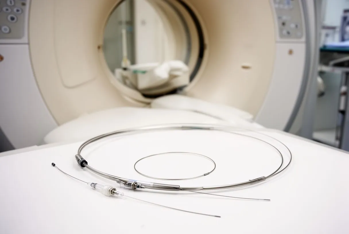
The terminology untangled. Cardiac catheterization is the umbrella term for any procedure that uses a catheter inside the heart or its vessels; coronary angiography is the imaging part of that procedure. So "cardiac catheterization vs angiography" is really one test described from two angles. Invasive angiography differs from Coronary CT Angiography, a scan performed with a CT scanner that shows the arteries from the outside without any catheter at all.
Each has its place. Coronary CT Angiography is excellent as a first-line test: it can rule out Coronary Artery Disease when suspicion is moderate, and a Coronary Calcium Score — a quick, non-invasive CT measurement of calcified plaque — helps estimate risk in patients without symptoms. But CT images can overestimate or underestimate how tight a narrowing truly is. When the question is whether a specific narrowing is blocking flow, or when a decision about a stent or surgery must be made, invasive angiography remains the definitive answer. The two studies work together rather than compete.
Which test will you be offered? The choice depends on your symptoms, risk profile, and what previous tests have shown. A sensible sequence is often: calcium score or CT angiography first in lower-risk patients, then invasive angiography when a treatment decision is on the table. Your cardiologist should explain why the chosen test fits your situation — and if that explanation never happens, it is fair to ask for it.
Coronary Angiography Risks and Benefits
Benefits. The test answers the single most important question in coronary care: is there a blockage that needs treating, or not? It prevents both under-treatment — dangerous missed disease — and over-treatment — procedures that would not have helped. For patients with unstable symptoms, it directs care quickly and can be lifesaving.
Risks, honestly stated. The main risks are bleeding or bruising at the puncture site, temporary kidney effects from contrast dye, abnormal heart rhythms that settle on their own, and rarely, more serious events such as heart attack or stroke. Contrast allergy is possible but preventable with premedication. A very small study published in the Journal of Thoracic and Cardiovascular Surgery even documented an incidental aortic arch mass discovered during conventional angiography (Incidental Finding of a Large Mobile Aortic Arch Mass during Conventional Angiography) — a reminder that the test sometimes reveals important findings beyond the coronary arteries themselves, which is a benefit as much as a curiosity.
The evidence base. Coronary angiography has decades of safety data behind it and continues to evolve with wrist access, lower dye volumes and better risk stratification. Large-scale studies of cardiovascular testing, such as the INCAPS COVID 2 research on cardiac testing in Asia (Open Heart), show how essential these procedures remained even during the pandemic — a measure of how central they are to cardiac care. Broader research published by Nature continues to refine how imaging findings translate into treatment decisions.
Choosing a Heart Team for Your Angiography
What to look for. Because the angiogram can lead directly to a stent or to surgery, the team reading it matters as much as the test itself. Look for a multidisciplinary group — interventional cardiology and cardiac surgery together — that explains results in plain language and presents every option with its trade-offs.
Dr Alejandro Molina trained in cardiothoracic surgery at the Universidad de Guadalajara, in cardiac, minimally invasive, robotic and structural surgery at the University of Montreal, and in structural cardiology and TAVR at CHU Bordeaux; the team also trained at Jena, Bad Berka and Buffalo, and performed the first robotic mitral valve repair in the Ángeles Health System and the first robotic heart surgery in Guadalajara. That range matters when an angiogram opens more than one door — valve disease, Coronary Artery Disease, aortic conditions and structural heart problems can all be reviewed by one team rather than bounced between specialists.
In the consultation we take the time to explain what the studies actually show, in words you can repeat to your family, and to answer every question before any decision is made. If you have been told you need an angiography, or you already have results you do not fully understand, we invite you to book a consultation — start with the free video consultation, and let the team help you decide what comes next.
The takeaway: a coronary angiography procedure is a precise, well-tolerated test with a short recovery that turns suspicion into certainty. Prepare by hydrating and reviewing your medications, expect a half-day at most, and insist on a team that explains the images and every option before you commit to any treatment. Discuss your own situation with a qualified cardiac team rather than self-diagnosing — the right next step is a conversation, not a guess.