

Coronary Angiography
Learn what coronary angiography is, how it detects heart blockages, and what to expect.
What is coronary angiography?
Coronary angiography is a specialized medical imaging test that allows your cardiologist to see exactly how blood flows through the blood vessels of your heart. It is considered the gold standard for evaluating the coronary arteries. By providing a clear, real-time map of your heart's blood supply, this diagnostic procedure helps your medical team determine the exact location and severity of any blockages. Understanding the coronary angiography procedure begins with understanding its purpose: it is designed to give your surgical team the precise information needed to plan your treatment safely.
Difference between angiography and angioplasty — Patients often confuse these terms, and it is important to clarify the distinction before undergoing treatment. Coronary angiography is strictly a diagnostic test used to take pictures of your heart's arteries. Angioplasty, on the other hand, is a therapeutic intervention where a balloon is inflated to widen a narrowed artery, often followed by the placement of a stent to keep it open. Sometimes both are performed during the same visit, but the imaging test itself is simply a picture-taking process.
Role of cardiac catheterization — The foundation of this imaging test is a process called cardiac catheterization. A thin, flexible tube called a catheter is gently guided through a small access point in your wrist or groin directly to your heart. This tube acts as a safe pathway to deliver a contrast dye into the coronary arteries. Once the dye is injected, an X-ray machine captures highly detailed moving images, illuminating the path of blood and highlighting any areas of narrowing.
Why is coronary angiography recommended?
Doctors recommend this test when they suspect a patient has coronary artery disease or when they need to evaluate damage after a heart attack. If you have been experiencing symptoms like chest pain, shortness of breath, or unexplained fatigue, your cardiology team needs to know if your heart muscle is receiving enough oxygen-rich blood. This imaging test provides the definitive answer. It is a critical step in diagnosing blocked arteries and determining whether surgical intervention or medication is the best course of action for your heart health.
Symptoms that lead to heart catheterization — Patients are typically referred for this test after reporting angina or chest tightness, especially during physical exertion or emotional stress. Abnormal results from a non-invasive stress test or an electrocardiogram also frequently prompt a doctor to order an angiogram heart test. By identifying the exact physiological cause of these warning signs, your surgeon can create a highly targeted plan to restore proper blood flow and prevent future cardiac events.
Planning for coronary stent placement — If previous scans suggest a severe blockage, angiography is used to confirm the precise shape and length of the narrowing. This mapping is absolutely essential before a surgeon can place a stent. The detailed images allow the surgical team to measure the diameter of your native artery and select the exact size of the medical device required to prop it open. It removes the guesswork from interventional cardiology.
How to prepare for coronary angiography
Preparing for coronary angiography requires clear communication with your medical team to ensure your safety throughout the process. Your surgeon will give you specific instructions tailored to your medical history, current medications, and overall physical condition. Following these guidelines precisely is one of the most important steps you can take to ensure a smooth experience and accurate test results.
Medication and fasting guidelines — You will likely be asked to stop eating and drinking for several hours before your appointment to prevent complications during the imaging. It is especially important to tell your doctor about any blood thinners or diabetes medications you are currently taking, as these may need to be adjusted or temporarily paused. Your surgical team will review your full medication list and provide a detailed schedule exactly when to take your morning pills.
Arranging for post-procedure care — Because you will receive mild sedation to keep you comfortable and relaxed during the test, you will not be allowed to drive yourself home afterward. It is vital to arrange for a trusted family member or friend to accompany you to the clinic and escort you back safely. You should also plan to have someone stay with you for the first twenty-four hours of your recovery period to monitor your wellbeing.
Coronary angiography procedure step by step
Knowing exactly what happens during the coronary angiography procedure can significantly reduce the anxiety that naturally comes with heart testing. The entire process is carefully orchestrated by a highly trained team of cardiologists, specialized nurses, and technicians in a sterile cardiac catheterization laboratory. From the moment you arrive, every step is taken with your comfort and safety as the absolute highest priority.
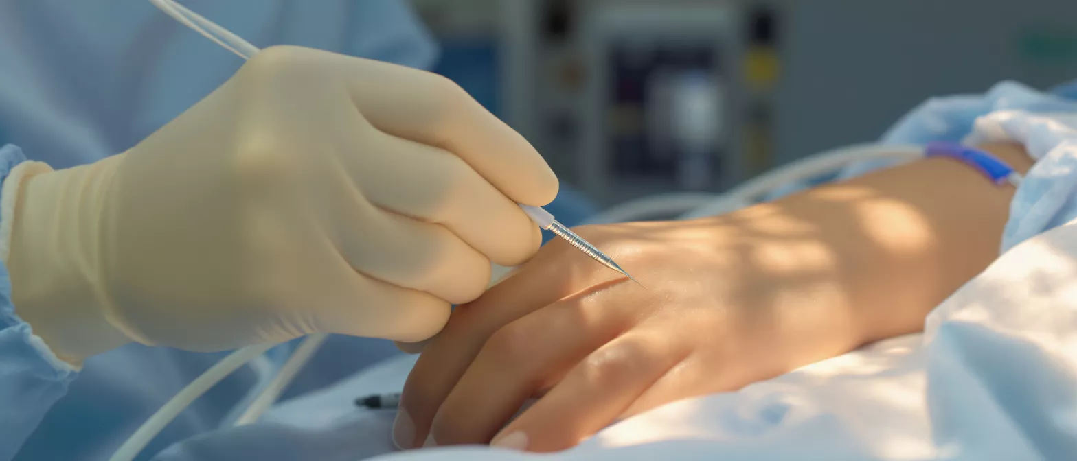
Local anesthesia and catheter insertion — The procedure begins by cleaning the insertion site, usually the radial artery in your wrist, with an antiseptic solution. The doctor then injects a small amount of local anesthetic to numb the area, ensuring you do not feel sharp pain. You may only feel a bit of pressure as the catheter is gently threaded through the blood vessel toward your heart. Throughout this time, the medical staff will continually check in with you to ensure you are resting comfortably.
Contrast dye and X-ray imaging — Once the catheter reaches the correct position near the heart, a specialized contrast dye is injected through the tube. This dye is what makes your blood vessels visible on the live X-ray screen, illuminating the entire network of coronary arteries. You might experience a brief warm sensation spreading through your chest for a few seconds when the dye is released. The camera captures rapid images from multiple angles, giving your surgeon a comprehensive view of your heart's blood supply.
- Continuous monitoring: Your heart rate, blood pressure, and oxygen levels are closely watched throughout the entire procedure.
- Duration: The actual imaging typically takes only twenty to thirty minutes, though preparation adds extra time.
- No pain receptors: The inside of your blood vessels and heart cannot feel pain, so you will not feel the catheter moving.
What coronary angiography images reveal
The images captured during a coronary angiography are incredibly detailed and provide your medical team with a precise map of your cardiovascular system. The live X-ray feed shows exactly where blood is flowing freely and where it is being restricted. For a patient awaiting answers, these images represent the first clear picture of what is happening inside their heart, translating complex cardiac physiology into a visual roadmap for treatment.
Identifying blocked artery diagnosis — The contrast dye highlights the inner walls of your arteries, making buildups of cholesterol and plaque clearly visible as dark spots or narrowings in the vessel. This allows your doctor to perform an accurate blocked artery diagnosis. They can see not only how much of the artery is blocked, but also the character of the blockage, determining if it is a smooth, stable narrowing or a more complex, hardened lesion that requires surgical attention.
Evaluating heart muscle function — Beyond just looking at the arteries, the angiogram also allows the medical team to assess how well the heart muscle itself is pumping. By observing the movement of the heart walls during the injection of the dye, cardiologists can identify areas of the heart that may have been damaged by a previous heart attack. This comprehensive view helps ensure that any surgical intervention targets the right areas to restore healthy circulation.
- Location of narrowing: Pinpoints exactly which branch of the coronary artery tree is affected.
- Severity of blockage: Measures the percentage by which the artery diameter is reduced.
- Collateral vessels: Shows if the heart has naturally grown new, small bypass vessels to route blood around a blockage.
Is coronary angiography safe?
Safety is the most common concern for patients facing any heart test, and it is completely normal to ask, are coronary angiography risks significant? The short answer is that this procedure is widely considered a very safe and routine diagnostic tool in modern cardiology. Advancements in medical imaging, catheter technology, and sterile techniques have dramatically reduced the likelihood of complications over the past few decades.
Understanding the safety profile — Millions of these tests are performed globally every year with excellent outcomes. Because it is a minimally invasive approach that avoids large incisions, the trauma to the body is minimal compared to traditional open surgery. The sedation used is very light, meaning you remain responsive enough to breathe and cooperate without the deeper risks associated with general anesthesia. Your medical team is highly trained to anticipate and manage any unexpected reactions immediately.
Allergic reactions and contrast dye — One of the primary considerations is your body's reaction to the contrast dye used to illuminate the arteries. While modern dyes are very safe, some patients may experience mild allergic reactions. It is vital to inform your doctor of any prior allergies to iodine or contrast materials. To provide a safe and clear diagnostic environment, the clinical team typically uses sterile contrast agents for angiography that are specifically formulated to minimize kidney strain and allergic responses.
Coronary angiography risks and complications
While the procedure is highly safe, a responsible medical team must always discuss potential coronary angiography complications with their patients. Informed consent is a critical part of the surgical process, ensuring you understand both the benefits and the rare risks. By knowing what to watch for, you become an active partner in your own recovery and can alert your care team if anything unexpected occurs.
Bleeding and vascular issues — Because a small tube is inserted into an artery, the most common complications involve the insertion site. You might experience minor bruising, tenderness, or slight bleeding at the wrist or groin where the catheter was introduced. In very rare cases, a small blood vessel tear or a pseudoaneurysm may occur. The clinical staff applies firm pressure and a specialized compression bandage after the tube is removed to ensure the artery seals properly and prevent these vascular issues.
Kid function and contrast considerations — The contrast dye is filtered out of your body by your kidneys. For patients with pre-existing kidney disease, there is a slight risk of strain on these organs. Your doctor will evaluate your kidney function through blood tests before the procedure and may provide intravenous fluids to help flush the dye safely out of your system. To support recovery, IV hydration for cardiac procedures is standard practice for maintaining optimal kidney function after the test.
Coronary angiography recovery and aftercare
Coronary angiography recovery is generally quick, allowing most patients to return home the same day. Because the test is minimally invasive and requires only a small puncture rather than a surgical incision, the physical healing time is remarkably short. However, following your doctor's specific aftercare instructions is vital to ensuring the insertion site heals correctly and that your overall heart health continues to improve.
Immediate post-procedure monitoring — After the test is finished, you will be moved to a recovery room where nurses will monitor your heart rate and blood pressure closely. You will need to lie flat and keep your arm or leg still for a few hours to allow the artery to seal completely. The nursing staff will frequently check the insertion site for any signs of bleeding and ensure you are comfortable. Most patients are cleared to go home within four to six hours after the test is completed.
Returning to daily activities — Once you are home, you should rest for the remainder of the day and avoid heavy lifting or strenuous exercise for at least a week. It is important to keep the insertion site clean and dry, watching for any sudden swelling or prolonged bleeding. Your doctor will schedule a follow-up appointment to discuss the results of the angiography and outline the next steps, which may include medication adjustments or planning for advanced coronary stent placement if a severe blockage was found.
Coronary angiography vs other cardiac tests
Patients often wonder why their doctor chose this specific test over others. There are many ways to look at the heart, but a coronary angiography offers a unique level of anatomical detail that non-invasive scans simply cannot match. Understanding how it fits into the broader landscape of cardiac diagnostics helps clarify its essential role in creating a definitive treatment plan for heart surgery patients.
Comparing with non-invasive scans — Tests like stress echocardiograms or CT calcium scoring are excellent for initial evaluations and screening. However, while a CT scan can show plaque build-up, it does not provide the same dynamic, real-time flow assessment as cardiac catheterization. Angiography is specifically designed to show exactly how blood moves through the tiniest branches of the heart. When a doctor needs to make a definitive decision about surgery, they require the unmatched visual clarity that only a live angiogram can provide.
When to choose direct visualization — If non-invasive tests yield unclear results, or if you are experiencing acute chest pain that suggests a severe blockage, your cardiologist will bypass other tests and go straight to angiography. It is the most definitive method for blocked artery diagnosis. By directly visualizing the coronary anatomy, the surgical team can accurately measure the exact degree of narrowing and immediately perform an intervention if necessary, saving valuable time in critical situations.
Coronary angiography cost and accessibility
Understanding the practical aspects of accessing this procedure is an important part of your healthcare journey. The coronary angiography cost can vary widely depending on the facility, the specific equipment used, and the length of your stay in the recovery unit. However, it is important to focus on the value of the precise diagnostic information it provides, which ultimately saves lives and prevents far more expensive emergency interventions down the line.
Navigating financial considerations — The total expense typically includes the facility fee, the cardiologist's professional fee, and the cost of the specialized imaging equipment and contrast dye. We believe that financial clarity is just as important as medical clarity, and we are dedicated to ensuring you understand all aspects of your care before proceeding.
We combine advanced medical technology with a deeply compassionate, patient-first approach. Trusting your heart to an experienced surgical team ensures that your diagnostic journey is safe, accurate, and focused entirely on your long-term health.






