

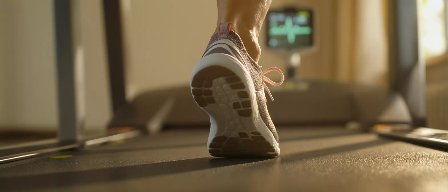
Stress Echocardiogram
Learn how a stress echocardiogram evaluates heart function under exertion.
What is a stress echocardiogram
A stress echocardiogram is a specialized diagnostic test that combines ultrasound imaging with a monitored period of physical exertion to evaluate how well your heart functions when it is working its hardest. For patients preparing for cardiac surgery or recovering from a heart event, this test provides critical information about blood flow to the heart muscle. The term "stress" refers to the controlled physical or pharmacological challenge used to make your heart beat faster and pump harder.
By capturing ultrasound images of your heart both before and immediately after this exertion, your cardiologist can directly compare your heart's resting performance against its peak effort. This comparison is vital for identifying areas of the heart muscle that may not be receiving enough oxygen-rich blood, a condition known as ischemia. For a frightened patient or family member, it helps to know that this is a non-invasive, painless way to gather highly detailed, real-time visuals of the heart's pumping chambers and valves under pressure.
Why cardiac patients need this specific test: A standard resting echocardiogram shows the heart's structure and function while you are lying still. However, many heart problems only manifest when the heart is under increased demand. The stress echocardiogram bridges this diagnostic gap. It reveals hidden blockages in the coronary arteries that might only cause symptoms during physical activity, ensuring your surgical team has a complete picture of your cardiovascular health before proceeding to the operating room.
How does a stress echo test work
The stress echo test works by leveraging the fundamental principle that the heart requires more oxygen when it works harder. During the test, small electrodes are placed on your chest to continuously monitor your electrocardiogram (ECG), tracking your heart's electrical activity. Simultaneously, a sonographer uses a handheld ultrasound transducer to take moving pictures of your heart's walls and valves while you are at rest.
Once the resting images are recorded, you will be asked to walk on a treadmill or ride a stationary bicycle. The intensity of the exercise gradually increases, forcing your heart to beat faster. When you reach your target heart rate, the exercise stops, and the sonographer quickly captures a second set of ultrasound images. If you cannot exercise due to physical limitations, a medication will be administered intravenously to safely mimic the effects of exercise on your heart.
How exercise echocardiography captures ischemia: When a coronary artery is narrowed, it may deliver enough blood to the heart muscle at rest, but it cannot deliver the extra blood required during exertion. This lack of blood flow causes the affected part of the heart muscle to contract abnormally. By comparing the resting ultrasound images to the peak-effort images, your cardiologist can see exactly which segments of the heart are struggling, pinpointing the compromised blood supply.
Stress echocardiogram preparation guide
Proper preparation ensures the test yields accurate results and proceeds safely. Your care team will provide specific instructions, but general guidelines apply to most patients undergoing this evaluation. You will typically be advised to avoid eating or drinking anything for at least three hours before the appointment to prevent nausea during exertion. It is also important to wear comfortable, loose-fitting clothing and supportive walking shoes, as you will be engaging in physical activity.
Medication adjustments before the test: You must discuss your current medications with your cardiologist well in advance. Certain drugs, particularly beta-blockers or other heart rate-controlling medications, may need to be paused for 24 hours before the test. These medications keep your heart rate low, which can prevent you from reaching the target heart rate needed for the test to be diagnostically useful. Never stop taking medications on your own without explicit instructions from your physician.
- Fasting: Avoid food and heavy liquids for three hours prior to the test.
- Attire: Wear comfortable exercise clothes and rubber-soled shoes.
- Medications: Bring a list of all medications; confirm which to take or skip.
What to expect during the stress echocardiogram procedure
Understanding the procedure can significantly reduce anxiety for patients and their families. When you arrive, a technician will prepare your chest by shaving small areas if necessary to ensure the ECG electrodes stick properly. A small amount of ultrasound gel will be applied to your chest, and the sonographer will move the transducer over your heart to capture the resting images. This gel is cool but harmless.
Next, you will begin the exercise portion on a treadmill. The belt will start slowly and remain relatively flat, then gradually increase in speed and incline every few minutes. A physician or specialized nurse will be present the entire time, monitoring your ECG, blood pressure, and asking how you feel. You will be instructed to exercise until you are too tired to continue or until you reach your maximum safe heart rate, at which point the exercise will stop.
Immediately after you stop, you will quickly lie back down on the examination table. The sonographer will take the second set of ultrasound images while your heart is still beating rapidly. The entire process, from resting images to peak images, usually takes about 45 to 60 minutes.
Stress echocardiogram vs stress test differences
Patients are often confused about the difference between a standard cardiac stress test and a stress echocardiogram. A regular stress test involves walking on a treadmill while your ECG is monitored, but it does not include the ultrasound imaging component. The doctor relies solely on the electrical signals and your symptoms to determine if there is a problem with blood flow to the heart.
Diagnostic accuracy of ultrasound imaging: While a standard stress test is useful, it can sometimes produce inconclusive or false-positive results. A stress echocardiogram adds a layer of anatomical and functional precision. By actually watching the heart muscle move on the ultrasound screen, the physician can see exactly which arteries might be blocked. This makes the stress echo much more sensitive and specific for detecting ischemia than an ECG alone.
In some cases, a patient's resting ECG is abnormal due to previous heart attacks or specific medications, making a standard stress test difficult to interpret. In these instances, a cardiac stress echo is preferred because the ultrasound images provide clear visual data independent of the baseline electrical abnormalities. This ensures that the doctor is making decisions based on direct visual evidence of heart function, not just electrical surges.
Stress echo interpretation and normal results
Interpreting a stress echocardiogram requires specialized training to analyze the moving ultrasound images. The cardiologist looks closely at the movement of the left ventricle, the heart's main pumping chamber. In a normal, healthy heart, the muscle walls thicken and move inward vigorously with each heartbeat, both at rest and under stress. A normal result means all areas of the heart are contracting normally and receiving adequate blood flow.
What abnormal stress echo results look like: If the heart muscle moves normally at rest but fails to contract properly during peak exercise, this is a strong indicator of ischemia. The specific wall segments that stop moving tell the cardiologist which coronary artery is likely narrowed. If a segment does not move normally at rest or during stress, it may indicate a previous heart attack where scar tissue has formed.
The results of this test directly influence your surgical planning. If the stress echo shows significant ischemia, it may prompt further investigations, such as a coronary angiogram, or accelerate the timeline for surgery. For patients seeking clarity on their heart health, discussing your stress echocardiogram results thoroughly with your cardiologist will provide peace of mind and a clear path forward for any necessary interventions.
Are stress echocardiogram tests safe
Yes, a stress echocardiogram is considered a very safe and routine diagnostic procedure. Because it is performed in a hospital or clinical setting under the direct supervision of a physician, risks are minimal. The test is carefully controlled, and the medical team monitoring your vital signs will stop the exercise immediately if they detect any dangerous changes in your heart rhythm or blood pressure.
Some patients worry about having a heart attack during the test, but the protocol is designed to provoke symptoms safely, not cause cardiac events. The team will ask you to report any chest pain, severe shortness of breath, or dizziness immediately. The most common complaints are minor and related to the physical exertion, such as muscle fatigue in the legs or mild joint discomfort from walking on a steep treadmill incline.
For patients who cannot exercise, the pharmacological stress echo carries slightly different, but still minimal, risks. The medication used to stimulate the heart can cause temporary flushing, palpitations, or a brief headache. These side effects wear off quickly once the infusion is stopped and an antidote is administered if needed. The clinical staff is highly trained to manage these reactions instantly.
Stress echocardiogram risks and contraindications
While complications are rare, it is important to understand the potential stress echocardiogram risks. The most significant, though highly uncommon, is the triggering of an irregular heart rhythm, known as an arrhythmia. In extremely rare cases, the test could provoke a heart attack if a severely blocked plaque ruptures. However, the continuous ECG monitoring ensures that any dangerous changes are caught and treated instantly.
Contraindications for the test: There are specific situations where a stress echo should not be performed. If a patient is currently experiencing unstable angina, has severe symptomatic heart failure, or has a recent history of a heart attack without stabilization, the exertion could be dangerous. Additionally, physical limitations like severe arthritis or vascular disease in the legs might prevent treadmill use, necessitating a pharmacological approach instead.
- ECG Monitoring: Continuous electrical tracking catches arrhythmias instantly.
- Contraindications: Unstable angina or recent acute heart attack.
- Physical Limits: Severe arthritis may require medication instead of exercise.
Stress echo cost and finding a stress echocardiogram near me
The stress echo cost can vary widely depending on your geographic location, the specific facility, and your insurance coverage. Generally, because it requires specialized ultrasound equipment and direct physician supervision, it is more expensive than a standard resting echocardiogram but significantly less costly than more invasive procedures like a coronary angiogram. Most insurance plans will cover the test if it is deemed medically necessary by your referring physician.
When looking for a facility, it is crucial to choose a center with experience in performing and interpreting these complex studies. A hospital or clinic with a dedicated echocardiography laboratory will have the most skilled sonographers and cardiologists.
Always discuss financial concerns with the hospital's billing department beforehand. They can provide estimates and help navigate insurance pre-authorization. Finding a reliable center is essential; searching for a stress echocardiogram near me should lead you to accredited cardiac facilities where the staff can comfortably and safely guide you through the entire testing process.
Understanding your stress echo heart test outcome
Ultimately, the goal of the stress echo heart test is to give you and your surgical team confidence in your diagnosis and treatment plan. Understanding your outcome empowers you as a patient. A normal result is highly reassuring, indicating that your heart muscle is strong and your blood supply is adequate during physical activity. This might mean surgery can be delayed or approached differently.
Conversely, an abnormal result is not a cause for immediate panic; rather, it is a roadmap for action. It tells your surgeon exactly where the weak points are, allowing for a targeted surgical approach. This test takes the guesswork out of cardiac care, replacing it with concrete visual evidence of how your heart behaves under pressure. Facing heart surgery is daunting, but precise diagnostics like the stress echo ensure your treatment is based on the most accurate information available.






