

Right Heart Catheterisation
Discover how right heart catheterisation diagnoses heart conditions and measures cardiac pressures.
What is right heart catheterisation?
Right heart catheterisation is a specialised cardiac diagnostic procedure used to evaluate how well the right side of your heart is pumping and to measure the pressure within your heart and blood vessels. If your doctor has recommended this test, it is natural to feel anxious, but understanding the process can bring significant peace of mind. Unlike standard imaging that shows the structure of the heart, this test provides dynamic, real-time data about its function.
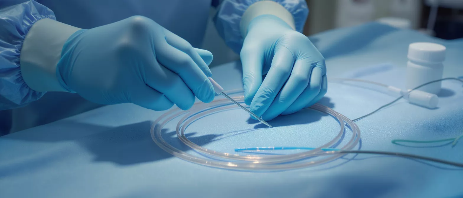
During the procedure, a thin, flexible tube called a catheter is gently guided through your veins directly into the right side of your heart and the pulmonary artery. This allows your surgical team to assess the exact pressures and oxygen levels in these specific chambers. It is a crucial tool for determining the severity of various heart conditions and ensuring you receive the most effective care.
For many patients facing heart surgery, this test acts as a vital roadmap. By clearly identifying how blood flows and what pressures exist within the heart, your surgical team can plan the safest, most effective approach to your treatment. It is typically performed in a hospital setting by a highly trained cardiologist, ensuring you are monitored closely and safely throughout the entire process.
Why is a heart catheterisation test performed?
Your doctor may suggest a heart catheterisation test for several important reasons. It is primarily used to diagnose and evaluate conditions like pulmonary hypertension, heart failure, and certain congenital heart defects. By measuring the exact pressures inside the heart, your care team can understand why you might be experiencing symptoms such as shortness of breath, dizziness, or fluid retention.
Differentiating heart failure types: Right heart catheterisation is essential for determining whether heart failure is primarily driven by reduced pumping strength or by stiff, non-compliant heart muscles. This distinction is critical because the treatment strategies for each type are vastly different. The data gathered ensures you are not prescribed medications that could worsen your specific condition.
Assessing treatment efficacy: If you have already been diagnosed with a heart or lung condition, this test helps your doctor see if your current medications are working effectively. It provides concrete, measurable data that guides adjustments to your treatment plan. For a patient preparing for cardiac surgery, it confirms whether the heart is strong enough to tolerate an operation.
How does the cardiac catheterisation procedure work?
The cardiac catheterisation procedure is meticulously designed to be as minimally invasive and painless as possible. You will typically receive a mild sedative to help you relax, and a local anaesthetic is applied to the insertion site. The doctor then makes a tiny needle puncture, usually in a vein in your neck or groin, to safely access the vascular system without making large incisions.
Once the venous access is established, a thin guidewire is carefully advanced through the veins. The catheter is then threaded over this wire, moving smoothly toward the right atrium, through the right ventricle, and finally into the pulmonary artery. Your progress is watched in real-time on an X-ray screen, ensuring the catheter is positioned perfectly to capture the most accurate measurements.
As the catheter moves through the heart chambers, it continuously records pressures and takes blood samples to check oxygen saturation. The entire process usually takes less than an hour. Because the heart muscle itself does not have pain receptors, you will not feel the catheter moving inside you, though you may sense slight pressure at the insertion site.
What to expect during a right heart cath procedure?
Knowing what to expect during a right heart cath procedure can alleviate much of the anxiety surrounding the test. You will remain awake throughout the process, allowing you to communicate with the medical team. The nursing staff will constantly monitor your heart rate, blood pressure, and oxygen levels, ensuring your absolute safety and comfort.
- Continuous monitoring: ECG leads track your heart rhythm, ensuring immediate response if any irregularity arises.
- Local numbing: The insertion site is thoroughly numbed, so you only feel light pressure, not sharp pain.
- Flat resting: Afterward, you must lie flat for a few hours to prevent bleeding from the puncture site.
During the procedure, the doctor might ask you to take deep breaths, cough, or shift slightly. These simple actions help obtain the most accurate pressure readings under different physiological conditions. Your cooperation directly assists the surgical team in gathering the comprehensive data needed to plan your ongoing cardiac care.
What does right atrial pressure measurement show?
A right atrial pressure measurement is a key component of right heart catheterisation. The right atrium is the top chamber of the right side of the heart, receiving blood from the body. Measuring the pressure here tells your doctor how effectively blood is returning to your heart and how well the right ventricle is pumping that blood onward to the lungs.
Fluid status evaluation: Elevated right atrial pressure can indicate fluid overload, often seen in worsening heart failure. It helps your doctor determine if diuretics or other medications are needed to reduce the strain on your heart. For surgical patients, understanding fluid dynamics is critical to ensuring stable anaesthesia and postoperative recovery.
Ventricular function clues: The measurement also provides indirect clues about the function of the tricuspid valve and the right ventricle. Abnormal waveforms on the pressure monitor can suggest valve leakage or stiffening of the heart muscle. This allows your surgical team to anticipate potential complications before you enter the operating room.
How is pulmonary hypertension diagnosed?
Pulmonary hypertension is a condition characterised by abnormally high blood pressure in the arteries of the lungs. Right heart catheterisation is the gold standard for pulmonary hypertension diagnosis because it is the only test that can directly and accurately measure the pressure inside the pulmonary artery. Non-invasive tests like echocardiograms can only estimate this pressure.
During the catheterisation, the doctor will measure the mean pulmonary artery pressure and the pulmonary vascular resistance. If these numbers are elevated, it confirms the diagnosis. Furthermore, the procedure allows the medical team to test how your blood vessels respond to specific medications, which is vital for prescribing the most effective targeted therapy.
For patients awaiting heart or lung surgery, diagnosing pulmonary hypertension is a critical safety step. High pulmonary pressures significantly increase the risks of major surgery, so knowing these numbers allows the anaesthesiologist and surgeon to tailor their approach. It helps them prepare specialised medications to protect your heart during the operation.
Are there right heart catheterisation risks?
While right heart catheterisation is generally safe, it is important to understand that all medical procedures carry some risk. Right heart catheterisation risks are relatively low, especially when performed by an experienced cardiac team in a controlled hospital environment. Your doctor will only recommend the test if the diagnostic benefits clearly outweigh the potential complications.
Common localized risks: The most frequent complications are related to the insertion site, such as minor bleeding, bruising, or localized pain. In rare cases, a small hematoma can form where the catheter was inserted. These issues are typically minor and resolve quickly with simple pressure and bed rest.
Rare cardiac events: Because the catheter passes through the heart, there is a very small risk of triggering arrhythmias, which are irregular heartbeats. These usually resolve on their own once the catheter is repositioned. Extremely rare but serious risks include infection, blood clots, or damage to the heart muscle or valves, which is why the procedure is continuously monitored.
What is right heart catheterisation recovery like?
Right heart catheterisation recovery is usually quick and straightforward. Most patients are able to go home the same day, though some may need to stay in the hospital overnight for observation, particularly if they are already hospitalised or preparing for surgery. You will be asked to lie flat for a few hours immediately following the test to ensure the puncture site seals completely.
Insertion site care: You will be given specific instructions on how to keep the puncture site clean and dry. It is normal to experience mild tenderness or a small bruise at the site for a few days. However, you should watch closely for any signs of excessive bleeding, increasing pain, or signs of infection like redness and warmth.
Activity limitations: Your doctor will advise you to avoid heavy lifting and strenuous exercise for a few days. Gentle walking is encouraged to promote healthy blood circulation. Following these guidelines carefully ensures the vein heals properly and prevents any unnecessary post-operative vascular care supplies from being needed. Most people return to their normal daily activities within 24 hours without issue.
Can right heart catheterisation complications be prevented?
Medical teams take extensive precautions to ensure that right heart catheterisation complications are prevented. Sterile techniques are strictly followed to eliminate the risk of infection. The insertion site is carefully sterilised, and the surgical team wears sterile gowns and gloves to maintain a completely clean environment.
- Ultrasound guidance: Using ultrasound to locate the vein reduces the risk of accidental arterial puncture.
- Continuous imaging: Fluoroscopy ensures the catheter follows the correct path without damaging heart walls.
- Heparin flushing: The catheter is frequently flushed with saline or heparin to prevent blood clots from forming inside the tube.
Before the procedure, your doctor will review your medical history to identify any bleeding disorders or allergies to local anaesthetics. By taking a proactive approach to these potential issues, the cardiac team ensures the test is as safe as possible. Your transparency about your medical history is a vital part of this preventative strategy.
Understanding heart pressure measurement results
Heart pressure measurement results provide a detailed picture of your cardiovascular health. The numbers obtained during right heart catheterisation are compared against established normal ranges. Normal pressures indicate that blood is flowing smoothly through the right side of your heart and into your lungs without any unwarranted resistance or blockages.
If the pressures are higher than normal, it points to specific areas of concern that need addressing. Elevated pressures in the pulmonary artery, for example, confirm pulmonary hypertension, while high right atrial pressures point toward right-sided heart failure. Discussing cardiac diagnostic reports with your cardiologist will help clarify exactly what these numbers mean for your specific situation.
Ultimately, the data gathered from this procedure empowers your surgical and medical team to make the best decisions for your treatment. Whether it dictates the need for a specific medication, a change in lifestyle, or a surgical intervention, the information is invaluable. Trust that this test is a vital step toward restoring your heart health and ensuring a safer surgical outcome.






