

Cardiac Magnetic Resonance
Cardiac magnetic resonance imaging provides safe, detailed diagnostics for surgery planning.
What is cardiac magnetic resonance?
Understanding your diagnosis is a critical step in preparing for heart surgery, and medical imaging plays a vital role in that journey. Cardiac magnetic resonance imaging, commonly referred to as a cardiac MRI, is a non-invasive medical test that uses powerful magnets and radio waves to create detailed pictures of your heart. Unlike standard X-rays, this technology does not rely on ionizing radiation, making it a remarkably safe option for evaluating complex cardiovascular structures.
For a patient preparing for cardiac surgery, this imaging test provides your surgical team with an unparalleled, three-dimensional view of the heart's anatomy. It allows your cardiologist and surgeon to see the size of your heart chambers, the thickness of your heart muscle, and the exact function of your valves in real-time. By offering a clear, unobstructed window into the chest cavity, it ensures your medical team can plan the safest, most effective surgical approach tailored to your specific condition.
Receiving clear answers about your upcoming procedure helps reduce anxiety for both you and your family. When your physician recommends a cardiac magnetic resonance imaging test, they are seeking the highest level of diagnostic accuracy to inform your care plan. Knowing exactly what the test is and why it is being used allows you to approach your treatment with confidence and clarity.
How does cardiac MRI work?
The technology behind cardiac resonance imaging is both fascinating and highly precise. The machine contains a large, tube-shaped magnet that creates a strong magnetic field around your body. This field temporarily realigns the hydrogen atoms naturally present in your body's water molecules. When radio waves are pulsed into the body, these atoms emit faint signals that a computer captures and translates into highly detailed, cross-sectional images of your beating heart.
How the scanner captures a beating heart: A unique feature of this scan is its ability to synchronize with your heartbeat. The machine uses an electrocardiogram (ECG) to monitor your heart rhythm, taking rapid snapshots only between contractions. This synchronization ensures the resulting images are crisp and free of motion blur, allowing doctors to evaluate your heart muscle function and blood flow with exceptional precision.
Understanding the mechanics of the scanner can demystify the experience, easing the minds of families waiting outside. The equipment is entirely external; nothing touches the heart directly during the scan. By relying on magnetic fields rather than radiation, it provides a safe, repeatable method for gathering the critical data your surgeon needs before entering the operating room.
Why is cardiac resonance imaging used for heart disease?
When evaluating complex cardiac conditions, physicians turn to this advanced imaging modality for its unmatched diagnostic accuracy. It is particularly effective at distinguishing between different types of heart tissue. For instance, if a tumor, scarring from a previous heart attack, or a thickened heart muscle is suspected, the scan provides definitive visual evidence that simpler imaging tools, like standard ultrasounds, cannot.
Evaluating heart muscle viability: For patients scheduled for coronary artery bypass or valve repair, your surgeon needs to know exactly which parts of the heart muscle are healthy and which are damaged. The scan reveals microscopic changes in tissue composition, showing your medical team if the heart muscle can recover after a surgical intervention.
The decision to proceed with major heart surgery is never taken lightly. Physicians use cardiac MRI for myocarditis and various cardiomyopathies to ensure that surgery is truly the best and most necessary course of action. By delivering exact measurements of ventricular volume and blood output, it prevents unnecessary operations and confirms the precise surgical target.
Cardiac magnetic resonance vs CT scan?
Patients are often confused when presented with multiple imaging options, but understanding the difference is crucial for your care. The primary distinction between a cardiac magnetic resonance and a CT scan lies in the technology used. An MRI employs magnetic fields and radio waves, whereas a CT scan utilizes X-rays to create its cross-sectional images. Both are valuable tools, but they excel at revealing different aspects of cardiac anatomy.
A CT scan is typically faster and is the gold standard for looking at the coronary arteries, particularly to check for calcium deposits that indicate blockages. Conversely, an MRI is the superior choice for evaluating the soft tissues of the heart, such as the myocardium and the valves. It provides unmatched detail regarding tissue viability, inflammation, and pumping function without exposing the patient to radiation.
- MRI: Best for soft tissue evaluation, muscle scarring, inflammation, and valve function without radiation.
- CT Scan: Best for quickly visualizing coronary arteries, calcium scoring, and evaluating bony structures.
- Clinical Choice: Your cardiologist will choose the modality that best answers their specific diagnostic questions before surgery.
What to expect during a heart MRI scan procedure?
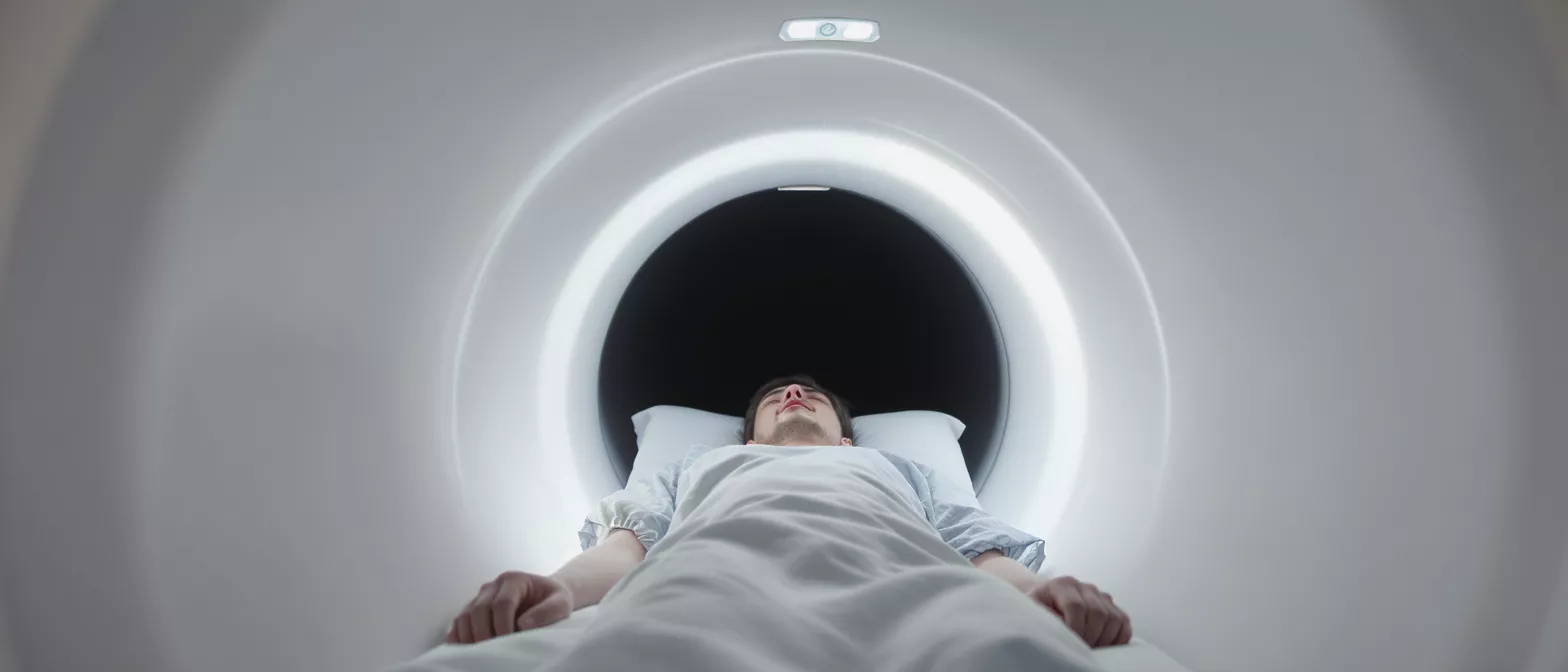
Feeling prepared for the actual scan day significantly reduces patient stress. When you arrive for your heart MRI scan procedure, you will change into a hospital gown to ensure no metallic objects interfere with the magnetic field. A nurse or technician will attach small, sticky pads to your chest to monitor your heart rhythm. You will then lie down on a specialized table that slides smoothly into the center of the MRI machine.
Staying still inside the scanner: The most important instruction you will receive is to remain completely still during the scan. Any movement can blur the highly detailed images. You will also be given specific breathing instructions to hold your breath for short intervals, ensuring the technician captures perfectly clear images of your heart's structure.
The inside of the machine can be noisy, with loud thumping and humming sounds as the machine operates. You will be provided with earplugs or headphones to ensure you remain comfortable. The procedure typically takes between forty-five to ninety minutes, during which the technician remains in an adjoining room and speaks to you through an intercom, ensuring your safety and comfort at all times.
How to prepare for a cardiac MRI scan?
Proper preparation helps the imaging center capture the best possible pictures for your surgical team. You will typically be advised to avoid eating or drinking for a few hours before the test, especially if a contrast agent will be used. It is essential to inform your medical team about any implants, pacemakers, or metal fragments in your body, as these can be affected by the powerful magnet.
Managing pre-scan anxiety: If you experience claustrophobia or fear enclosed spaces, inform your physician before scheduling the appointment. They can provide a mild sedative to help you relax during the scan.
Leaving all jewelry, watches, and hairpins at home is critical, as metal can become dangerous projectiles in the magnetic field. Additionally, some cosmetics contain trace amounts of metal, so arriving with a clean face is often recommended. Following these guidelines meticulously ensures a smooth, uninterrupted scan that yields the accurate data your surgeon needs.
How safe is cardiac magnetic resonance?
Safety is a primary concern for any patient facing a heart procedure, and medical imaging protocols are strictly enforced. Cardiac magnetic resonance safety is exceptionally high because the technique does not use damaging ionizing radiation. The magnetic fields and radio frequencies used in this test have no known long-term harmful effects on the human body, making it a preferred method for both initial diagnoses and long-term follow-ups.
Understanding contrast agent safety: In many cases, a contrast dye called gadolinium is injected into a vein to highlight specific areas of the heart. While this is generally very safe, it is filtered through the kidneys. Your doctor will order a quick blood test beforehand to ensure your kidney function is strong enough to clear the dye efficiently from your system.
The most significant safety considerations involve metallic implants. Modern surgical clips, artificial valves, and stents placed during heart surgeries are typically MRI-safe. However, older devices or certain pacemakers may require special protocols or alternative imaging methods, which your cardiologist will carefully review before the appointment is finalized.
Understanding cardiac resonance results interpretation?
Once the scan is complete, the raw data is sent to a specialized radiologist who is trained in cardiovascular imaging. Cardiac resonance results interpretation requires a highly trained eye to analyze the hundreds of cross-sectional images captured during your scan. The radiologist looks for subtle anomalies in the heart muscle, evaluates the thickness of the ventricular walls, and assesses the competence of the heart valves.
Translating imaging for your surgeon: The findings are compiled into a comprehensive report that acts as a roadmap for your surgeon. By quantifying the exact degree of muscle damage or valve leakage, the report dictates whether a repair, replacement, or alternative intervention is most appropriate. This critical step often incorporates data from a comprehensive cardiovascular screening panel to ensure all physiological metrics align.
Your cardiologist will sit down with you to discuss these results in plain, understandable terms, explaining how the findings impact your surgical plan. Having this detailed anatomical information prior to surgery drastically reduces the time spent in the operating room and significantly improves overall surgical outcomes. The images ensure the surgical team faces no unexpected anatomical surprises when the procedure begins.
Cardiac MRI for myocarditis and inflammation?
Inflammation of the heart muscle is a serious condition that can often mimic other cardiac diseases or require urgent surgical intervention. Cardiac MRI for myocarditis is widely considered the gold standard for non-invasive diagnosis. By utilizing specialized contrast techniques, the scan can highlight areas of the heart muscle that are actively inflamed, allowing doctors to see the exact extent of the tissue damage.
Detecting tissue scarring: The scan can differentiate between acute inflammation and permanent scarring from a previous heart attack. This distinction is vital for a cardiac surgeon. If the muscle is merely inflamed, medical therapy and rest may be prescribed instead of an invasive procedure. If the tissue is irreversibly scarred and at risk of rupturing, surgical intervention becomes a necessity.
For patients experiencing unexplained chest pain or heart failure symptoms, this imaging modality offers definitive answers without the need for a surgical biopsy. This ability to rule out dangerous conditions without making an incision is a cornerstone of modern, patient-centered cardiac care, offering immense peace of mind to frightened patients and their families before any surgical decisions are made.
Summary of non-invasive cardiac imaging
Cardiac magnetic resonance has revolutionized how surgeons prepare for complex heart operations. By utilizing safe, powerful magnets instead of radiation, it delivers high-resolution, three-dimensional images of the heart's anatomy and function. This comprehensive visibility allows your medical team to precisely plan your surgery, minimizing risks and improving your overall recovery trajectory.
Understanding what to expect during the procedure, from the loud thumping sounds to the breathing instructions, helps alleviate the natural anxiety associated with medical tests. Knowing that this scan can definitively diagnose conditions like myocarditis, evaluate tissue viability, and differentiate between various heart diseases empowers you to make informed decisions about your healthcare journey.
As you prepare for your operation at Cirugía de Corazón, remember that this advanced imaging is a vital tool designed to protect your health. It provides your surgical team with the exact, detailed blueprint they need to perform your procedure with the highest level of skill and safety possible.






