

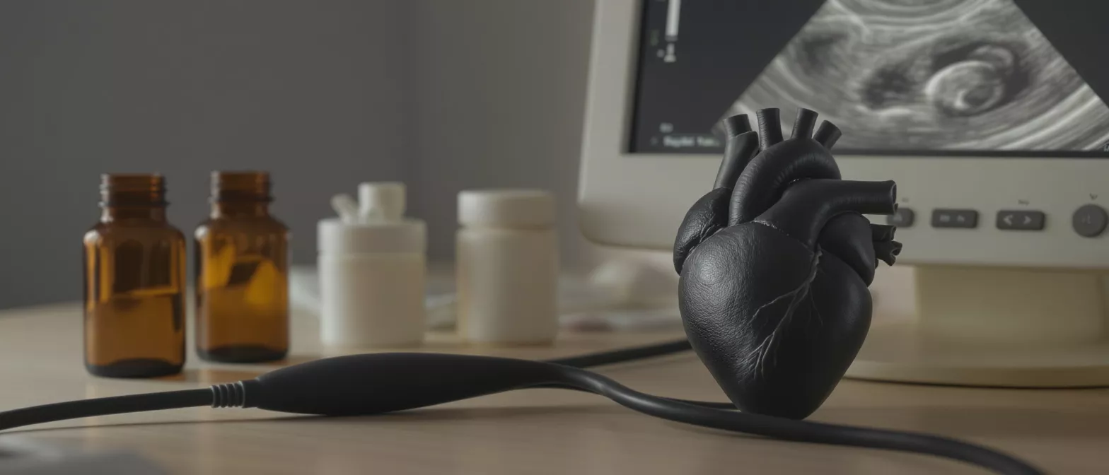
Surveillance Imaging
Learn how surveillance imaging monitors heart health after surgery.
What Is Surveillance Imaging in Cardiac Care?
Surveillance imaging refers to the scheduled, non-invasive medical scans performed after a heart operation to continuously monitor the structural integrity and function of the cardiovascular system. Rather than waiting for physical symptoms to signal a complication, these advanced visual diagnostics provide a direct look at how well the surgical repairs, artificial valves, or implanted grafts are performing over time.
For patients recovering from procedures performed by specialists like Dr Alejandro Molina at Cirugia de Corazón, these routine scans act as a crucial window into the healing process. By capturing detailed, real-time pictures of the heart chambers, valves, and major blood vessels, your medical team can verify that the intricate surgical work is enduring the continuous stress of daily life. This proactive approach ensures that the heart maintains optimal blood flow and functional capacity during the vulnerable recovery period.
Ultimately, the goal of post-operative surveillance imaging is to detect any subtle mechanical changes or physiological shifts long before they manifest as dangerous clinical symptoms. Whether monitoring tissue healing around an incision, checking the velocity of blood flow through a repaired valve, or assessing the overall pumping strength of the heart muscle, these diagnostic tests provide immense reassurance. They confirm that the complex, life-enhancing cardiac surgery remains a durable success.
Why Is Cardiac Surveillance Imaging Necessary After Surgery?
Even the most flawlessly executed cardiac surgery alters the natural plumbing of the cardiovascular system, and the body must adapt to these internal changes during the weeks and months of recovery. Surveillance imaging allows your surgical team to verify that the heart tissue is healing properly, that internal sutures are holding securely, and that no fluid buildup is compressing the heart muscle or lungs. Monitoring surgical site healing: Deep imaging scans confirm that the delicate tissues manipulated during surgery are bonding correctly, effectively preventing potential complications like localized infections or structural weakening that might compromise the heart's natural rhythm.
Another critical reason for these routine cardiovascular scans is to establish a reliable baseline of your new, post-surgical heart anatomy. Each time a scan is performed, the gathered images are directly compared against previous results, allowing clinicians to chart a highly precise trajectory of your internal recovery. Evaluating valve and graft function: Regular surveillance imaging ensures that replaced valves are opening and closing fully without leaking, and that bypass grafts are delivering robust blood flow to the heart muscle without any unexpected blockages or narrowing.
Finally, these scheduled imaging appointments serve as an essential early warning system for potential, treatable issues that could otherwise silently undermine the surgical outcome. Issues such as a gradual thickening of an artificial valve, a slow-developing fluid collection around the pericardium, or an unexpected dilation of a repaired aorta can be identified early. Preventing long-term complications: By identifying these silent, structural shifts during a routine heart surveillance scan, your medical team can intervene conservatively and effectively before a minor anomaly escalates into a medical emergency.
How Does Surveillance Imaging Work for Heart Monitoring?
The process relies on sophisticated medical technology designed to capture high-resolution, dynamic images of the cardiovascular system without the need for additional surgical incisions. High-frequency sound waves, powerful magnetic fields, or low-dose X-rays are transmitted harmlessly through the chest wall, and the returning signals are computationally translated into detailed cross-sectional or three-dimensional pictures of the beating heart. This allows your cardiologist to observe the heart's mechanical function in real-time, analyzing how the muscle contracts and how the blood flows through the chambers.
During these sessions, technicians apply specialized sensors or guide you into advanced imaging machines that systematically map the anatomy of the heart and surrounding blood vessels. Real-time hemodynamic assessment: By watching the heart beat in real-time, clinicians can measure precise pressure gradients across valves, calculate the volume of blood pumped with each contraction, and verify that the natural electrical conduction is producing synchronized, efficient muscular movement. This functional data is just as important as the static structural images, giving a complete picture of cardiovascular health.
The imaging equipment is carefully calibrated to distinguish between the repaired structures, native tissues, and circulating blood, clearly highlighting any abnormalities in the healing process. The resulting data is immediately digitized and stored in your medical record, creating a highly detailed visual timeline of your recovery journey.
Types of Surveillance Imaging for Cardiovascular Follow-up
Cardiac specialists utilize several distinct types of imaging technology, each specifically chosen for its unique ability to highlight different aspects of cardiovascular anatomy and surgical healing. The choice of which modality to use depends entirely on the specific surgical procedure performed, the structures involved, and the exact clinical information the surgical team needs to extract at that particular stage of recovery.
- Echocardiography: The most common first-line approach, using advanced sound waves to assess heart valve function, chamber size, and overall pumping efficiency without any radiation.
- Cardiac MRI: Utilizing powerful magnetic fields to produce highly detailed, three-dimensional tissue images, ideal for evaluating complex structural repairs and identifying scarring or inflammation.
- XCT Cardiac CT Scans: Providing precise, high-resolution images of the coronary arteries, implanted grafts, and structural implants to verify clear, unobstructed blood flow.
By selecting the appropriate combination of these diagnostic modalities, Dr Molina and your medical team can comprehensively evaluate the structural integrity of the surgical work, ensuring that every internal component is functioning in perfect physiological harmony.
Echocardiogram Surveillance for Post-Operative Heart Monitoring
The echocardiogram remains the absolute cornerstone of post-operative cardiac monitoring due to its speed, safety, and incredible ability to show the heart in real-time motion. By placing a small handheld transducer against the chest wall, a specialized technician sends painless sound waves into the chest that bounce off the dense structures of the heart and return as highly detailed moving images. This painless test immediately reveals how well the heart muscle is squeezing and relaxing, and how efficiently the blood is moving through the chambers.
This specific type of surveillance imaging is particularly vital for evaluating the function of newly repaired or replaced heart valves. Assessing valvular integrity: The ultrasound can precisely measure the speed of blood traveling through a mechanical or biological valve, detecting even microscopic amounts of backward leakage that could indicate a structural failure. It also allows the team to safely check the pressures inside the heart chambers, ensuring the new plumbing has successfully reduced the strain on the cardiovascular system.
Because it uses no ionizing radiation, echocardiogram surveillance can be safely repeated as frequently as clinically necessary, making it the perfect tool for closely tracking the rapid changes that occur during the early stages of surgical recovery. It is often performed at the bedside in the recovery room, during outpatient clinic visits, and as an annual check-up tool.
Cardiac MRI Surveillance for Detailed Tissue Imaging
When a deeper, more detailed view of the heart's intricate architecture is required, cardiovascular specialists turn to Cardiac MRI surveillance. This advanced technology uses powerful magnetic fields and radio waves to generate exquisitely detailed, three-dimensional cross-sectional images of the heart muscle and surrounding structures. Unlike standard X-rays, MRI does not use ionizing radiation, making it a remarkably safe option for comprehensive, long-term surveillance of complex structural heart repairs.
Cardiac MRI is particularly unmatched in its ability to differentiate between healthy heart tissue and scar tissue, damaged muscle, or areas of inflammation. Evaluating complex repairs: If a patient has undergone complex congenital defect repair, aortic root replacement, or intricate tissue reconstruction, the MRI provides the exact anatomical map needed to verify that the structural integrity is holding up over time. It accurately measures the thickness of the heart walls and the precise volume of blood the heart pumps with every beat.
This level of intense scrutiny is invaluable for detecting subtle, slow-developing complications that might be completely invisible on less sensitive diagnostic tests. Identifying tissue inflammation: The highly detailed images can reveal early signs of internal inflammation, microscopic tissue damage, or abnormal tissue thickening around surgical implants. By catching these silent warning signs early, the surgical team can implement protective therapies before serious clinical symptoms ever emerge.
What to Expect During Imaging After Heart Surgery
Knowing what to expect during your scheduled heart surgery monitoring can significantly reduce anxiety, allowing you to approach the appointment with confidence and calm. Most post-operative cardiovascular scans are completely painless and require no needles, incisions, or special medications. You will typically be asked to change into a hospital gown to ensure clear access to your chest, and a technician will guide you through each step of the process, ensuring you are comfortable and perfectly positioned.
For an echocardiogram, you will simply lie on your left side on a padded examination table while the technician places a small amount of warm gel on your chest and moves the transducer around to capture different internal angles of the heart. The process is highly interactive, and you may be asked to hold your breath for a few seconds at a time to produce clearer, more stable images. The entire procedure usually takes less than an hour, and you can return to your normal daily activities immediately afterward.
If your specific surgical surveillance protocol requires a cardiac MRI or CT scan, you will lie flat on a specialized table that gently slides into a large, circular scanning machine. The machine will make rhythmic, humming sounds while it operates, and you must remain perfectly still for several minutes at a time to prevent the high-resolution images from blurring. In some cases, a specialized contrast dye may be injected into a vein in your arm to highlight the heart's blood vessels, ensuring the medical team gets the clearest possible diagnostic picture.
Are Cardiac Surveillance Imaging Tests Safe?
For the vast majority of cardiac surgery patients, post-operative imaging follow-up tests are exceptionally safe and carry very minimal clinical risk. These non-invasive or minimally invasive procedures are specifically designed to gather critical medical information without causing physical trauma, additional stress, or pain to the recovering heart. The absolute diagnostic benefits of closely monitoring the surgical repairs heavily outweigh any theoretical risks associated with the scanning technology itself.
Echocardiograms use harmless sound waves and carry zero radiation risk, making them entirely safe for absolutely everyone, including patients with pacemakers or implantable defibrillators. Cardiac MRI is also radiation-free, though patients must inform their medical team if they have certain older metallic implants or surgical clips, as the powerful magnetic field can interact with ferrous metals. However, many modern implants used in cardiac procedures today are MRI-safe or MR-conditional, so screening before the scan is still required.
If a surveillance protocol requires a CT scan, the test does involve a minimal, carefully controlled dose of ionizing radiation. Radiation safety protocols: Cardiologists meticulously weigh the diagnostic necessity of this scan against the tiny, cumulative radiation dose, utilizing modern low-dose machines to keep exposure to an absolute minimum. If a contrast dye is used to enhance the images, the clinical team will first check your kidney function to ensure your body can safely process and clear the contrast material.
Surveillance Imaging Protocol and Follow-Up Schedule
The specific surveillance imaging protocol for any given patient is highly personalized, tailored precisely to the exact type of cardiac surgery performed, the individual patient's overall health, and the immediate post-operative recovery trajectory. There is no universal, one-size-fits-all schedule; instead, your surgical team will design a customized timeline that ensures the most critical monitoring occurs during the most vulnerable phases of the healing process.
- Immediate Post-Op Phase: Frequent bedside echocardiograms in the intensive care unit to verify immediate surgical success and rule out early complications.
- Short-Term Follow-Up: A comprehensive outpatient scan at 4 to 6 weeks post-discharge to assess initial tissue healing and early cardiovascular adaptation.
- Long-Term Surveillance: Annual or bi-annual cardiac MRIs or echocardiograms to ensure long-term durability of the surgical repairs and monitor for slow-developing structural changes.
Following the initial, intensive recovery period, most patients transition to a long-term maintenance schedule that requires cardiovascular scans only once a year or once every few years. Following this personalized follow-up schedule is one of the best ways to support the long-term success of your cardiac procedure, so that any future mechanical wear or biological aging of the repair can be detected and managed early.
Understanding Your Surveillance Imaging Results
Once your heart surveillance scan is complete, the highly detailed images are thoroughly reviewed by a specialized cardiologist who compares them against your previous scans and the original surgical reports. The interpreting physician looks for stable, healthy healing, ensuring that chamber sizes remain normal, that valves open fully without restricting blood flow, and that the heart muscle is pumping forcefully and synchronously. A stable report indicates that the surgical work is holding up perfectly under the daily stress of life.
If the imaging results reveal any areas of concern, such as a slight decrease in pumping efficiency or a minor alteration in valve function, this does not necessarily mean the surgery has failed. Interpreting structural changes: Many early post-operative changes are completely expected as the heart muscle adapts to its new geometry and the surrounding tissues heal and remodel. Your surgical team will explain exactly what the findings mean in the context of your specific operation, distinguishing between normal physiological adaptation and true mechanical failure.
Ultimately, the information gathered from your imaging after heart surgery empowers you and your medical team to make highly informed, proactive decisions about your ongoing cardiovascular care. Whether the results confirm robust health or highlight the need for a minor medication adjustment, this precise diagnostic data is the foundation of a confident, healthy recovery. By maintaining a strict surveillance imaging schedule, you actively participate in protecting your long-term heart health, ensuring the brilliant surgical work performed at Cirugía de Corazón lasts a lifetime.






