

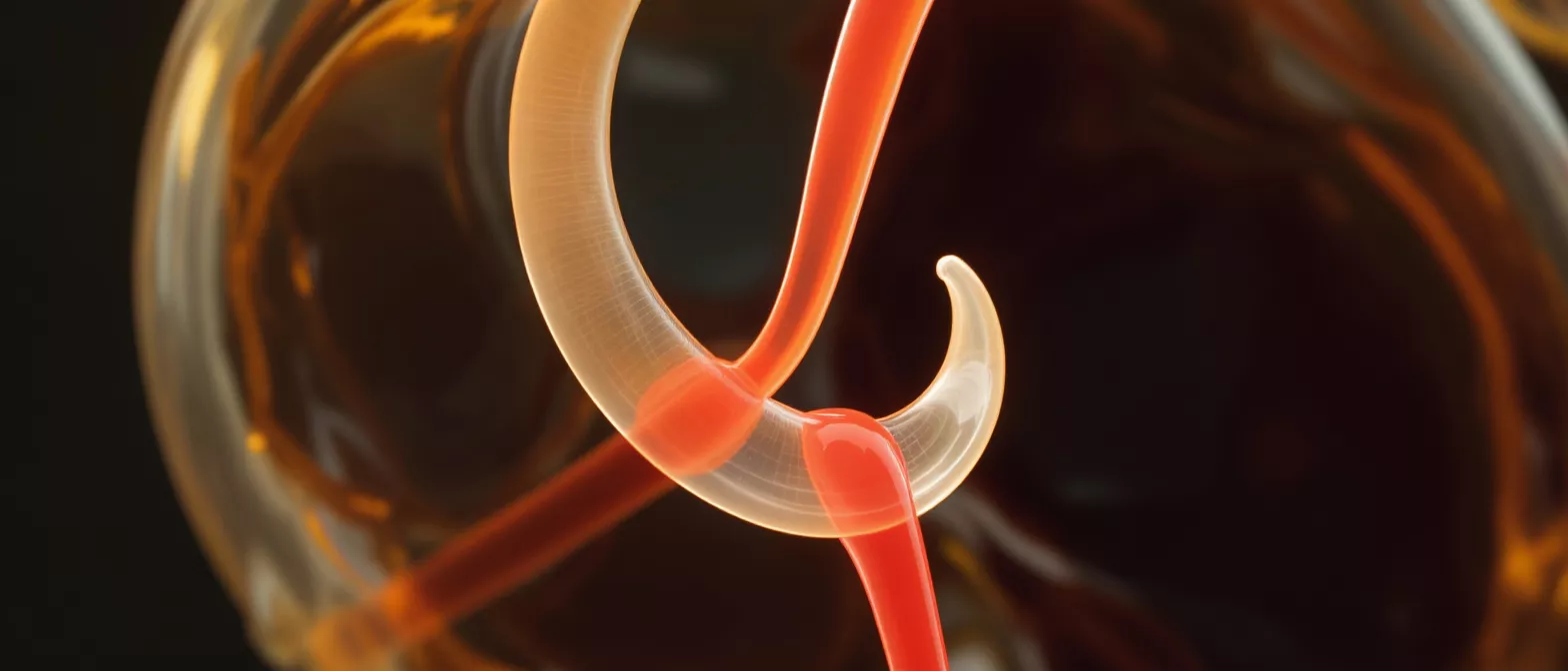
Valve Cusp
Learn what a valve cusp is, how heart valve leaflets function, and why cusps matter for cardiac health.
What is a valve cusp
A valve cusp is a thin, flexible flap of tissue inside the human heart that ensures blood flows in exactly one direction. When you hear the phrase "heart valve leaflet," it refers to these same vital structures. Each time your heart beats, these cusps open to let blood pass through the heart's chambers and snap tightly shut to prevent any backward leakage. They are the essential doors of the circulatory system, operating flawlessly billions of times throughout a person's life.
Valve cusp definition in everyday terms: Imagine a one-way door that only pushes open when blood pushes against it, then falls instantly back into a sealed frame when the pressure reverses. This is exactly how a healthy valve cusp behaves. The tissue is incredibly strong yet delicate, designed to withstand the constant mechanical stress of the cardiac cycle without tearing or stretching.
Understanding heart valve anatomy provides tremendous comfort when facing cardiac surgery. Knowing the mechanical logic of how your heart moves blood demystifies the surgical process. Your surgeon's goal is to ensure these tiny doors open fully and close completely, and modern techniques for repairing them are precise and highly successful.
How does valve cusp function work
Valve cusp function relies entirely on pressure changes within the heart's chambers. When the heart muscle contracts, pressure rises behind the cusps, forcing them open so blood can move forward. When the heart muscle relaxes and pressure drops, the blood naturally tries to fall backward. This backward flow catches the curved edges of the cusps, pushing them together to form a perfect, watertight seal.
How cusps maintain unidirectional blood flow: The edges of each valve cusp meet at a precise angle. When pressure reverses, the tissue edges overlap slightly, ensuring no blood can slip backward. This rapid, seamless opening and closing is what produces the familiar "lub-dub" sound of a heartbeat, and it is entirely driven by passive fluid dynamics rather than active muscle control.
This mechanical elegance is why the heart is so incredibly efficient. The valve cusps themselves do not contain muscle fibers; they respond only to the physical forces of the blood. Because they are passive structures, when a cusp becomes diseased or stiff, surgeons can often repair or replace just the faulty valve without touching the living heart muscle itself.
Types of heart valve leaflets
There are four valves in the human heart, each defined by the number and shape of their cusps. The way these cusps are arranged depends entirely on the specific pressure requirements of the heart chambers they separate. By understanding these variations, patients can better appreciate the complexity of valve cusp anatomy and the surgical approaches required for each.
- Aortic valve: Has three cusps and handles the highest pressure in the heart, pushing oxygen-rich blood out to the entire body.
- Mitral valve: Has only two cusps, a design perfectly suited to handling the high-pressure return of blood from the lungs.
- Pulmonary and tricuspid valves: Feature three cusps each, managing the lower-pressure blood flow entering the lungs and returning from the body.
Each type of heart valve leaflet is uniquely shaped to handle its specific workload. The aortic valve cusps must be exceptionally strong and thick to withstand the highest pressure, while the tricuspid valve cusp is thinner and more delicate due to the lower pressure in the right side of the heart. Surgeons must adapt their repair techniques to match the structural realities of each specific valve.
What is aortic valve cusp anatomy
The aortic valve cusp anatomy is specifically designed to handle the most demanding job in the heart: holding back the entire volume of blood the body relies on. The aortic valve normally has three crescent-shaped cusps that open to allow blood into the aorta and close immediately to prevent it from draining back into the heart's main pumping chamber. These cusps are thick, tough, and anchored to a structural support called the aortic root.
Why the aortic valve cusps face the most pressure: Every time the left ventricle contracts, it throws blood against the aortic valve cusps with massive force. To withstand this, the tissue is reinforced with dense collagen fibers. When these specific cusps become diseased, they often calcify, turning flexible tissue into a rigid shell that restricts blood flow—a condition known as aortic stenosis.
Because the aortic valve cusps operate under such extreme mechanical stress, they are the most common site for age-related valve disease. Surgical correction here requires extreme precision to ensure the newly repaired cusps can withstand a lifetime of high-pressure cycles.
Mitral valve cusp and tricuspid valve cusp differences
Unlike the aortic valve, the mitral and tricuspid valves operate differently and feature a unique structural support system. A mitral valve cusp is attached to thin, tough strings called chordae tendineae, which connect to tiny papillary muscles inside the heart. This parachute-like design prevents the cusps from blowing backward under pressure, ensuring they seal perfectly when the heart squeezes.
The tricuspid valve cusp works similarly but is located on the right side of the heart, managing blood returning from the body before it heads to the lungs. It features three leaflets rather than two, and its structure is slightly thinner. Because the right side of the heart operates under much lower pressure than the left, these cusps are more delicate but follow the same mechanical principles.
When mitral or tricuspid valve cusps fail, they often stretch or tear at the margins, causing blood to leak backward. Reconstructive surgery for these valves often involves tightening the supportive strings rather than replacing the cusp tissue itself. Patients recovering from this specific reconstruction may benefit from exploring post-surgery circulation aids to support healthy blood flow during the healing process.
What causes a damaged valve cusp
A damaged valve cusp can result from several different conditions, but the most common cause is age-related calcification. Over decades, calcium deposits build up on the cusps, turning them stiff and preventing them from opening fully. This restricts blood flow and forces the heart to work harder with every beat. Another common cause is wear-and-tear degeneration, where the tissue simply stretches or weakens over time.
What leads to a damaged or floppy valve cusp: Structural weakness, often genetic, can cause the cusps to become floppy and fail to seal properly. This condition, known as prolapse, allows blood to leak backward. Other causes include congenital defects where a patient is born with only two aortic cusps instead of three, or infections like endocarditis that directly attack the cusp tissue.
Recognizing these underlying causes helps patients understand why a specific surgical intervention is recommended. If the tissue is merely stretched, a surgeon can often tighten it. If the tissue is heavily calcified or infected, replacing the damaged valve cusp entirely may be the safest and most durable solution.
Signs of valve cusp regurgitation
When a valve cusp fails to close completely, blood leaks backward—a condition known as valve cusp regurgitation. Because the heart must work harder to pump the same volume of blood forward while constantly recirculating the leaked blood backward, patients begin to experience a distinct set of symptoms. The body struggles to get enough oxygen-rich blood to meet its demands.
Recognizing valve cusp regurgitation symptoms: The most common signs include unusual shortness of breath, especially when lying flat, chronic fatigue, and a feeling of heavy exhaustion after light activity. Some patients develop heart palpitations, a sensation that the heart is fluttering or beating irregularly. In more advanced cases, fluid builds up in the lungs or legs, causing swelling and persistent coughing.
If you experience these symptoms, your cardiologist will use an echocardiogram to examine your heart valve leaflets closely. This painless ultrasound allows the medical team to see the exact point of leakage and measure the severity. Catching valve cusp regurgitation early allows for careful surgical planning before the heart muscle is permanently overworked.
How is valve cusp repair performed
Valve cusp repair is a surgical procedure designed to fix a faulty valve without removing it. Instead of replacing the entire valve, the surgeon works directly on the damaged tissue. If a cusp is stretched, the surgeon may trim away the excess tissue and stitch the edges back together to create a tight seal. If the supportive strings are loose, artificial supports can be added.
How surgeons fix valve cusp regurgitation safely: During the operation, the heart is temporarily stopped, and a bypass machine takes over the work of pumping blood. The surgeon opens the heart chamber to access the diseased valve. By reshaping the valve cusp tissue and reinforcing its structural anchors, the surgeon restores normal blood flow while preserving the patient's natural anatomy.
Preserving your natural valve cusp tissue is always preferred when possible, as it reduces the need for long-term blood thinners. The precision of modern cardiac surgery makes successful repair highly likely for most structural defects.
Are valve cusp repairs safe
Valve cusp repairs are highly established, routine procedures with an excellent safety profile in modern cardiac surgery. Because heart surgery technology has advanced dramatically over the past few decades, the risks associated with stopping the heart and putting it on bypass are lower than ever. Surgeons perform these operations daily with incredibly high rates of complete functional recovery.
Risks and safety of valve cusp repair: As with any major operation, there are inherent risks, including infection, bleeding, or adverse reactions to anesthesia. However, the risk of leaving a severely leaking valve untreated is far greater, as heart failure becomes inevitable without surgical intervention. Your surgical team will evaluate your specific heart valve anatomy and overall health to minimize any potential complications.
Patients often find tremendous reassurance in understanding the safety protocols of modern operating rooms. The care team continuously monitors every aspect of heart function, and the bypass machine ensures your brain and organs receive steady oxygen throughout the procedure. Recovery is closely managed to ensure the repaired cusps heal properly and function immediately.
Supporting recovery after heart valve surgery
Recovering from valve cusp repair or replacement requires time, patience, and a commitment to following medical guidance. The heart muscle itself needs several weeks to heal from the surgical incisions, even if the valve cusp function is immediately restored. Patients usually stay in the hospital for close monitoring for about a week before transitioning to a carefully guided at-home recovery phase.
What to expect during heart valve recovery: Initially, you will feel tired and will need to avoid heavy lifting or strenuous activity. A structured cardiac rehabilitation program will slowly reintroduce safe physical exercise. Your cardiology team will check the valve function through follow-up echocardiograms to ensure the repaired cusps are maintaining a perfect seal.
The ultimate goal of the surgical journey is to return you to a fully active, healthy life without the breathlessness and fatigue of a damaged valve. With the repaired heart valve leaflets working correctly, the heart's efficiency is restored. By following your recovery plan and attending all check-ups, you can look forward to decades of strong, unimpeded heart function.






