

Endocarditis: when the heart fights to survive
Endocarditis is a serious heart infection.
An infection inside the heart — who is at risk, how the Duke criteria establish the diagnosis, how it is treated, and why dialysis patients are especially vulnerable.
Endocarditis is an infection inside the heart. Here is what it is, who is most at risk, how it is diagnosed with the Duke criteria, how it is treated, and why patients on haemodialysis face particular danger.
In a single breath, life can change. We know that well enough — but when that breath involves an infection inside your heart, it is life itself that can hang by a thread.
Endocarditis is a very serious clinical situation. If you are reading this, it may be that you or someone you love has been diagnosed with it. I understand how stressful that can be. Perhaps you simply want to learn a little more about the subject. That is why I wrote this piece, to help you and your family have a better idea of what you are dealing with.
What is endocarditis?
Endocarditis is an uncommon but devastating disease affecting the inner lining of the heart, known as the endocardium.
The disorder usually begins with bacteria or other microorganisms entering the bloodstream, which then adhere to the heart valves or to damaged surfaces of the heart.
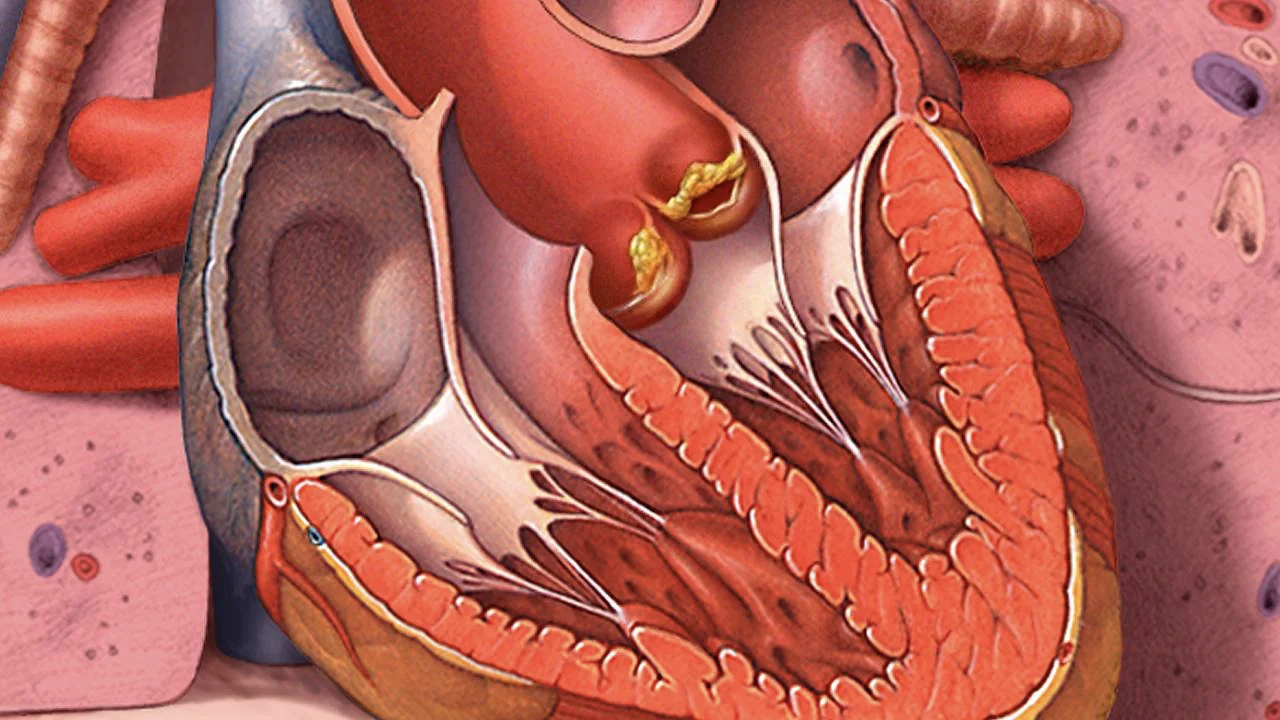
As these bacteria grow and multiply in the heart, they can cause inflammation, damage to the valves and, in serious cases, lead to heart failure, stroke or even death.

Causes and who is most at risk
Endocarditis can affect anyone, but certain risk factors increase the likelihood of contracting it:
- People with pre-existing heart disease
- People with damaged heart valves or congenital heart defects
- Those with a history of previous endocarditis — that is, who have already had an infection inside the heart and recovered
- Intravenous drug users
- People with valve prostheses, who have already had a heart valve replaced
- Those who have undergone dental or surgical procedures without adequate antibiotic prophylaxis
- People with kidney failure, especially those requiring haemodialysis
How is endocarditis diagnosed?
Diagnosing endocarditis is a complex process generally involving a combination of clinical assessment, laboratory studies and imaging.
Clinical history and physical examination. Your doctor will gather information about your symptoms, your medical history, previous cardiac procedures and risk factors. They will also carry out a physical examination looking for signs of the disease, such as heart murmurs, fever, marks on the skin and other signs of infection.
Blood tests are a fundamental part of the diagnosis:
- Blood cultures. Blood samples are taken to look for bacteria or microorganisms that may be causing the infection.
- General laboratory tests. These include full blood counts, liver and kidney function tests, and markers of inflammation such as C-reactive protein (CRP) and the erythrocyte sedimentation rate (ESR).
- Serological tests. These look for specific antibodies against microorganisms that could cause endocarditis, such as antinuclear antibodies (ANA) or antineutrophil cytoplasmic antibodies (ANCA).
Echocardiogram. The echocardiogram is a fundamental tool in diagnosing endocarditis, and can be of two types:
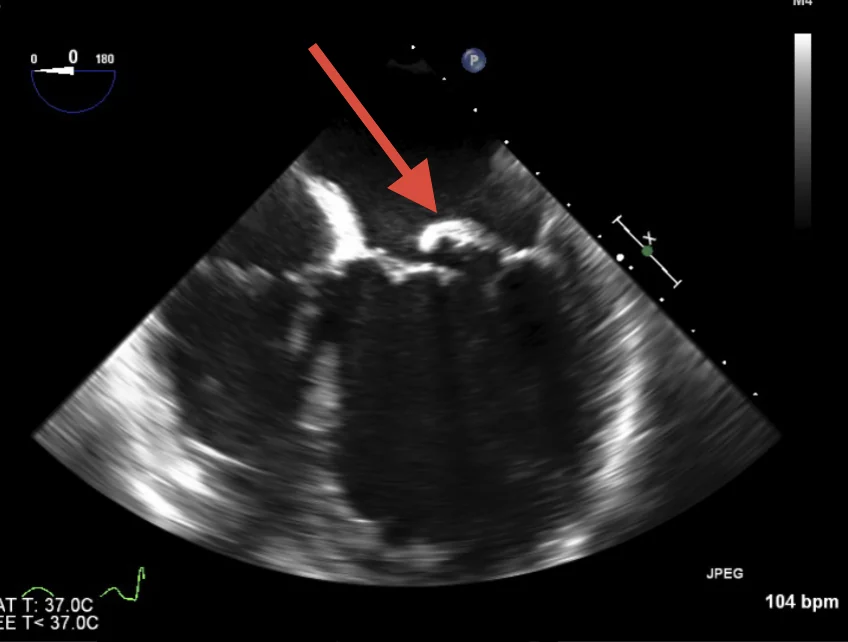
- Transthoracic echocardiogram (TTE). Uses sound waves to create images of the heart from outside the body — an ultrasound of the heart. It can show vegetations, accumulations of bacteria and tissue, on the heart valves.
- Transoesophageal echocardiogram (TOE). Performed by passing a probe through the oesophagus to obtain more detailed images of the heart. It is especially useful for detecting small vegetations on the valves.
Cardiac catheterisation. This study is used to see the coronary arteries — the ones responsible for heart attacks. If they are damaged or obstructed they may require an intervention or bypass surgery.
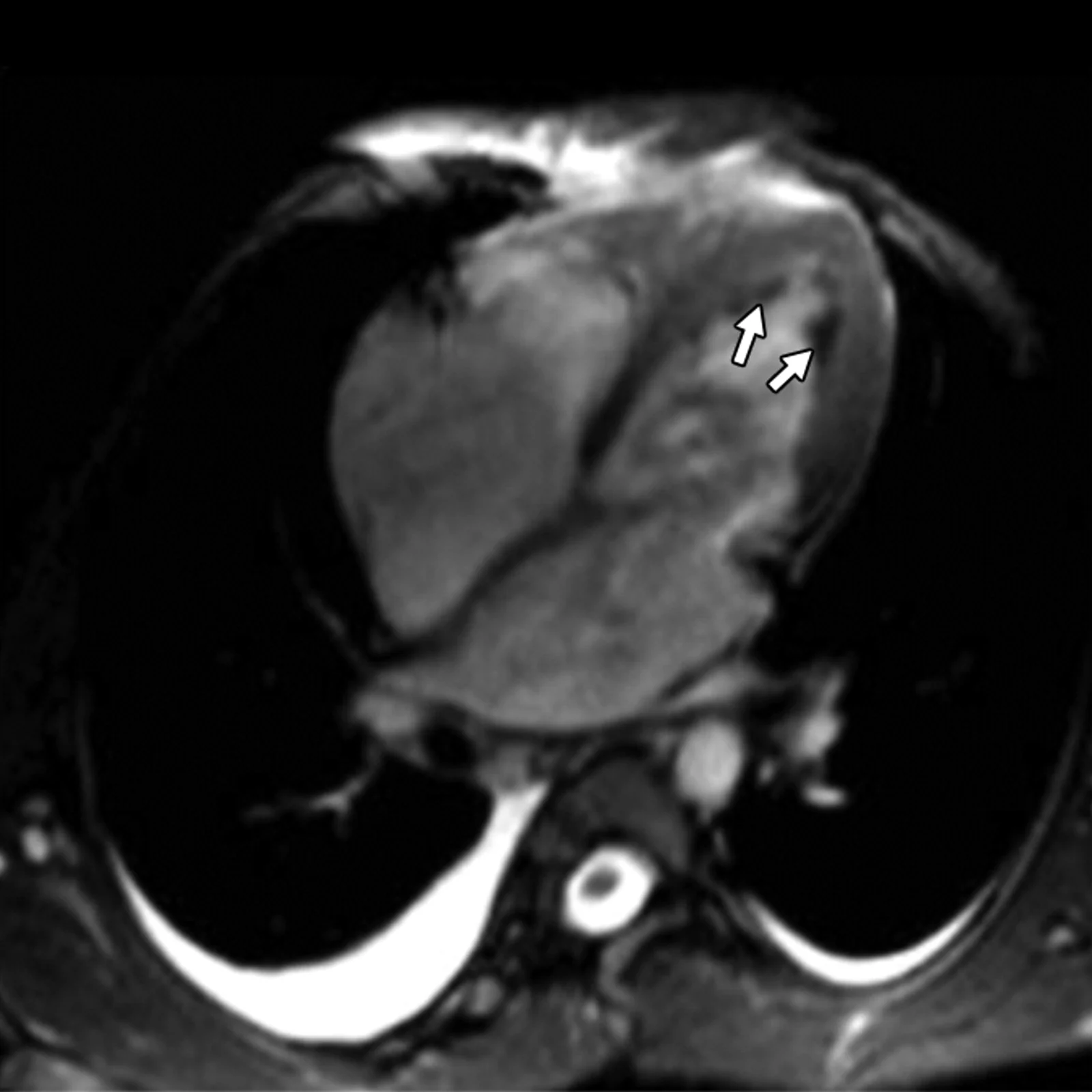
Additional imaging. In some cases other studies such as cardiac MRI or cardiac CT can be performed to assess cardiac damage and detect complications.
Duke criteria. Specific clinical and laboratory criteria, known as the Duke criteria, are used to help in the diagnosis. These consider clinical findings, blood culture results and imaging.
The Duke criteria, and what they are for
The Duke criteria are a set of clinical and laboratory criteria used to help diagnose infective endocarditis.
They were developed to give doctors structured guidance and to help identify patients with endocarditis, particularly where the clinical presentation is ambiguous. They have been revised and updated over time to improve their accuracy.
This is genuinely technical material designed for doctors. Even so, it is important that you know diagnosis and treatment here is a challenge even for the most experienced.
Major criteria
- Blood cultures positive for organisms typical of endocarditis. Persistently positive blood cultures for bacteria that commonly cause endocarditis, such as Streptococcus viridans, Streptococcus bovis, Streptococcus gallolyticus, Enterococcus, Staphylococcus aureus, or the HACEK organisms (Haemophilus spp., Aggregatibacter spp., Cardiobacterium spp., Eikenella spp., Kingella spp.).
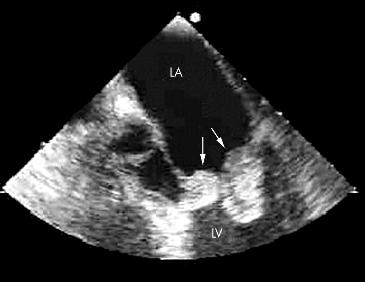
- Evidence of endocarditis on echocardiogram. Vegetations detected on transthoracic or transoesophageal echocardiography, or intracardiac abscesses.
- New regurgitation of a heart valve. Evidence of regurgitation — backward flow — through a heart valve on echocardiogram that was not present previously.
Minor criteria
- Fever. Body temperature of 38.0°C (100.4°F) or above.
- Vascular evidence. Vascular phenomena such as arterial emboli, pulmonary infarcts, conjunctival haemorrhages, Roth spots (retinal haemorrhages), Osler nodes (painful nodules on the fingers), and Janeway lesions (painless lesions on the palms and soles).
- Immunological phenomena. Glomerulonephritis, subcutaneous nodules, Osler nodes, Janeway lesions.
- Microbiological evidence. Positive blood cultures that do not meet the major criteria, or positive serology for organisms causing endocarditis.

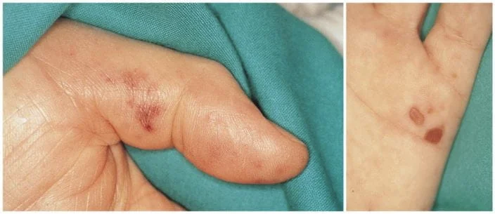
How the criteria are applied
Definite endocarditis: two major criteria; or one major and three minor; or five minor criteria.
Possible endocarditis: one major and one or two minor criteria; or three minor criteria.
It is important to stress that these criteria are a useful tool, but not infallible. Clinical assessment, imaging results and other medical findings also play a crucial part. Endocarditis can have a variable presentation, which makes its diagnosis challenging at times.
I have been diagnosed with endocarditis. What treatment options do I have?
Medical treatment of endocarditis, also known as infective endocarditis, is a fundamental part of managing this disease. It usually requires a great deal of time and several specialists.
The main aim is to eliminate the bacterial infection affecting the heart valves and to prevent serious complications such as heart failure, stroke and the spread of infection to other organs. It is usually an enormous challenge.
- Antibiotics. Antibiotics are the basis of medical treatment. The choice depends on the type of bacteria causing the infection, so blood cultures must be taken to identify the organism responsible. Treatment is usually long-term, often for four to six weeks or more. Antibiotics may be given intravenously in hospital and, in some cases, orally at home once the patient is stable.
- Dose and duration. These vary with the severity of the endocarditis, the type of bacteria and other individual factors. It is fundamental to complete the full course of antibiotics as prescribed, even if symptoms improve before the treatment ends.
- Treatment in hospital. Many patients with endocarditis require admission to receive intravenous antibiotics and to have their condition monitored. During the stay, the response to treatment is followed closely and laboratory tests and imaging are carried out to assess progress.
- Surgery. In some cases, especially where there are serious complications such as heart failure, abscesses in the heart or valves damaged beyond repair, open-heart surgery may be necessary. Surgery can involve repairing or replacing the affected valves.
- Preventing complications. During treatment, measures are taken to prevent complications such as embolism — fragments of vegetation breaking off and travelling to the brain or other organs — and anticoagulants or antiplatelet drugs may be prescribed according to individual need.
- Medical follow-up. After discharge the patient must continue with follow-up to ensure the infection has been eradicated completely and there is no relapse. This can include regular echocardiograms to assess cardiac health.
- Education and prevention. It is important that patients and their families receive education about endocarditis and how to prevent it in future, particularly where risk factors exist. This can include proper oral hygiene and antibiotic prophylaxis before certain dental or medical procedures.

Endocarditis in people on haemodialysis
A person with kidney failure requiring the replacement treatment known as haemodialysis can face additional challenges. They usually need several sessions a week and are connected to various lines and catheters in order to carry out dialysis.
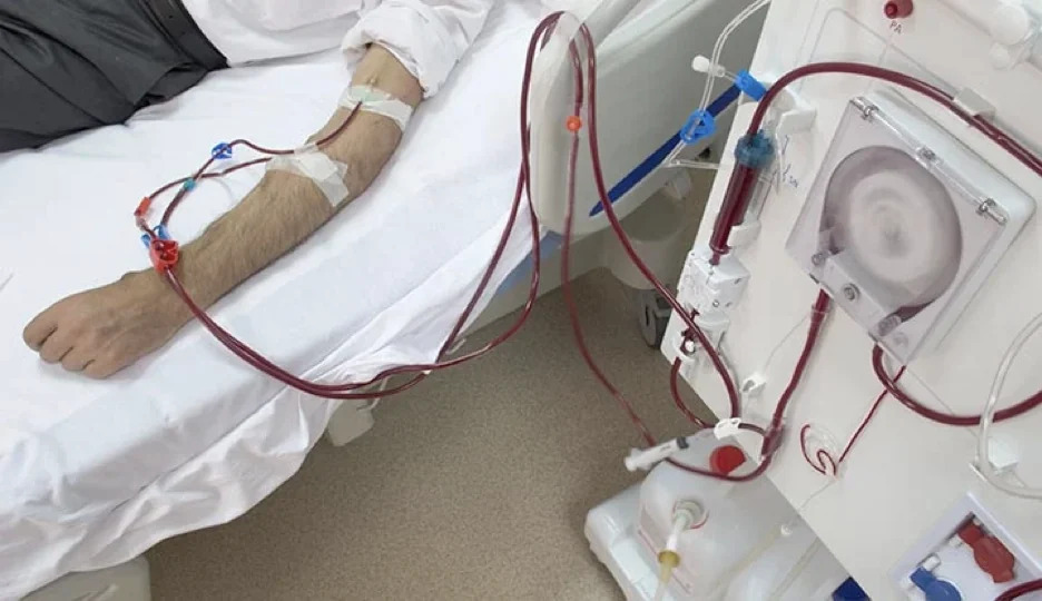
That puts them at additional risk of developing infections of all kinds, endocarditis being one of the most dangerous of all.
The need for constant vascular access can increase the risk of infection, and antibiotic therapy may require specific adjustments to avoid complications.
Close collaboration between nephrologists, cardiologists and cardiovascular surgeons is crucial to managing this condition effectively in this vulnerable population.
Treatment often includes open-heart surgery, minimally invasive surgery or endovascular treatment, according to each person's presentation and characteristics. The context is plainly more severe in these patients and the risk of surgery can increase considerably.
In this group, complications can range from the relatively simple — a clot or a small vegetation in the heart — through to more complex scenarios such as destruction of the valves and heart failure.
That is why it matters to be well informed and surrounded by a team that has earned your trust to follow up any situation with your heart.
What does surgery for endocarditis involve?
Open-heart surgery is the most widely used technique, with an incision made in the middle of the chest and the sternum to reach the heart. An artificial heart–lung machine is required to "put the heart to sleep" so we can work inside it.

The procedures performed vary enormously from patient to patient, since endocarditis can affect different structures in each person.
They may be valve replacements with biological prostheses, or repair of the valves in cases where that is feasible without replacing them. Corrections of fistulas, dilatations or perforations of the heart can also be carried out, among many others as the situation demands.
Despite being a complex and sometimes high-risk procedure, surgery can be the only effective treatment in many patients where everything else has already failed.
What alternatives exist for treatment?
Minimally invasive cardiac surgery. Where it is indicated and patients are well selected, they can benefit enormously from these procedures, particularly through faster recovery, less pain and fewer blood transfusions.
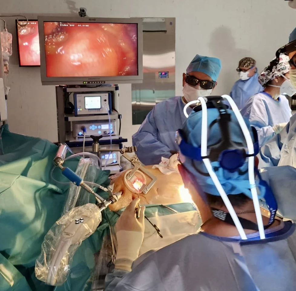
Medical treatment. In some patients this is sufficient and no more invasive treatment is required. In others, where the risk of surgery is too high, this can be considered.
Endovascular treatment. In recent years new transcatheter valve replacement techniques have been developed which avoid open surgery — TAVR, MitraClip, transcatheter mitral valve and others.
It is important to mention that in most cases of endocarditis these techniques are contraindicated, because of the infective and inflammatory process in the heart.
Life after treatment
Recovery after endocarditis can be a long and challenging road. In some people it is far shorter than in others for individual reasons; even so, many people can lead a full life after successful treatment, whether surgical or more conservative.
Adherence to medical therapy and regular follow-up are essential to ensuring an optimal recovery.
Endocarditis can have a variable presentation, which makes its diagnosis challenging at times. Its diagnosis and treatment should therefore be carried out by a medical team experienced in infective cardiac disease, to ensure appropriate and timely care.
To take home
On this journey towards recovery from endocarditis, it is fundamental to remember that you are not alone. The doctor–patient relationship, based on trust and mutual respect, is a powerful force in the fight against this disease. As science advances and treatments evolve, the future is more hopeful than ever.
So do not let fear overwhelm you. Face it with the certainty that there is a team of health professionals committed to your wellbeing.
Together, as patients and doctors, we can overcome the challenges and build a path towards a full and healthy life.

About the author
Dr Alejandro Molina is a cardiovascular surgeon practising in Guadalajara, Querétaro and Hermosillo. He trained in cardiothoracic surgery at the Universidad de Guadalajara, completed advanced training in cardiac surgery, minimally invasive surgery, robotic surgery and structural interventions at the University of Montreal, and a fellowship in structural cardiology and TAVR at CHU Bordeaux, France. He performed the first robotic mitral valve repair in the Ángeles Health System and the first robotic heart surgery in Guadalajara.
Medical degree: Universidad Autónoma de Guadalajara, Cédula Profesional 5757260 · Cardiothoracic surgery: Universidad de Guadalajara, Cédula de Especialidad 10768871 · Current certification, Consejo Nacional de Cirugía del Tórax, no. 447
Talk to a specialist
Request a free video consultation to clear up your questions and make a better-informed decision.





