

Transcatheter heart valve replacement: a medical revolution
Discover how transcatheter valve replacement transforms heart care without open-heart surgery.
How the field began in 2002, which valves need intervention most, the aortic, mitral and tricuspid devices available now, and how the procedures are performed.
Replacing heart valves by catheter, without open-heart surgery: how the field began, which valves most often need intervention, the devices available today, and where it is heading.
Welcome. You are about to take a leap into a subject that is fascinating, and vital for many patients and their loved ones: the new technologies for replacing heart valves through a catheter, that is to say without the need for open-heart surgery.
Gone are the days when invasive surgery was the only option. Medicine has taken a step forward with less invasive procedures, and that has transformed the lives of countless people around the world. Procedures that seemed to come out of a science fiction film have today become routine in catheterisation laboratories everywhere.
A little anatomy
To understand better how the heart valves work and how they affect the circulation, it helps to know something about the anatomy of these vital structures. The human heart has four main chambers: two atria, right and left, and two ventricles, right and left. The heart valves sit between these chambers and are responsible for ensuring blood flows in one direction only as the heart beats.
The main valves of the heart:
- Tricuspid valve. Sits between the right atrium and the right ventricle. It has three leaflets that open when blood flows from the atrium into the ventricle during filling, and close when the ventricle contracts to pump blood towards the lungs.
- Mitral (bicuspid) valve. Sits between the left atrium and the left ventricle. It has two leaflets that open to allow blood to flow from the atrium into the ventricle as the heart relaxes (diastole), and close when the ventricle contracts to send blood to the rest of the body.
- Pulmonary valve. Sits between the right ventricle and the pulmonary artery, which carries blood to the lungs for oxygenation. It has three leaflets that open when the right ventricle contracts and close when it relaxes, preventing blood flowing back into the ventricle.
- Aortic valve. Sits between the left ventricle and the aorta, the main artery carrying oxygenated blood from the heart to the rest of the body. It has three leaflets and opens when the left ventricle contracts to let blood flow into the aorta, then closes to stop blood returning to the ventricle.
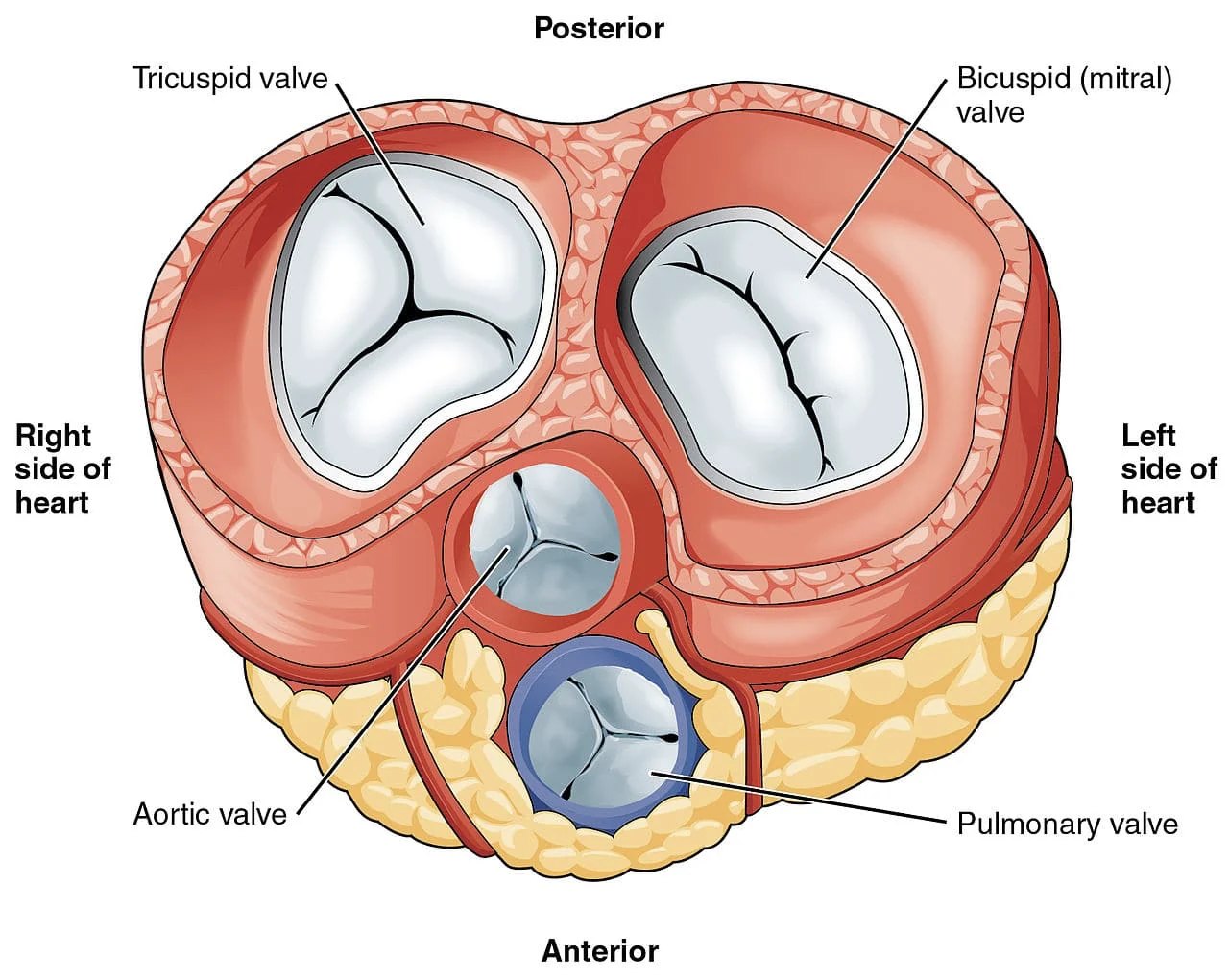
These valves are essential to the proper working of the heart and therefore of the whole circulatory system. Their coordinated opening and closing ensures blood circulates effectively and efficiently, providing oxygen and nutrients to the body's tissues and removing waste products.
Any alteration in the structure or function of these valves can result in cardiac problems such as stenosis (narrowing), regurgitation (leakage) or prolapse (bulging of the valve). Early diagnosis and treatment of valve conditions are crucial to ensuring optimal cardiac function and a good quality of life. Advances in valve replacement technology have been fundamental in providing effective solutions to those with valve disease.
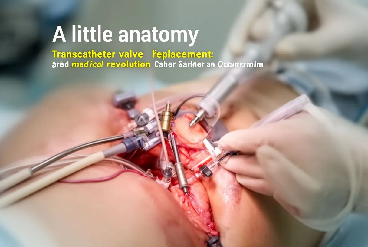
Where did this begin?
The history of transcatheter heart valve replacement goes back to the first decades of the twenty-first century. Although traditional surgery had already demonstrated its high effectiveness over many years, those procedures carried significant risks for a certain number of patients and sometimes prolonged recovery times.
Many patients were considered "inoperable" because of their frailty and their multiple cardiovascular risk factors, and unfortunately were left without any treatment that was genuinely effective.
It was then that a new hope emerged for those who needed a heart valve replaced but were not ideal candidates for open-heart surgery.
In 2002 the first successful transcatheter aortic valve replacement was performed in a high-risk patient in France. The medical world initially received the news with notable scepticism. From that moment on, however, the technology has advanced rapidly, and what was once a limited option has become a viable alternative for many patients with heart disease. This innovative technique has allowed heart valves to be implanted through a catheter introduced generally via the femoral artery, avoiding the need to open the chest.
The international guidelines and their recommendations have been modified constantly by multidisciplinary expert groups. Those guidelines have tried to keep pace with the almost unreachable speed at which the technology has been moving in recent years. It is no exaggeration to say the challenge has been enormous.
These procedures were initially indicated for patients considered at high surgical risk. But with advances in the technology and the growth of our experience placing these devices, we have moved steadily towards a much wider range of patients — younger, with surgical risk from mild through to severe.
Which valves most often require intervention?
Transcatheter heart valve replacement is used mainly in two contexts:
- When a valve is very narrowed (stenosis) or calcified and does not allow adequate blood flow through it.
- When the valve is regurgitant. Regurgitation occurs when the valve does not close properly, allowing blood to flow backwards instead of out of the heart, which places an additional load on the heart's work. The longer this goes unaddressed, the more complications can appear — dilatation of the chambers, arrhythmias and heart failure.
Among the valves requiring intervention most frequently, the aortic and mitral stand out. These are essential structures for the correct working of the heart and, over time, can degenerate or become damaged through ageing, heart disease, infection or congenital defects.
Transcatheter replacement devices available today
There are currently several devices used for replacing heart valves through a catheter, but two main types have proved highly effective:
- Transcatheter aortic valve (TAVR). This procedure involves inserting a new aortic valve into position over the damaged valve. Once deployed, the new valve takes over its function and restores adequate blood flow.
- Transcatheter mitral valve (TMVR). Similar to TAVR, TMVR involves implanting a mitral valve into position over the affected valve, allowing proper closure and preventing backflow of blood.
Transcatheter aortic valve (TAVR)
TAVR is a highly effective technique for replacing the aortic valve in patients with severe aortic stenosis, or in those considered at high risk for open-heart surgery. Among the most widely used devices:
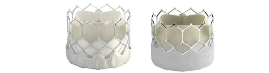
Sapien / S3, Edwards Lifesciences. One of the best-known devices in the TAVR field. It uses a bovine pericardial valve mounted on an expandable stainless steel stent. It is introduced through the femoral artery or via a small incision in the chest and positioned over the damaged aortic valve.
CoreValve / Evolut R / Evolut Pro, Medtronic. This valve uses a self-expanding stent design implanted over the diseased aortic valve. It is made with porcine pericardium and is suitable for different anatomies.
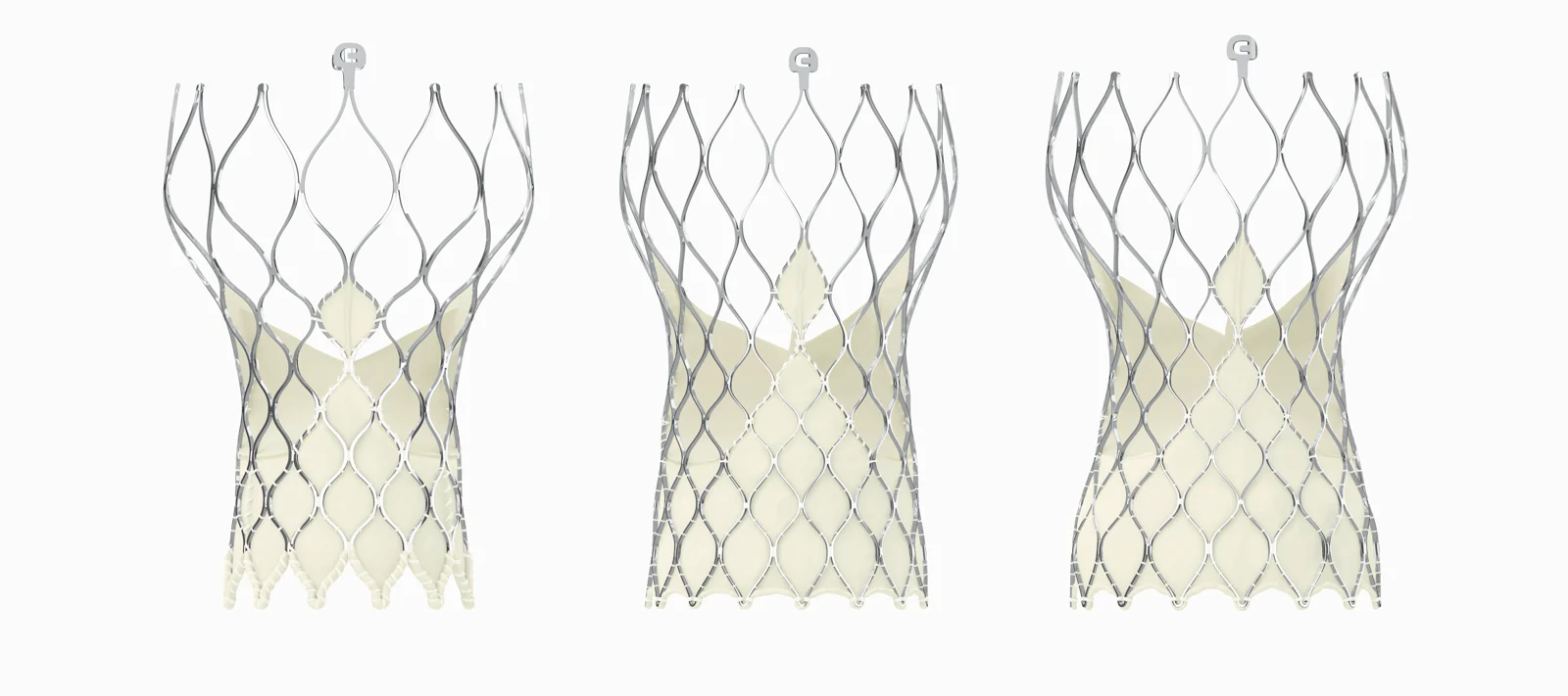
There are several other transcatheter valves from other manufacturers, but these two are the most used and the ones we have most experience with so far. Each has its own advantages and its own implantation characteristics.
Transcatheter mitral valve (TMVR)
TMVR is a less common procedure than TAVR, but it has proved a promising option for certain patients with significant mitral regurgitation. Notable devices include:
MitraClip, Abbott. Approved by the FDA in 2013, the MitraClip is a device used to repair the mitral valve without the need for open-heart surgery. It is implanted by catheter and holds the leaflets of the mitral valve together to reduce the backflow of blood into the left atrium.
Pascal, Edwards Lifesciences. The Pascal system is another device intended to repair the mitral valve using a set of clips. These clips are fitted to the leaflets of the valve, restoring proper function.
New tricuspid valves
Although transcatheter replacement of tricuspid valves is at an earlier stage of development than aortic and mitral valves, significant advances have been made. Tricuspid valves are used to treat tricuspid regurgitation, a condition in which the tricuspid valve does not close correctly and allows blood to flow back into the right atrium. Devices and techniques under study include:
- Cardioband, Edwards Lifesciences. Rather than being a valve itself, Cardioband is a device implanted around the tricuspid annulus to narrow it and improve the function of the valve.
- Forma, Edwards Lifesciences. Based on a transcatheter annuloplasty technique, using an implant placed in the tricuspid annulus to improve its function.
- Trialign, Mitralign. Similar to the MitraClip, Trialign uses a catheter to apply an adjustable suture around the tricuspid annulus to reduce the backflow of blood.
It is important to stress that although these devices have proved effective in many cases, every patient is unique and requires an individual assessment and treatment plan from their medical team.
Research and innovation in transcatheter heart valve replacement continue to advance rapidly, which offers the hope of better treatment options and still more promising results in future. Several valve replacement options are being studied and will shortly be available on the market.
How are they placed?
These procedures must be carried out by highly qualified staff with specific training in them. There must be a cardiovascular surgeon and an interventional cardiologist present during the intervention. Make sure you have this conversation with your doctor before proceeding.
General characteristics
- Less invasive. One of the main benefits is that these procedures are less invasive than traditional open-heart surgery. They are performed through a small incision or puncture in the skin, generally in the groin (femoral artery) or in the chest (transapical or transaortic access).
- Image-guided. During the procedure, imaging technology such as echocardiography and fluoroscopy (X-ray) is used to guide the doctors through the vascular system to the heart. That allows precise positioning of the replacement device.
- General anaesthesia or deep sedation. Most procedures are carried out under general anaesthesia or deep sedation to ensure the patient is comfortable and safe throughout.
- A multidisciplinary team. These procedures require the collaboration of interventional cardiologists, cardiovascular surgeons, anaesthetists and specialist nursing staff.
The implantation process
- Preparation. Before the procedure the patient is assessed thoroughly with diagnostic tests such as echocardiograms, CT scans and cardiac catheterisation, to determine the anatomy and the viability of a transcatheter procedure.
- Vascular access. Access to the femoral artery, the chest or the vena cava is obtained through a small incision or puncture. Through this access a catheter — a flexible tube — is inserted, which will guide the device to the heart.
- Positioning the device. Once inside the heart, the device is carefully positioned in the correct location, whether at the aortic, mitral or tricuspid valve, depending on the type of procedure.
- Deployment and fixation. Once the device is in the desired position it is deployed and secured in place, either by expanding a stent or by using clips or adjustable sutures to fix it to the valve annulus.
- Assessment and adjustment. A careful assessment is carried out to be sure the device is working properly and that there are no leaks or obstructions to blood flow.
- Withdrawing the catheter and closing. Finally the catheter is withdrawn, the incision or puncture is closed, and pressure is applied to prevent bleeding.
How long does the procedure take?
The duration varies with the type of valve and the complexity of the case, but generally ranges between one and four hours. Some procedures may require more time because of the complexity of the patient's anatomy or underlying health conditions. Time for preparation and postoperative recovery also has to be taken into account.
It is important to stress that although these procedures are less invasive than open-heart surgery, they remain serious and complex medical interventions. Recovery time afterwards varies with the patient's condition, but in general patients experience a faster recovery and a shorter hospital stay than with traditional open-heart surgery. As always, the medical team will assess each case carefully to ensure the best treatment and care for each individual patient.
Where are we heading?
The future of cardiovascular medicine is exciting and promising. The technology is expected to keep evolving, allowing more patients to become candidates for transcatheter heart valve replacement. New materials and valve designs are also being investigated, which will further improve the durability and adaptability of these devices.
Transcatheter heart valve replacement has demonstrated its effectiveness in improving patients' quality of life and, more importantly, has given hope to those who previously had few options. As research advances, it is encouraging to think that more lives will be touched and transformed by this pioneering medical technology.
Replacing heart valves through a catheter has been an impressive journey from its beginnings to the present day. These less invasive procedures have revolutionised how we approach heart disease, providing solutions to patients who previously faced an uncertain future. And as we look to the horizon of medicine, we are confident this field will continue to flourish, offering new hope and opportunities for a fuller, healthier life.

About the author
Dr Alejandro Molina is a cardiovascular surgeon practising in Guadalajara, Querétaro and Hermosillo. He trained in cardiothoracic surgery at the Universidad de Guadalajara, completed advanced training in cardiac surgery, minimally invasive surgery, robotic surgery and structural interventions at the University of Montreal, and a fellowship in structural cardiology and TAVR at CHU Bordeaux, France. He performed the first robotic mitral valve repair in the Ángeles Health System and the first robotic heart surgery in Guadalajara.
Medical degree: Universidad Autónoma de Guadalajara, Cédula Profesional 5757260 · Cardiothoracic surgery: Universidad de Guadalajara, Cédula de Especialidad 10768871 · Current certification, Consejo Nacional de Cirugía del Tórax, no. 447
Talk to a specialist
Request a free video consultation to clear up your questions and make a better-informed decision.





