Valve Thrombosis: Clots on a Replaced Valve
Valve thrombosis treatment saves replaced heart valves blocked by clots.
Valve thrombosis treatment exists for one reason: a clot can form on an artificial heart valve, block its movement, and turn a successful valve replacement into a sudden emergency. If you or a family member has had a valve replaced, you already know the surgery was the hard part. What surprises many patients is that the years afterward carry their own watch list. The most important thing to know is that clot on a replaced valve is treatable, and outcomes are best when it is found early. This guide explains what valve thrombosis is, how it is spotted, how it is treated, and how you can lower your risk.
What Is Valve Thrombosis on a Replaced Heart Valve?
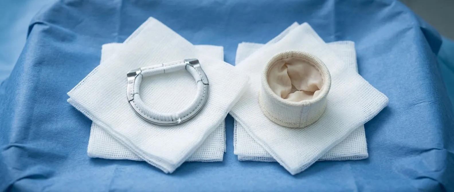
Every prosthetic valve — the medical term for an artificial or animal-tissue replacement valve — is foreign to your blood. Blood is designed to clot when it touches anything that is not your own tissue, and a replaced valve is exactly that. When platelets and clotting proteins build up on the valve surface, the result is prosthetic valve thrombosis: a clot that interferes with how the valve opens and closes.
Why mechanical valves are most affected. Mechanical valves, made of metal and carbon, require lifelong blood thinners precisely because they clot more easily. Tissue valves, including a Porcine Valve (made from pig tissue) or a Bovine Pericardial Valve (made from cow pericardium), clot less often, but they are not immune. Clot can also form on the small fabric ring that holds a tissue valve in place.
The subtle form: leaflet thrombosis. Not every clot is a dramatic blockage. A thin layer of clot can stiffen the Valve Cusp — the leaflet or door of the valve — restricting how far it opens without fully stopping it. This is called bioprosthetic valve thrombosis, and it often causes no symptoms at first. It is one reason routine follow-up imaging matters even when you feel well. Over time, it can contribute to Structural Valve Deterioration, the gradual wearing out of a tissue valve.
What Are the Symptoms of Mechanical Valve Thrombosis?
Mechanical valve thrombosis symptoms usually appear suddenly, which is what makes them frightening — and what makes fast action so effective. A valve that was working yesterday can be partly blocked today. The symptoms depend on which valve is affected and how much the clot restricts blood flow.
- Breathlessness that comes on quickly: the valve cannot open fully, so the heart struggles to move blood forward, and simple activities like walking to the kitchen feel exhausting.
- New or irregular heartbeat: many patients feel palpitations or a racing pulse as pressure builds behind the blocked valve.
- Signs of a stroke or embolism: weakness on one side, slurred speech, or sudden vision loss can occur if a piece of clot breaks off and travels to the brain or limbs.
- Coughing up blood or fainting: these suggest a serious obstruction and need emergency care immediately.
How do you know if your tricuspid valve is getting worse? For valves on the right side of the heart, such as after a Tricuspid Valve Repair, the warning signs are different. Look for swelling of the ankles and legs, abdominal bloating from fluid, and a full, heavy feeling in the neck veins. These come from blood backing up rather than from the valve failing to open, and they tend to develop more gradually.
The key message is this: a sudden change in how you feel after valve replacement is never something to wait out. A replaced valve that has worked well for years does not quietly stop mattering. Any new breathlessness, palpitation, or neurological symptom deserves same-day medical attention.
How Is Valve Thrombosis Diagnosed?
Valve thrombosis diagnosis starts with a story: a patient with a replaced valve describing sudden breathlessness or a change in symptoms. From there, the workup is straightforward and non-invasive in most cases.
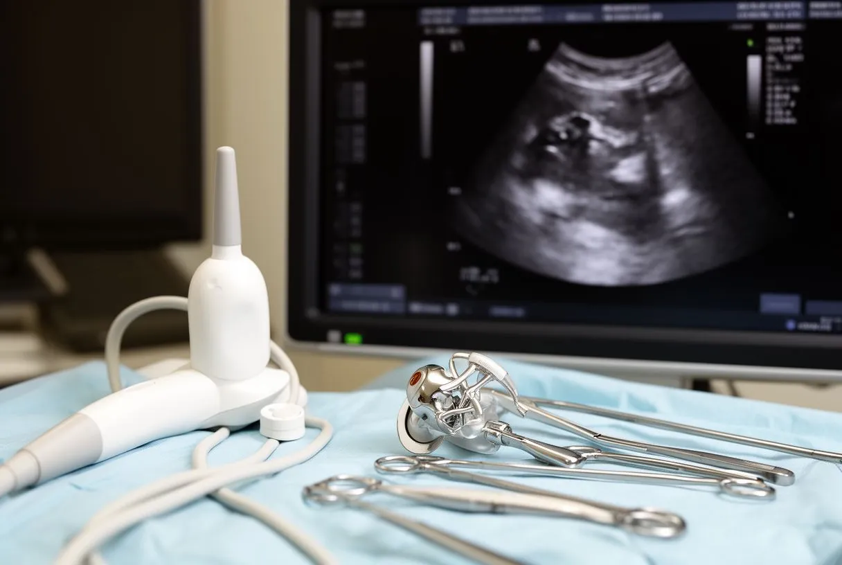
Echocardiography is the first test. An echocardiogram uses ultrasound to watch your valve move in real time. It shows whether the valve leaflets open fully, how fast blood flows through the valve, and whether the Valve Area — the opening space blood passes through — has shrunk. A narrowed valve area with restricted leaflet motion strongly suggests clot. Transesophageal echocardiography, where the probe is passed down the throat, gives a much clearer view than a chest scan and is usually the next step.
Fluoroscopy and CT imaging. Fluoroscopy is a moving X-ray that shows the valve's metal components opening and closing in rhythm. Reduced movement of one or both leaflets is a hallmark finding. A CT scan adds detail, especially for transcatheter valves, and can distinguish clot from other causes of valve dysfunction.
Ruling out the mimics. Not every blocked valve is a clot. Pannus — an overgrowth of scar tissue — can look identical on screening tests, and the treatment is different. Blood tests, including INR monitoring (a measure of how thin your blood thinner is working), help complete the picture. This is why an experienced valve team, not a single test, makes the diagnosis.
What Are the Main Valve Thrombosis Treatment Options?
There are three broad approaches to heart valve clot treatment, and choosing between them depends on the type of valve, how severe the blockage is, and how stable the patient is. In our practice, every case is reviewed by the multidisciplinary team against international practice guidelines before a recommendation is given, and the patient receives a clear proposal with the alternatives — the final decision is always theirs.
- Intensified anticoagulation: for mild or slow-onset clot, doctors adjust and closely monitor blood thinners, sometimes adding antiplatelet medication, and watch the valve with repeat imaging.
- Thrombolysis: clot-dissolving drugs delivered through an intravenous line can break up the obstruction without surgery — powerful, but with bleeding risk.
- Surgery or catheter intervention: for severe obstruction, emergency surgery to clean or replace the valve, or a catheter-based procedure to clear it, offers the most direct fix.
Stable versus urgent cases. If the clot is small and the patient is stable, many teams begin with medication and observe. If the patient is in heart failure, has had a stroke, or the valve is barely moving, waiting is dangerous. The rule most guidelines follow: the sicker the patient and the more obstructed the valve, the more aggressive the treatment.
What we see in practice. In the consultation we take the time to explain what the studies actually show, in plain words, and to answer every question before any decision. We encourage second opinions and treat seeking one as a normal part of deciding about heart surgery — especially when the choice is between urgent intervention and medical management.
When Is Thrombolysis Used for a Heart Valve Clot?
Thrombolysis means dissolving the clot with medication — usually a tissue plasminogen activator given through an intravenous line over hours. It avoids open-heart surgery, which matters enormously for patients who are elderly, frail, or too unstable for an operation.
The right patient for thrombolysis. It works best on clots that formed recently, on right-sided valves such as the tricuspid, and in patients who cannot safely undergo surgery. The published literature shows success rates that make it a legitimate first option in selected cases, and some studies of transcatheter valve dysfunction have compared medical, catheter, and surgical approaches head to head, including the EVAL registry comparing balloon-expandable and self-expanding valve designs in severe aortic stenosis (Journal of Clinical Medicine).
The trade-off is bleeding. Thrombolysis thins the blood throughout the whole body, not just at the valve. The main risks are bleeding, including bleeding around the heart or a stroke. For a patient with recent surgery, recent bleeding, or a brain at risk, this risk can outweigh the benefit.
What happens during treatment. Patients are monitored continuously in an intensive care setting while the drug is infused. Echocardiography is repeated to see whether the valve is opening better. If the clot does not respond, or bleeding occurs, the plan switches to surgery or catheter treatment. In our team's experience, deciding between these routes is exactly the kind of judgment that benefits from a multidisciplinary review rather than a single opinion.
When Does Valve Thrombosis Need Surgery?
Surgery is the most definitive valve thrombosis treatment, and some situations leave little choice about it.
Clear indications for emergency surgery. Guidelines and expert consensus point to surgery when the patient is in cardiogenic shock — the heart cannot pump enough to keep organs working — when the valve is almost completely blocked, when a clot keeps coming back despite medication, or when a floating clot threatens to break off and cause a stroke. Left-sided valves with large, mobile clots are the classic surgical case.
What the operation involves. The surgeon opens the heart, removes the clot, inspects the valve, and either keeps the existing valve if it is intact or replaces it. When replacement is needed, redo valve surgery carries higher risk than the first operation because of scar tissue, but outcomes in experienced centers are good. Alternatives to the original valve include an Intra-annular Valve, a Supra-annular Valve, or a Stentless Valve, each with different fit and flow characteristics — a conversation for a surgeon who knows your anatomy.
Minimally invasive alternatives. Not all surgical answers mean a full sternotomy. Our team performs conventional, minimally invasive, robotic and transcatheter procedures, and we recommend the less invasive route only where the evidence supports it for that patient. Dr Molina trained in cardiothoracic surgery at the Universidad de Guadalajara, in cardiac, minimally invasive, robotic and structural surgery at the University of Montreal, and in structural cardiology and TAVR at CHU Bordeaux — training that shapes how we weigh open surgery against catheter options for each individual.

How Does Anticoagulation After Valve Replacement Prevent Clots?
Anticoagulation after valve replacement is the single most important preventive measure a patient controls. Anticoagulants — blood thinners such as warfarin — slow the clotting process so a valve's surface does not accumulate clot.
Warfarin valve management and INR targets. Warfarin dosing is guided by the INR, a blood test that measures how long your blood takes to clot. Mechanical valves generally need a higher INR target than tissue valves. Too low, and clot risk rises; too high, and bleeding risk rises. This balance is why warfarin valve management is a lifelong partnership between patient and clinic, not a fixed prescription.
INR monitoring prosthetic valve routines. Patients check their INR regularly — weekly after any dose change, and on a stable schedule once therapeutic. Keep a written log. Bring it to every appointment. Skipping tests is a common, avoidable risk factor in patients who later present with valve clot.
What about newer blood thinners? Direct oral anticoagulants are convenient, but for mechanical valves and certain other situations, warfarin remains the standard because trials have raised safety concerns with the alternatives. Never stop or swap a blood thinner without your cardiac team's guidance. Johns Hopkins Medicine notes that anticoagulation management is a central part of long-term care after valve replacement (Johns Hopkins Medicine).
What Are the Big 5 Complications of TAVR?
Many patients receive their valve through TAVR — transcatheter aortic valve replacement — where a new valve is delivered through a catheter, usually from the groin, without opening the chest. TAVR has transformed care for aortic stenosis, but like every treatment it has complications worth knowing.
The five that matter most. The major risks are: stroke, from debris or clot dislodged during the procedure; vascular complications at the catheter entry site; bleeding; conduction disturbances that may require a pacemaker; and valve dysfunction, including transcatheter valve thrombosis. None of these are common in modern practice, but all are monitored.
Transcatheter valve thrombosis specifically. Clot on a transcatheter valve often behaves differently from clot on a mechanical valve: it is frequently the subtle leaflet-thrombosis type, seen on CT follow-up scans before it ever causes symptoms. Research programs, including studies comparing a balloon-expandable Valve with a Self-expanding Valve design, have examined how valve type and anticoagulation strategy influence this risk in severe aortic stenosis. Depending on the finding, treatment ranges from intensified anticoagulation to catheter or surgical intervention.
Follow-up protects your investment. Patients who had a Stented Valve, a Valve-in-valve procedure, or a Transcatheter Aortic Valve Implantation (the same procedure as TAVR) benefit from scheduled imaging. In our practice, the first contact is a free video consultation: we get to know the patient, talk through the condition, and say which studies are still needed so no unnecessary ones are ordered. Face-to-face follow-up at our consulting rooms in Guadalajara, Querétaro or Hermosillo follows when the patient decides to go ahead.
Can a Heart Valve Problem Be Fixed? Redo Surgery and Long-Term Outlook
The question patients ask most is simple: can a heart valve problem be fixed? For valve thrombosis, the honest answer is yes — in most cases the valve is either cleared of clot or replaced, and patients return to good quality of life. The outlook depends on how quickly treatment begins and on the patient's overall condition.
Redo valve surgery. A second or third valve operation is more complex than the first because scar tissue makes dissection harder. Still, outcomes in dedicated centers are consistently good, and for patients with a completely blocked valve, surgery can be lifesaving. Some patients are candidates for a valve-sparing approach — a Valve-sparing Root Replacement preserves the patient's own valve and root when the anatomy allows, avoiding prosthesis issues entirely.
Choosing the right prosthesis matters for the future. The differences between valve types — mechanical versus tissue, stented versus stentless, balloon-expandable versus self-expanding — affect both clot risk and future options. A mechanical valve demands strict lifelong anticoagulation. A tissue valve offers freedom from warfarin for many patients but carries the longer-term risk of structural deterioration. When we review a case, we match the prosthesis to the patient's age, lifestyle, and ability to manage blood thinners — not the other way around.
Life expectancy questions. Patients also ask about calcification of their own valve, such as what the outlook is with aortic valve calcification. Calcification progresses slowly in many people and only requires intervention when the valve becomes severely narrowed and symptomatic — decisions driven by guidelines, not fear. If your own valve is calcified but not yet severe, surveillance rather than surgery is often the right call, and a proper evaluation will tell you which side of that line you are on.

Living With a Replaced Valve: Prevention and When to Seek Help
Prevention is where patients hold the most power over valve replacement complications. The habits that protect a replaced valve are unglamorous but proven.
The daily disciplines that count.
- Take your anticoagulant exactly as prescribed and never miss or double doses without medical advice.
- Keep every INR appointment and record your results in a log your team can review.
- Tell your dentist and any doctor about your valve before procedures — antibiotics to prevent Prosthetic Valve Endocarditis may be recommended, and infection on a valve makes clot more likely.
- Report new symptoms the same day — sudden breathlessness, palpitations, or any stroke-like sign.
When to seek emergency help. Go to an emergency department immediately for sudden severe breathlessness, fainting, chest pain, coughing up blood, or any stroke symptoms such as facial droop or one-sided weakness. Time matters enormously with a blocked valve: hours make a difference between medication alone and needing major surgery. In these situations, do not wait until morning.
A word about your team. Our multidisciplinary team of cardiac surgeons, led by Dr Alejandro Molina, reviews every case together before making a recommendation, because valve decisions are rarely one-specialty decisions. If you have a replaced valve and are unsure about your follow-up plan, or if you have symptoms that concern you, we invite you to book a consultation — it starts with a free video visit where we listen first. Valve thrombosis is treatable, and the best results belong to patients who know the warning signs and act on them.