

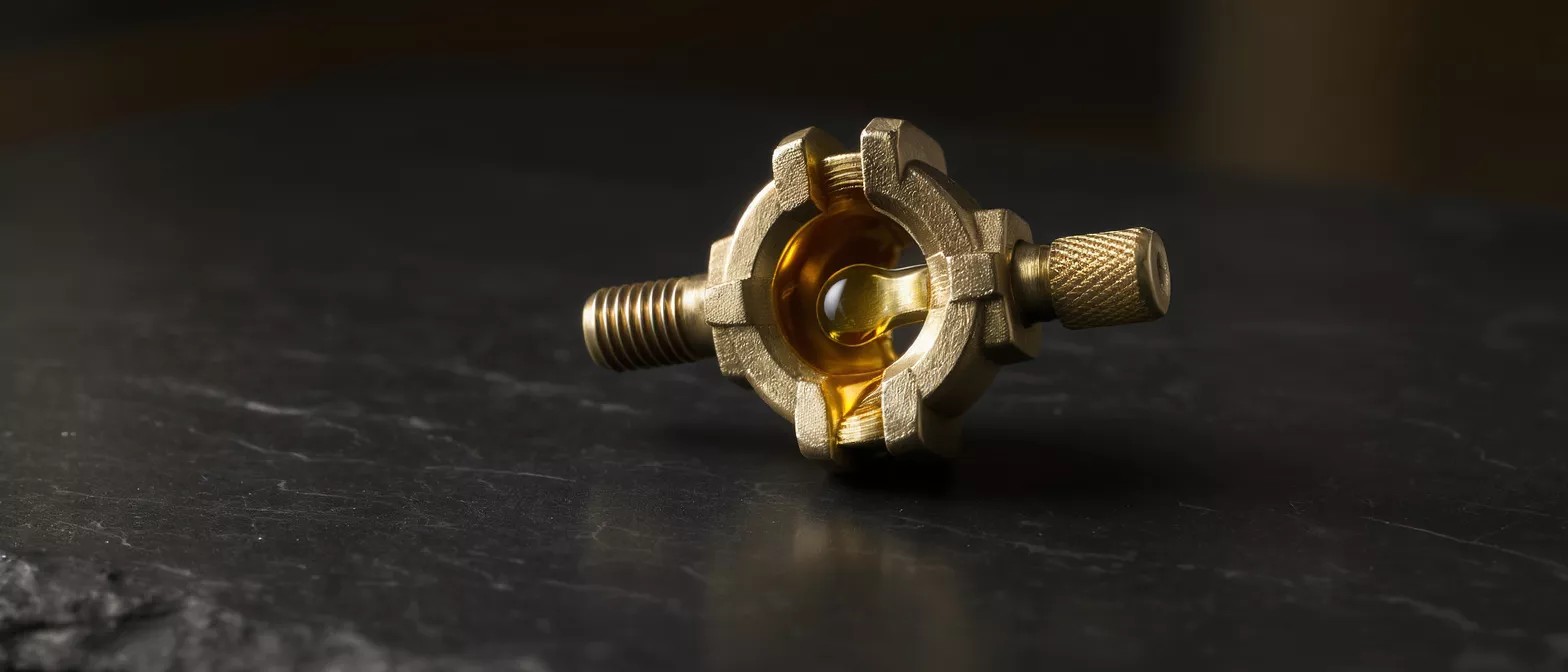
Valve Thrombosis
Valve thrombosis can complicate recovery after heart surgery.
What is Valve Thrombosis?
Valve thrombosis occurs when a blood clot forms on or near one of the valves inside your heart. This can happen on a natural heart valve, but it is most commonly discussed in the context of prosthetic valve thrombosis, where a clot develops on a surgically implanted replacement valve. When a clot restricts the movement of the valve leaflets, it prevents the valve from opening and closing normally, which disrupts healthy blood flow. For patients recovering from heart surgery, understanding this condition is a critical part of long-term cardiac care.
Normally, blood flows smoothly through the heart's chambers and valves without clotting. However, artificial materials used in mechanical or biological replacement valves can sometimes trigger the body's natural clotting cascade. If a clot begins to form on the valve structure, it can gradually obstruct the blood flow or, in severe cases, break loose and travel to other parts of the body. This is why continuous medical monitoring and proper medication are essential after a valve replacement procedure.
It is important to know that a thrombosed heart valve is a recognized complication that cardiologists and surgeons are specifically trained to prevent and manage. By maintaining regular follow-ups and following your prescribed medication plan, the risk of a dangerous clot forming is significantly reduced. Understanding the mechanics of this condition helps patients recognize the importance of their ongoing cardiac care.
Why Valve Thrombosis Matters After Heart Surgery
Impact on prosthetic valves: Following a valve replacement, your heart is adapting to a new mechanical or biological structure. Because the body recognizes artificial valves as foreign surfaces, blood cells are more prone to stick to them, making valve thrombosis after valve replacement a primary concern for cardiac surgeons. Without proper management, this biological response can compromise the success of the surgery.
Effect on blood flow dynamics: When a clot forms on a prosthetic valve, it physically blocks the valve from opening fully or closing tightly. This obstruction forces the heart to work much harder to pump blood, which can lead to heart muscle strain, fluid buildup in the lungs, and reduced oxygen delivery to the rest of the body. Restoring normal blood flow quickly is the central goal of valve thrombosis management.
Understanding these risks does not mean you should live in constant fear. Instead, it highlights why your cardiac team emphasizes strict medication adherence and routine echocardiograms. When patients understand the direct relationship between their daily care routine and the longevity of their prosthetic valve, they are better equipped to ensure a successful, long-term recovery.
Recognizing Valve Thrombosis Symptoms
Identifying valve thrombosis symptoms early can be life-saving. Because a clot restricts blood flow through the heart, the body often presents clear warning signs that something is wrong. Patients may notice a sudden change in their exercise tolerance, feeling unusually breathless during activities that were previously easy. This shortness of breath, known as dyspnea, is often one of the first indicators of a restricted valve.
Other common symptoms include a sudden onset of fatigue, dizziness, or a noticeable change in the sound of your heartbeat, sometimes described as a new or different murmur. If you have a mechanical valve, you might notice a change in the familiar clicking sound it makes. In more severe cases, if a piece of the clot breaks off and travels to the brain, it can cause symptoms of a stroke, such as sudden weakness or numbness on one side of the body.
Because these symptoms can overlap with other cardiac or respiratory conditions, sudden changes in your health should never be ignored. If you experience any acute shortness of breath, chest pain, or neurological symptoms, seeking valve thrombosis emergency care immediately is crucial to preventing permanent damage.
- Shortness of breath: Sudden difficulty breathing, especially during routine activity.
- Neurological changes: Sudden weakness, numbness, or difficulty speaking, which may indicate a traveling clot.
- Audible changes: A muffled or altered clicking sound from a mechanical valve.
Valve Thrombosis Risk Factors
Inadequate anticoagulation: The single most significant risk factor for mechanical valve thrombosis is failing to take blood thinners exactly as prescribed. Anticoagulants, often called blood thinners, are specifically designed to prevent clots from forming on the artificial materials of the valve. Missing doses or taking incorrect amounts leaves the valve vulnerable to clot formation.
Underlying medical conditions: Certain health conditions can increase the likelihood of blood clots developing. Atrial fibrillation, an irregular heart rhythm, can cause blood to pool in the heart chambers, increasing clotting risks. Similarly, conditions that affect blood clotting naturally, such as genetic thrombophilias, or lifestyle factors like smoking and dehydration, can significantly raise the risk of a thrombosed heart valve.
Recognizing your personal risk factors allows you and your medical team to tailor your care plan effectively. By managing underlying conditions and maintaining strict adherence to your medication schedule, you can actively participate in mitigating valve thrombosis risk factors and protecting your new heart valve.
How Valve Thrombosis Diagnosis Works
When valve thrombosis is suspected, time is of the essence, and accurate imaging is critical. Valve thrombosis diagnosis almost always begins with an echocardiogram, which uses sound waves to create detailed images of the heart's structures. This non-invasive test allows cardiologists to visualize the valve in real-time, assessing how well it opens and closes and identifying any obstructing masses.
Depending on the location of the valve and the clarity of the images, your doctor may order a transthoracic echocardiogram (TTE) performed on the chest, or a transesophageal echocardiogram (TEE) where a specialized probe is guided down the esophagus to get a closer look at the heart. A TEE provides highly detailed images and is often the gold standard for confirming the presence of a clot on a prosthetic valve. In some cases, advanced imaging like a CT scan or fluoroscopy may be utilized to assess valve mobility.
The diagnostic process is designed to be as swift and precise as possible. Once the presence, size, and location of the clot are confirmed, the cardiac team can immediately pivot to valve thrombosis management, determining whether medical treatment or surgical intervention is the safest approach.
Anticoagulation for Valve Thrombosis
When a clot is detected early and is not causing severe obstruction, doctors often opt for medical valve thrombosis management using anticoagulation for valve thrombosis. This approach involves administering fast-acting blood thinners, typically intravenously in a hospital setting, to help the body naturally dissolve the clot over time. This therapy requires close monitoring to ensure the blood thins enough to dissolve the clot without causing bleeding complications.
For patients with mechanical valves, long-term anticoagulation is a standard part of life, usually managed with medications like warfarin. If a clot forms despite this therapy, the medical team will reassess the dosage, potentially switching to a more aggressive anticoagulation protocol or utilizing intravenous heparin to stabilize the valve. The goal is to restore normal blood flow safely without requiring additional surgery.
Medical management is highly effective for smaller, non-obstructive clots. During this treatment phase, your care team may recommend a continuous heart rhythm monitor to ensure your heart is maintaining a safe and stable pace as the clot resolves. This careful, non-surgical approach often allows patients to recover without the need for repeat operations.
Surgical Valve Thrombosis Treatment
Emergency surgical intervention: When a clot is large, severely obstructs blood flow, or fails to dissolve with anticoagulation therapy, surgical valve thrombosis treatment becomes necessary. In acute cases, surgeons may perform an emergency operation to physically remove the clot from the valve, a procedure known as a thrombectomy. This approach is often required if the patient is experiencing severe heart failure or shock.
Valve replacement surgery: In situations where the prosthetic valve itself has been damaged by the clot or cannot be safely cleaned, the surgeon will replace the thrombosed valve entirely. This decision is typically made in the operating room, where the cardiac team assesses the structural integrity of the valve once they have direct visual access. A specialized cardiac surgical consultation is vital to determine if a new mechanical or biological valve is the best choice for the patient's lifestyle.
While the prospect of a repeat surgery is daunting, modern surgical techniques have made these procedures highly effective. Your surgical team will carefully evaluate the risks and benefits, ensuring that the chosen treatment path offers the best long-term outcome for your cardiac health and overall quality of life.
Valve Thrombosis Prevention
Preventing a thrombosed heart valve is always preferable to treating one. The cornerstone of valve thrombosis prevention is strict adherence to your prescribed anticoagulation therapy. Taking your blood thinners at the exact same time every day, never skipping a dose, and attending all scheduled blood tests ensures your medication levels remain in the safe therapeutic range. This daily routine is your primary defense against clot formation.
Beyond medication, lifestyle factors play a significant role in prevention. Staying well-hydrated helps maintain healthy blood viscosity, while avoiding smoking is critical, as tobacco use increases blood clotting risks. Your cardiologist will also provide specific dietary guidelines, as certain foods high in vitamin K can interfere with medications like warfarin, altering its blood-thinning effects.
Integrating these daily habits protects your valve and your heart. For patients looking to support their overall cardiovascular wellness, exploring a complete cardiovascular wellness support routine can be an excellent complement to medical therapies, helping to maintain optimal blood flow and heart strength.
Valve Thrombosis Complications
If left untreated, valve thrombosis complications can be severe and life-threatening. A completely obstructed valve prevents blood from exiting the heart, leading to acute heart failure, a dangerous buildup of fluid in the lungs, and cardiogenic shock. This is why prompt recognition of valve thrombosis symptoms and immediate medical intervention are so critical to survival.
Another major risk is embolization, where a piece of the clot breaks away from the valve and travels through the bloodstream. This traveling clot can lodge in the brain, causing an ischemic stroke, or block blood flow to other vital organs, causing significant damage. The risk of these complications underscores the necessity of regular monitoring and immediate reporting of any new symptoms to your cardiac care team.
Fortunately, with vigilant care, these severe outcomes are rare. Understanding the potential complications reinforces the importance of taking your cardiac health seriously and trusting your medical team to guide your recovery.
- Acute heart failure: The heart cannot pump effectively due to the obstructed valve.
- Thromboembolic stroke: A traveling clot blocks blood flow to the brain.
- Cardiogenic shock: A life-threatening state where the heart suddenly fails to pump enough blood.
Valve Thrombosis Emergency Care and Follow-Up
Navigating life after a valve replacement requires a commitment to ongoing cardiac care. If you ever suspect valve thrombosis emergency care is needed, do not wait to see if symptoms improve. Immediate transportation to an emergency department, particularly one with a specialized cardiac unit, ensures that rapid diagnosis and treatment can begin without delay.
Long-term follow-up is the safety net that keeps your heart functioning optimally. Routine valve thrombosis echocardiography allows your cardiologist to visualize the valve, ensuring it is moving freely and that no small, asymptomatic clots are beginning to form. These regular check-ins allow your medical team to adjust your medications long before a physical symptom ever appears.
By combining strict daily medication adherence, lifestyle awareness, and consistent medical follow-up, patients can live full, active lives after heart surgery. Valve thrombosis is a serious condition, but with the right knowledge and a dedicated cardiac team, it is one that can be effectively prevented and successfully managed.






