

Fluoroscopy
Learn how fluoroscopy uses real-time X-ray imaging to guide cardiac procedures.
What is fluoroscopy?
When facing a cardiac procedure, unfamiliar terminology can feel intimidating. Real-time X-ray guidance is the simplest way to understand this technology. Fluoroscopy uses a continuous X-ray beam passed through the body to create moving images on a monitor, much like an X-ray movie. Instead of taking a single static picture, it allows your surgical team to see your heart, blood vessels, and medical devices in motion. For patients undergoing treatment at Cirugía de Corazón, this tool is a fundamental part of modern cardiac care.
The technology works by sending X-rays through your body to a fluorescent screen or digital detector. Dense tissues like bone appear white, while softer tissues and blood vessels appear in varying shades of gray. Live cardiac visualization gives cardiologists and surgeons the ability to track the movement of catheters, guide wires, and stents with incredible precision. This continuous feedback is what separates it from standard imaging techniques.
For a frightened patient or family member, knowing that the surgical team can see exactly what is happening inside the heart provides immense reassurance. It transforms complex procedures from blind interventions into highly controlled, visual operations.
How fluoroscopy works in cardiac procedures
Tracking catheters in real time is the primary function of this imaging tool during heart interventions. A specialist will position a specialized camera, called a C-arm, over the patient's chest. This device can rotate around the operating table to capture the heart from multiple angles, feeding those live images directly to monitors in the surgical suite.
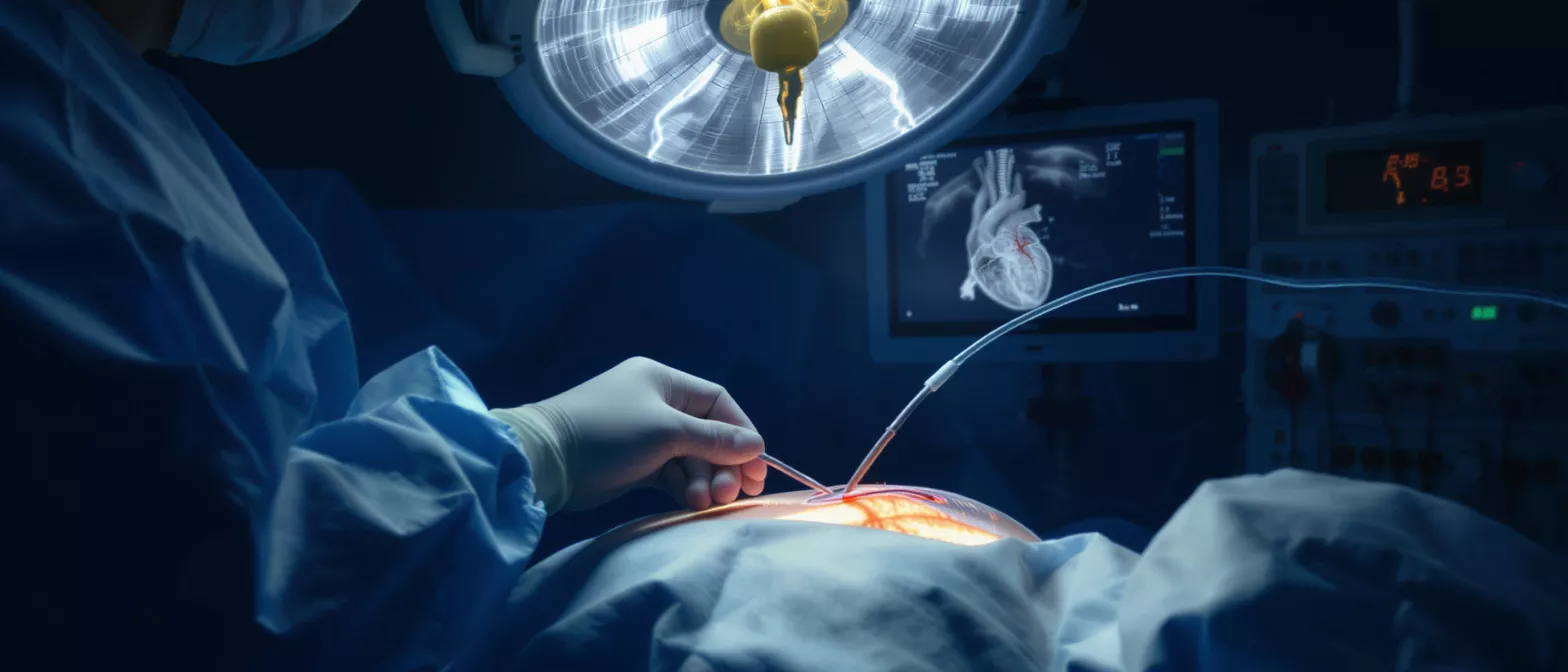
The procedure room is equipped with large displays that show the inside of the body as the surgery progresses. Guided surgical precision relies on this continuous visual feedback loop. If a surgeon is performing a coronary angiogram to look for blockages, they can watch the contrast dye flow through the arteries in real time, instantly identifying the exact location and severity of a narrowed vessel.
By providing a clear roadmap of the vascular system, the technology minimizes the need for larger, more invasive incisions. It allows doctors to navigate complex cardiac anatomy safely and effectively.
Why fluoroscopy matters for heart surgery patients
Minimally invasive heart care would not be possible without this advanced imaging technique. For heart surgery patients, the ability to see inside the body without making large surgical openings means smaller incisions, less pain, and typically faster recovery times. It is the cornerstone of transcatheter valve replacements and coronary interventions.
Ensuring accurate device placement is another critical benefit. When placing a stent to hold an artery open or deploying an artificial heart valve, the margin for error is incredibly small. Fluoroscopy provides the magnified, high-resolution view required to confirm that these life-saving devices are seated perfectly before the procedure concludes.
Ultimately, this tool bridges the gap between surgical intention and physical reality. It allows your medical team to execute delicate maneuvers with confidence, directly translating to better patient outcomes and a smoother healing process.
Fluoroscopy vs standard X-ray differences
Static images versus moving pictures is the easiest way to distinguish the two. A standard X-ray takes a single, stationary snapshot of an internal structure, much like a photograph. It is excellent for finding bone fractures or checking the static position of a device, but it cannot show how blood is flowing or how a heart valve is opening and closing.
Fluoroscopy, by contrast, captures continuous internal movement. Continuous vascular monitoring allows doctors to see the dynamic flow of contrast dye, identifying leaks, blockages, or abnormal blood flow patterns that a static image would entirely miss. To manage the unique physical stress of cardiac procedures, the medical team relies on specialized tools; similarly, patients preparing for surgery might benefit from a pre-surgery wellness booster.
While both use X-ray radiation, the delivery method and clinical purpose are distinct. The real-time capability is what makes live imaging indispensable for guiding tools through the delicate pathways of the cardiovascular system.
What to expect during a fluoroscopy procedure
Preparation for cardiac imaging is generally straightforward. If you are having a fluoroscopy-guided procedure, the clinical staff will position you on a specialized table. The camera may move around you, but you will not feel the X-rays passing through your body. The room might be dimmed so the medical team can see the monitors clearly.
Depending on the specific intervention, you may receive a local anesthetic or light sedation. Staying still during the scan is important, as sudden movements can blur the live images the surgeon relies on. The team will communicate with you throughout the process, letting you know when to hold your breath or stay perfectly still. Here are a few things to keep in mind:
- Communication: The surgical team will guide you step-by-step.
- Contrast dye: You might feel a brief warm sensation if dye is used.
- Movement: Remaining still ensures the clearest possible images.
Knowing what happens in the room removes the fear of the unknown. The sounds you hear and the movements of the equipment are all normal parts of capturing high-quality live images.
Is fluoroscopy safe for cardiac patients?
Monitoring radiation exposure is a top priority for any cardiac surgical team. Because this technology relies on X-rays, it does involve some level of radiation. However, modern equipment is highly optimized to deliver the lowest possible dose needed to achieve clear images. Your doctor will only use it when the clinical benefits far outweigh the minimal risks.
Balancing risks and surgical benefits is a careful calculation made by your cardiologist. The ability to safely navigate a catheter through your coronary arteries without opening your chest is a massive advantage that heavily tips the safety scale in favor of using live imaging. To minimize risk, the team uses lead shielding and precise collimation to narrow the X-ray beam strictly to the area of interest.
If you have concerns about radiation, it is entirely appropriate to discuss them with your physician before the procedure. They can explain exactly why the imaging is necessary and how they plan to keep your exposure to a minimum.
Understanding fluoroscopy radiation dose
Optimizing dose for heart patients involves sophisticated technology built directly into the imaging equipment. Modern cardiac fluoroscopy uses pulsed X-rays, which turn the beam on and off rapidly, significantly reducing the total radiation delivered compared to older continuous-beam machines. The dose is carefully tracked throughout the procedure.
Low-dose imaging protocols are standard practice in contemporary cardiac care. The radiation you receive during a typical catheterization or guided intervention is generally low. To put it in perspective, the exposure is often comparable to a few months of natural background radiation we all experience from the environment. Key safety measures include:
- Time: Keeping the X-ray beam active for the shortest duration necessary.
- Distance: Using precise zoom and positioning to target only the heart.
- Shielding: Protecting surrounding tissues with lead garments and barriers.
Your surgical team is highly trained in radiation safety. They constantly balance the need for crystal-clear visualization with the strict mandate to protect your long-term health.
Fluoroscopy in the operating room
Hybrid operating suites represent the pinnacle of modern cardiac surgical environments. These specialized rooms combine the sterile requirements of a traditional operating room with the advanced imaging capabilities of a cardiac catheterization lab. This integration allows surgeons to seamlessly switch between open surgical techniques and minimally invasive, image-guided interventions.
Surgical team coordination is vastly improved when live imaging is available in the operating room. The surgeon, anesthesiologist, and nursing staff can all view the same real-time images, ensuring everyone is perfectly aligned on the progress of the procedure. If an unexpected anatomical challenge arises, the team can immediately assess it without delaying the surgery.
Having this technology directly in the operating room means patients do not need to be transported to a different imaging department. It streamlines the entire surgical experience and enhances overall safety.
Contrast dye in fluoroscopy imaging
Highlighting blood vessels is the primary role of contrast dye in live X-ray imaging. Blood vessels are normally difficult to see clearly on an X-ray because they blend in with surrounding soft tissues. A special iodine-based dye is injected into the bloodstream, making the vessels appear starkly dark or bright against the anatomy, depending on the imaging mode used.
Coronary angiogram visualization relies entirely on this contrast material. As the dye flows through the coronary arteries, the fluoroscopy monitor captures its path, revealing exactly where blood flows freely and where blockages restrict the passage. This allows the surgeon to map the vascular system with absolute accuracy before placing a stent or performing a bypass.
It is important to tell your doctor if you have any kidney conditions or previous allergic reactions to contrast materials. They can take specific precautions to ensure your safety during the imaging process.
Fluoroscopy recovery and key takeaways
Post-procedure cardiac care focuses on the specific intervention you received rather than the imaging itself. Because the X-rays leave no residual radiation in your body, there is no specific recovery time required just for the fluoroscopy. You will recover from the sedation or the surgical incision, but the live imaging leaves no lasting physical trace.
Visualizing successful outcomes is the final benefit of this technology. Before concluding the procedure, the surgical team uses live imaging to confirm that blood flow is restored, a valve is functioning properly, or a device is securely in place. This immediate visual confirmation provides both the doctor and the patient with peace of mind, knowing the intervention was successful before leaving the operating suite.
Understanding the tools your cardiac team uses empowers you to approach your heart surgery with confidence. This real-time imaging is a vital, safe, and highly effective component of modern cardiovascular care, ensuring your procedure is guided by clear vision every step of the way.






