

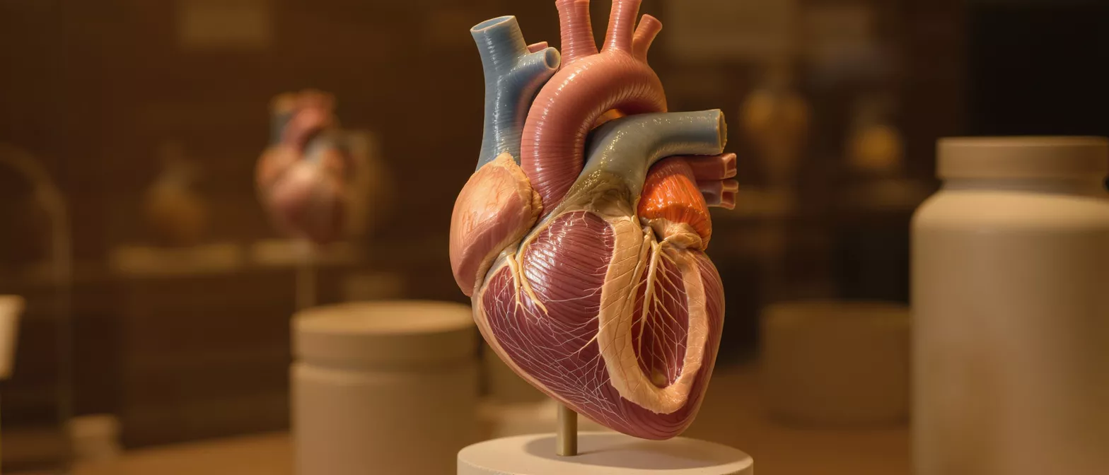
Papillary Muscle
Learn what a papillary muscle does for your heart valves, how dysfunction is diagnosed, and treatment options available.
What is a papillary muscle?
A papillary muscle is a small, pillar-like bundle of muscle tissue located deep inside the ventricles, which are the lower pumping chambers of the heart. When patients first hear this term while preparing for heart surgery, it can sound highly technical, but the concept is straightforward. These muscles act as sturdy anchors, securing the delicate strings that hold your heart valves in place. Understanding the papillary muscle definition is the first step in realizing how the heart maintains unidirectional blood flow.
By securing the valve flaps at exactly the right moment, they ensure that blood moves forward efficiently rather than washing backward. Recognizing what this structure does helps demystify why surgeons pay such close attention to it during valve operations and post-operative care.
Where is it located in the heart? These muscles project upward from the inner walls of the left and right ventricles. The left ventricular papillary muscle is particularly critical, as it supports the mitral valve under high pressure. They are connected to the valve leaflets by thin, fibrous threads called chordae tendineae. When you look at heart anatomy diagrams, these cords are often compared to the strings of a parachute, with the papillary muscle acting as the central anchor holding the canopy open and stable.
How does papillary muscle function support heart valves?
Papillary muscle function is entirely mechanical, relying on precise physical contractions to protect the heart's valves. Every time the ventricle squeezes to push blood out into the body, the pressure inside that chamber rises dramatically. Without a reliable anchor, the force of this pressurized blood would push the valve leaflets backward into the upper chamber, causing a dangerous backward leak. The muscle contracts exactly in sync with the rest of the ventricle, pulling the valve strings taut just before the pressure peaks, holding the leaflets firmly shut.
Why is timing so critical for heart valve support? The electrical system of the heart ensures the muscle tenses up a fraction of a second before the main pumping action pushes blood forward. This holds the valve tightly closed at the exact moment it experiences the highest force. If the muscle does not contract properly, the valve leaks, and blood flows backward, forcing the heart to work twice as hard to manage the same volume of blood.
Proper function keeps the circulatory system efficient, preventing unnecessary strain on the cardiac muscle and preserving healthy blood flow to the lungs and body.
What is papillary muscle anatomy in the left ventricle?
Papillary muscle anatomy in the left ventricle involves specific structural layouts that surgeons must carefully navigate during heart operations. There are typically two main groups of these muscles in the left ventricle, known as the anterolateral and posteromedial papillary muscles. Each group sends out a complex network of fibrous cords to the mitral valve leaflets, securing them against the high pressure generated by the left side of the heart.
The right ventricle also contains these muscles, usually grouped into three smaller bundles that support the tricuspid valve, though they are less commonly the focus of surgical intervention. The left ventricular papillary muscle is generally the primary focus in cardiac care because the left side of the heart generates significantly more pressure, making any structural weakness here far more impactful.
How does anatomy affect surgical planning? Knowing the exact layout of these muscles helps surgeons determine how to approach valve repairs, ensuring they preserve the natural anchoring system while restoring normal blood flow.
What causes papillary muscle dysfunction or ischemia?
Papillary muscle dysfunction usually occurs when the blood supply to the muscle is compromised, preventing it from contracting with enough strength to hold the valve closed. This lack of blood flow is known as papillary muscle ischemia, a condition where the heart tissue is starved of oxygen. The coronary arteries that supply these specific muscles are often smaller and more vulnerable to blockages than the main arteries feeding the rest of the heart wall.
Can ischemia cause valve leakage? Yes, when these muscles suffer from ischemia, they become temporarily weakened. Even a brief reduction in oxygen can cause the muscle to fail in its anchoring role, resulting in a condition called functional mitral regurgitation. The valve leaflets droop or fail to meet correctly, allowing blood to leak backward into the left atrium.
Addressing the underlying ischemia through medical management or revascularization procedures often restores normal oxygen delivery, allowing the muscle to regain its strength and function over time.
What is papillary muscle rupture after a heart attack?
Papillary muscle rupture is a severe but rare complication that can occur following a massive heart attack. If a blockage completely cuts off the blood supply to the muscle for an extended period, the tissue eventually dies. The muscle tissue weakens until it physically tears away from the ventricular wall. When the anchor breaks, the valve strings go slack, and the mitral valve instantly fails, causing severe, acute mitral regurgitation.
This failure leads to a sudden and catastrophic backward flow of blood, often resulting in severe shortness of breath and fluid pooling in the lungs. This situation requires immediate surgical intervention to repair or replace the valve.
How is a rupture treated? Surgeons address a rupture by repairing the torn muscle, reattaching it securely, or replacing the entire mitral valve with a mechanical or biological prosthesis. For patients recovering from severe valve complications, careful monitoring is essential, and a doctor might recommend a home blood pressure monitor for post-surgery care to keep precise records of cardiovascular stability during the healing process.
How does papillary muscle dysfunction appear on echocardiography?
Papillary muscle dysfunction echocardiography is the primary tool cardiologists use to visualize how well these muscles and their attached valves are working. An echocardiogram uses sound waves to create a real-time, moving picture of the heart, allowing physicians to observe the movement of the valve leaflets and the contraction of the ventricular walls.
What do doctors look for on the scan? Doctors look for specific signs of structural stress, such as leaflets that do not meet perfectly in the center or cords that appear unusually slack during the heartbeat. Doppler imaging highlights backward-moving blood flow, instantly revealing valve leakage.
This diagnostic test is entirely non-invasive and painless, providing critical insights without requiring any surgical intervention or prolonged hospital stay. It remains the gold standard for diagnosing valve issues related to muscle dysfunction.
What are papillary muscle abnormalities and symptoms?
Papillary muscle abnormalities range from congenital defects, where the muscle developed unusually before birth, to acquired conditions stemming from previous heart damage or chronic disease. These structural changes can alter the resting tension of the valve cords, leading to chronic leakage or improper valve opening. Some individuals may be entirely unaware of minor structural anomalies until a routine physical reveals a heart murmur.
When do symptoms appear? Symptoms usually present when the valve leakage becomes significant enough to disrupt normal circulation. Patients might experience unexplained fatigue, a rapid or irregular heartbeat, or shortness of breath, particularly when lying flat or exerting themselves physically. If the heart cannot push blood forward efficiently, fluid can build up in the lungs, causing a persistent cough or a feeling of breathlessness.
Recognizing these warning signs early is vital for preventing long-term damage to the heart muscle and maintaining overall cardiovascular health.
When is papillary muscle treatment or repair needed?
Papillary muscle treatment becomes necessary when dysfunction or structural damage severely limits the heart's ability to pump blood efficiently. For mild cases of ischemia, medications that reduce the workload of the heart or lower blood pressure may be sufficient to allow the muscle to heal and recover its function. When the damage is structural, surgical intervention is the only way to restore proper valve mechanics.
What does surgical repair involve? Surgeons evaluate the extent of the damage before deciding whether to repair the existing structure or replace the valve entirely. Reconstructive papillary muscle repair aims to tighten or shorten the existing cords, restoring the natural anchor points without removing the native tissue.
- Medication: Used for mild dysfunction to reduce cardiac workload and manage ischemia.
- Valve repair: Tightening or reattaching existing structures to preserve native anatomy.
- Valve replacement: Utilized when the muscle is irreversibly damaged or ruptured.
How long does recovery from papillary muscle repair take?
Recovery from papillary muscle repair depends heavily on the patient's overall health, the extent of the structural damage, and whether the surgery was minimally invasive or required a traditional open approach. In the first few weeks following the procedure, patients focus on resting, allowing the breastbone and chest incisions to heal. Breathing exercises and controlled physical movement help prevent fluid buildup and maintain lung capacity.
Full recovery generally takes several months, with a structured cardiac rehabilitation program to gradually rebuild cardiovascular strength under professional supervision.
What is the long-term outlook? The long-term outlook for patients who undergo successful repair is generally excellent, with most experiencing a return to normal, active lifestyles. Maintaining follow-up appointments ensures the repaired structures remain stable over time.
Understanding your heart health and next steps
Navigating a heart surgery diagnosis can feel overwhelming, especially when confronting complex anatomical terms and conditions. Understanding how the papillary muscle supports heart valves demystifies the surgical process and empowers you to engage actively in your own recovery. By recognizing the signs of ischemia, dysfunction, and structural abnormalities, you can better appreciate the careful planning your surgical team undertakes to restore your heart's natural mechanics.
How can family members support recovery? Family members play a crucial role in the post-operative journey by helping monitor symptoms, managing medications, and encouraging gentle physical movement. The emotional and physical support of loved ones significantly impacts the speed and quality of healing after cardiac intervention.
Equipped with this knowledge, you can approach your treatment journey with confidence, knowing that these structures, though small, play a monumental role in your long-term cardiovascular wellness.
If you have concerns about your valve function or recovery plan, always consult your cardiologist or surgical team for personalized guidance tailored to your specific medical history and current condition.






