

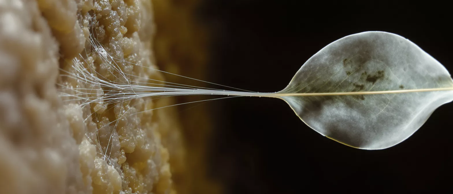
Chordae Tendineae
Discover chordae tendineae, the heart's critical cords anchoring valve leaflets.
What Are Chordae Tendineae?
The chordae tendineae are delicate, tendon-like cords inside the heart that connect the valve leaflets to the papillary muscles. When medical professionals talk about "heart strings," they are referring to these precise structures. Understanding the chordae tendineae definition helps patients grasp how the heart maintains unidirectional blood flow, ensuring that blood moves forward efficiently without leaking backward into the upper chambers.
Chordae tendineae medical term meaning: The phrase translates from Latin as "tendinous cords," reflecting their thin, tendon-like appearance. Despite their fragile look, these structures are incredibly strong and resilient, engineered by nature to withstand the high-pressure forces generated with every single heartbeat.
For patients facing cardiac surgery, knowing what these cords do provides peace of mind. They are the essential anchors of the heart's lower valves, playing a critical role in maintaining cardiovascular stability and preventing the mechanical failure of valve closure. Surgeons prioritize preserving or reconstructing these structures to maintain the heart's natural geometry.
How Chordae Tendineae Function in Heart Valves
The primary chordae tendineae function is to prevent the heart valve leaflets from prolapsing into the atria when the ventricles contract. As the lower chambers squeeze blood out to the lungs and body, the pressure pushes against the closed valves. The chordae act like the tethers on a parachute, holding the valve flaps tightly shut at the exact right moment to ensure a perfect seal.
Heart strings anatomy in motion: During ventricular contraction, the papillary muscles also contract, pulling the chordae taut. This synchronized mechanical event prevents blood from flowing backward. It is a perfectly timed, highly coordinated physical process that happens thousands of times a day without conscious effort.
When this tethering system works correctly, blood flows seamlessly from the atria to the ventricles, and then out to the rest of the body. If the cords fail, the valve fails, leading to significant cardiac stress and requiring prompt clinical attention to restore normal hemodynamics.
Chordae Tendineae Anatomy and Papillary Muscles
Chordae tendineae anatomy is directly linked to the papillary muscles, which are small muscular projections located on the inner walls of the ventricles. These muscles anchor the strings, providing the tension necessary to hold the valve leaflets in place. Without this muscular support, the cords would have no source of tension.
Papillary muscles and chordae tendineae synergy: The relationship between these two structures is entirely interdependent. The muscles generate the active force, while the tendons provide the passive structural connection to the valve. In cardiac surgery, maintaining this anatomical relationship is crucial for long-term valve repair durability. Surgeons use specialized annuloplasty repair rings to support the valve structure alongside the natural chords.
Understanding this anatomy helps patients see why surgical techniques focus so heavily on reconstruction rather than simple removal. Preserving the natural attachments keeps the heart functioning as closely to its original design as possible.
What Happens When Chordae Tendineae Rupture?
A chordae tendineae rupture occurs when one or more of these strong cords snaps, often due to trauma, infection, or age-related degeneration. When this happens, the valve leaflet is no longer tethered correctly. It will flop backward into the atrium during contraction, allowing blood to leak backward in the wrong direction.
Broken heart strings condition symptoms: Patients may suddenly experience shortness of breath, fatigue, or a rapid heart rate. This mechanical failure leads to a regurgitation of blood, meaning the heart has to work much harder to pump the same volume forward. The sudden onset of symptoms usually prompts immediate medical evaluation and echocardiography.
If left untreated, a rupture can lead to severe heart failure. The good news is that modern cardiac surgery offers highly effective ways to repair or replace the damaged structures, restoring normal blood flow and relieving the excess workload on the heart.
Mitral Valve Chordae Tendineae and Regurgitation
The mitral valve chordae tendineae are the most commonly affected by rupture and degeneration. The mitral valve sits between the left atrium and left ventricle, handling the highest pressure in the heart. When its cords break, it leads to mitral valve regurgitation, where oxygen-rich blood leaks backward into the atrium.
Chordae tendineae valve regurgitation impacts: This condition forces the heart to pump twice as hard to maintain adequate forward blood flow. Over time, the left ventricle enlarges, and the muscle weakens. Addressing mitral regurgitation early is critical to preventing permanent muscle damage and maintaining cardiovascular endurance.
Surgeons can often repair the mitral valve by replacing the broken cords with artificial ones or by resecting the damaged segment. This targeted approach avoids full valve replacement, keeping the patient's own native tissue functioning for as long as possible.
Tricuspid Valve Chordae and Right Heart Function
Tricuspid valve chordae operate on the right side of the heart, managing blood flow to the lungs. While they face lower pressure than the mitral valve, they are equally important. Rupture or dysfunction here leads to tricuspid regurgitation, causing blood to leak backward into the right atrium and leading to systemic congestion.
Right heart valve cord repair: Fixing the tricuspid cords often involves tightening the valve opening and reinforcing the existing chords. This surgical focus restores proper blood flow to the pulmonary circuit, preventing fluid buildup in the abdomen and lower extremities. When patients explore surgical options, a bioprosthetic tissue valve replacement may be discussed if repair is not structurally viable.
Managing tricuspid health is a vital part of comprehensive cardiac care. Surgeons evaluate these chords during any mitral surgery to ensure both sides of the heart will function efficiently post-operatively.
How Chordae Tendineae Repair Surgery Works
Chordae tendineae repair surgery is a highly specialized procedure where the surgeon reconstructs the broken or elongated cords. Instead of replacing the entire valve, the surgeon uses fine sutures or artificial chords to re-establish the proper tension on the leaflets. This maintains the native valve's natural mechanics.
Surgical reconstruction techniques: There are several approaches surgeons use to fix valve cords. These advanced techniques allow for durable repairs that last for decades.
- Artificial chord replacement: Surgeons attach specialized sutures to the papillary muscle and the valve leaflet to replace broken cords.
- Chordal shortening: Elongated cords are surgically shortened to restore proper tension and prevent prolapse.
- Leaflet resection: The damaged section of the valve supported by the broken cord is carefully removed to restore a tight seal.
These surgical strategies are tailored to the exact anatomy of the patient's heart, emphasizing preservation of native tissue over total mechanical replacement whenever clinically feasible.
Are Chordae Tendineae Safe to Repair?
Many patients ask, "Are chordae tendineae safe to repair?" The answer is a resounding yes. Valve repair surgery, particularly for the mitral valve, has a long track record of safety and success. Repairing the native chords carries a lower risk of blood clots compared to mechanical valve replacement, eliminating the need for long-term blood thinners.
Benefits of chordal preservation: Keeping the natural cords intact preserves the heart's ventricular geometry. This leads to better long-term heart function and a more natural physiological outcome than total valve replacement. It is the gold standard for treating structural valve failure.
Recovery from repair surgery is generally excellent. The surgical team evaluates the valve after repair using echocardiography to check for leakage before the chest is closed.
Diagnosing Broken Heart Strings Condition
Diagnosing a chordae tendineae rupture involves a combination of physical examination and advanced imaging. Doctors will listen for a new heart murmur, which is the sound of blood leaking backward through the damaged valve. This clinical sign prompts immediate further investigation.
Cardiac imaging and echocardiography: An echocardiogram is the gold standard for viewing the heart strings in action. This non-invasive ultrasound test allows doctors to see the broken cords, the prolapsing valve leaflet, and the severity of the blood leakage in real-time.
- Transthoracic echocardiogram (TTE): A standard chest ultrasound providing a clear initial view of heart function.
- Transesophageal echocardiogram (TEE): A specialized probe used during surgery for high-resolution, real-time structural assessment.
- Cardiac MRI: Used to measure ventricular volume and muscle function when evaluating complex valve issues.
These diagnostic tools provide the surgical team with a precise roadmap, ensuring they know exactly which cords need repair before entering the operating room.
Recovery After Chordae Tendineae Surgery
Recovery after chordae tendineae surgery involves a structured cardiac rehabilitation program. Patients spend a few days in the hospital being monitored for proper valve function and overall stability. Once home, the focus shifts to gradually rebuilding cardiovascular strength and protecting the healing sternum.
Post-operative expectations: It is completely normal to feel fatigued for the first few weeks. Patients are encouraged to walk daily, avoid heavy lifting, and follow their surgeon's guidelines closely. Regular follow-up appointments ensure the repaired cords and valve leaflets remain structurally sound.
Ultimately, the goal of this surgery is to restore a normal, active lifestyle. By repairing the broken heart strings, the heart regains its mechanical efficiency. Patients often return to their daily routines with more energy and without the breathlessness that accompanied the valve dysfunction, ensuring long-term cardiac health.






