

Papillary Muscle Rupture and Mitral Valve Surgery
Papillary muscle rupture causes sudden, life-threatening mitral regurgitation after a heart attack.
Inside every beating heart, small but crucial structures keep the mitral valve from failing under pressure. When one of them — a papillary muscle — tears, often in the days after a heart attack, the result can be sudden, life-threatening mitral regurgitation. This guide explains what these muscles do, how a rupture happens, which symptoms demand immediate attention, and what emergency heart surgery involves, so you and your family can act quickly and with confidence.
What Is the Papillary Muscle and What Does It Do?
A papillary muscle is a small, cone-shaped bundle of heart muscle that rises from the inner wall of the left ventricle, the heart's main pumping chamber. It is not a valve itself, but it is the anchor that keeps the mitral valve working. Thin, strong cords called the chordae tendineae connect the papillary muscle to the valve cusps — the leaflets that swing open and shut with every heartbeat.
When the ventricle contracts and blood is pushed toward the aorta, pressure inside the chamber rises sharply. Without the papillary muscle pulling downward on the chordae, the valve leaflets would be blown backward into the left atrium, like an umbrella turned inside out by wind. The muscle contracts at precisely the right moment to hold the leaflets closed and stop blood from leaking backward. This is its role in the functioning of the heart: it turns a floppy pair of leaflets into a sealed, one-way door.
The left ventricle normally contains two papillary muscles serving the mitral valve — one on the front wall and one on the back wall. A related muscle group serves the tricuspid valve on the right side of the heart. The mitral group bears the highest pressures, and it is the one whose failure most often needs surgery.
What Is Papillary Muscle Rupture?
A tear in the anchoring system. Papillary muscle rupture means one of these muscular pillars has torn, either partially or completely. Once the muscle breaks free, the chordae tendineae lose their anchor and the valve leaflets can no longer meet. Blood then surges backward from the left ventricle into the left atrium with every beat — a condition called acute mitral regurgitation.
Why it is so dangerous. The left atrium is a thin-walled chamber that is used to receiving blood at low pressure. A sudden, severe leak floods it, and the back-pressure quickly extends into the lungs. Patients develop sudden breathlessness and, in severe cases, cardiogenic shock — a state in which the heart can no longer pump enough blood to the body. This is not a slow leak that can wait; it is one of the most urgent emergencies in heart valve disease.
It helps to picture the anatomy: the valve, its chordae, and the papillary muscle form one continuous structure. Damage anywhere along that chain — a torn cord (chordae tendineae rupture), a torn muscle, or a diseased leaflet — produces leaking of a similar kind, but a ruptured muscle is the most abrupt and severe of the three.
Papillary Muscle Rupture Causes After a Heart Attack
Loss of blood supply is the main cause. The most common of all papillary muscle rupture causes is a heart attack (a myocardial infarction). The muscle is fed by small branches of the coronary arteries, and when a vessel closes, the muscle can be deprived of blood and its tissue can die. In the days that follow — typically between two and seven days after the infarct — the dead tissue weakens and can tear under the constant stress of every heartbeat.
Why the posteromedial muscle is the vulnerable one. The two mitral papillary muscles are supplied differently. The anterolateral papillary muscle has a dual blood supply from two coronary arteries, which gives it some protection. The posteromedial papillary muscle, on the back wall, usually depends on a single vessel, so a blocked artery in that territory leaves it exposed. This is why post-MI papillary muscle rupture most often involves the posteromedial muscle, and it can even occur after a relatively small heart attack — sometimes when the patient did not realize a heart attack was happening. When the rupture happens in this territory, cardiogenic shock can develop abruptly; it has been described even in patients whose coronary arteries were otherwise free of significant disease.
Rarer causes. Rupture can also follow chest trauma, infection of the valve (endocarditis), or inflammatory conditions, though these are far less common. In our practice, when we review a patient with this complication, the story almost always begins with an ischemic event — which is why we treat every recent heart attack with new breathlessness as a possible valve emergency until imaging says otherwise.
Papillary Muscle Rupture Symptoms to Recognize Fast
The change is usually sudden. A patient who was stabilizing after a heart attack — or who had only mild chest discomfort in the preceding days — abruptly deteriorates. Recognizing the pattern is what saves time, and time is heart muscle.
- Sudden severe breathlessness: the hallmark symptom, often worse when lying flat, caused by fluid backing up into the lungs within hours.
- Frothy, sometimes blood-tinged sputum: a sign of acute pulmonary edema, when the lungs fill with leaked fluid.
- Chest discomfort and palpitations: often the first warning, felt as a racing or irregular heartbeat as the heart struggles against the leak.
- Signs of shock: cold, clammy, pale skin; confusion; fainting or near-fainting; and very low blood pressure — this is cardiogenic shock and needs an emergency call, not a clinic visit.
- A new, harsh murmur: the doctor hears a loud noise of blood leaking through the failing valve at the apex of the heart.
Families matter here as much as patients. Because the deterioration can be fast, relatives who notice that someone recovering from a heart attack has suddenly turned gray, sweaty, and unable to speak in full sentences should call emergency services immediately, not wait until morning. The symptoms of acute mitral regurgitation from rupture overlap with those of a new heart attack, which is exactly why the patient must reach a hospital where imaging can tell the two apart.

How Papillary Muscle Rupture Is Diagnosed
Echocardiography is the key test. Diagnosis rests on an echocardiogram — an ultrasound of the heart. It shows the leaflets moving abnormally, the torn muscle head prolapsing into the left atrium, and, critically, the direction and severity of the leak. The scan also measures the heart's pumping function and the pressures in the lungs, which together tell the surgical team how urgent the situation is. No other test is needed to make the diagnosis; the rest of the workup serves the operation, not the diagnosis.
Confirming the cause. Because rupture is usually an ischemic event, the team will also look for evidence of a recent heart attack — ECG changes, blood tests for heart muscle injury, and a coronary angiogram to map the blocked artery. The angiogram matters because most patients need the blocked vessel treated at the same time as the valve is repaired, and the surgical plan depends on knowing the coronary anatomy. In our consultations, we take the time to explain what each of these studies actually shows, in plain words, before any decision is made — patients and families deserve to understand the images, not just be told the conclusion.
Distinguishing it from other causes of sudden leak. The echo also separates muscle rupture from chordae tendineae rupture and from a flail leaflet caused by degenerative disease. The distinction changes the operation: a torn muscle usually forces valve replacement, while a torn cord often allows repair. Getting this right before the operating room doors open is part of good surgical planning.
Why Papillary Muscle Rupture Is a Surgical Emergency
Medicine alone cannot fix a torn muscle. Medication can lower the blood pressure, reduce the workload of the heart, and buy time, but no drug can reattach a ruptured papillary muscle. The mortality of medically treated complete rupture in the first days is very high, and the published experience with emergency surgery — including off-pump repair techniques using artificial neochords in extreme situations — shows that closing the leak is what rescues the patient. This is why post-MI papillary muscle rupture treatment means emergency heart surgery in virtually every case.
Timing drives outcome. The longer the heart struggles against the leak, the more the kidneys, liver, and lungs suffer, and the higher the operative risk. When a patient arrives in shock, the team works to stabilize them — oxygen, medications to support the circulation, sometimes temporary mechanical support — while the operating room is prepared. But stabilization is a bridge to surgery, never a substitute for it.
Every case of this kind is reviewed by our multidisciplinary team against international practice guidelines before a recommendation is given, and the patient — or their family when the patient cannot decide — receives a clear proposal with the alternatives. We encourage second opinions, and treat seeking one as a normal part of deciding about heart surgery, though in a true rupture there is rarely time for anything but the fastest available operation.
Mitral Valve Surgery Options for Papillary Muscle Rupture
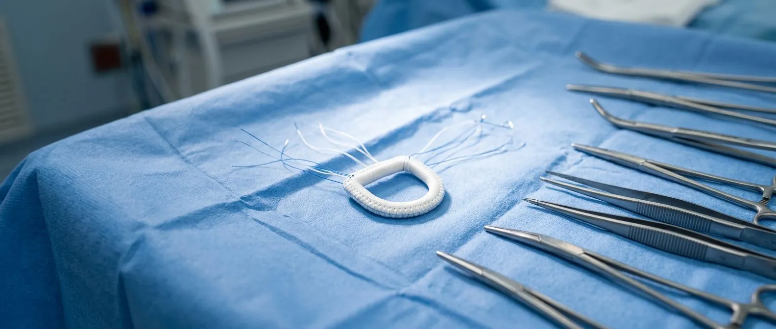
Repair versus replacement. Papillary muscle rupture surgery is done through an opening in the left atrium, on the beating-stopped heart supported by the heart-lung machine. The surgeon inspects the mitral valve and decides between mitral valve repair — rebuilding the anchoring system — and mitral valve replacement with a prosthetic valve. With a completely torn muscle, replacement is more commonly the safest option; when the tear is partial, reconstruction with artificial chordae or reimplantation of the muscle head may be possible.
Addressing the coronary blockage at the same time. Because the rupture follows a heart attack, the surgeon usually also bypasses the blocked coronary artery (coronary artery bypass grafting) in the same operation. Doing both at once treats the cause and the consequence in a single anesthetic. For most patients this means an aortic cross-clamp, cardioplegia to protect the heart, and a longer but comprehensive operation.
Less invasive routes exist for the right patient. Beyond the emergency scenario, our field has moved toward less invasive options. Minimally invasive mitral repair through a small right-chest incision, robotic mitral valve surgery, and catheter-based options such as edge-to-edge repair (TEER) for mitral regurgitation or, in selected degenerative cases, transcatheter approaches, all play a role — though these are generally for non-emergency, planned procedures. We recommend the less invasive route only where the evidence supports it for that patient; in an acute rupture, the conventional open operation remains the standard because speed and certainty matter most. Dr Molina performed the first robotic mitral valve repair in the Ángeles Health System and the first robotic heart surgery in Guadalajara, so when a planned case can be done robotically, the team has the experience to offer it.
Here is how the main decision points look to a family hearing them for the first time:
- Repair (valve preserved): keeps the native valve; preferred when anatomy allows; avoids lifelong anticoagulation if a repair is feasible.
- Replacement (biological or mechanical valve): the reliable choice when the muscle is destroyed; mechanical valves require lifelong blood thinners, biological ones may need reoperation years later.
- Coronary bypass at the same time: treats the blocked artery that caused the infarct, protecting the future of the repaired or replaced valve.
Recovery After Emergency Mitral Valve Surgery
The first days. After an emergency operation of this kind, the patient wakes in an intensive care unit, usually with a breathing tube for the first hours and monitoring lines in place. Because many arrive in shock, the first days can involve support for the kidneys and the circulation while the heart recovers from both the infarct and the operation. This phase is unpredictable, and families should expect honest updates rather than promises.
The first weeks. Most patients who survive the early period leave intensive care within a few days and the hospital within one to two weeks, depending on complications. Sternal precautions — no lifting, no driving, no pushing — protect the breastbone for six to eight weeks. Cardiac rehabilitation, a supervised program of gradual exercise, begins once the surgeon confirms the chest has healed and is one of the strongest predictors of a good long-term recovery.
Life afterward. A repaired or replaced mitral valve can support a full, active life. Follow-up echocardiograms track the valve's function; patients with replacement valves take anticoagulation as prescribed; and every patient continues treatment for the coronary disease that caused the rupture in the first place. Recovery from this operation is a marathon measured in months, not weeks, and the patients who do best are those who follow the rehabilitation plan and keep every follow-up appointment.

One practical note for international patients: if you are traveling to Guadalajara, Querétaro or Hermosillo for care, plan to stay locally through the early follow-up visits before returning home.
Papillary Muscle Dysfunction vs Rupture: How They Differ
Two different injuries to the same structure. Papillary muscle dysfunction means the muscle is alive but not working properly — it may be scarred from an old infarct, displaced by a remodeled ventricle, or too weak to hold the leaflets closed. The leak is usually gradual and moderate, and it often responds first to medication and careful follow-up. Rupture, by contrast, is a mechanical failure: the muscle is torn and the leak is abrupt and severe.
Why the distinction changes treatment. Dysfunction is typically managed medically, with drugs that reduce the workload of the heart, and — in chronic cases known as ischemic mitral regurgitation — with planned surgery when the leak becomes significant. Rupture almost always requires emergency operation. The difference between the two is the difference between a scheduled conversation and a 2 a.m. call to the operating room, which is why the echocardiogram's findings are so consequential.
Where prolapse fits in. Mitral valve prolapse — a degenerative condition in which the leaflets billow backward — can also cause chordae tendineae rupture and acute severe regurgitation, but it is a different disease with a different mechanism. In prolapse the muscle is usually normal and the leaflet tissue is the problem; in post-infarct rupture the muscle itself is the problem. Our team evaluates every case of severe mitral regurgitation against its true mechanism before recommending an approach, because the right operation depends entirely on what actually broke.
When to Seek a Heart Valve Surgeon in Mexico
Warning signs that cannot wait. New breathlessness after a recent heart attack, fainting, coughing pink frothy sputum, or cold clammy skin with chest discomfort — these are emergency symptoms and belong in an emergency department, not a waiting room. For planned situations — a known moderate leak, an echocardiogram showing prolapse, a question about whether surgery is needed — a specialist consultation with a heart valve surgeon is the right step, and sooner is better than later.
What a first consultation with us looks like. The first contact is a free video consultation: we get to know the patient, talk through the condition, and say which studies are still needed so no unnecessary ones are ordered. A face-to-face visit at our consulting rooms in Guadalajara, Querétaro or Hermosillo follows when the patient decides to go ahead. Our work covers valve disease, coronary disease, the aorta and structural heart conditions, with conventional, minimally invasive, robotic and transcatheter procedures all available — the choice is made patient by patient, and the final decision is always the patient's.
Patients traveling from abroad often ask about coordination. Cost, travel logistics and insurance are discussed openly at the first meeting — no family should face heart surgery with unanswered questions about money or logistics.
Key Takeaways About Papillary Muscle Rupture and Your Heart
The papillary muscle is a small structure with an enormous job: anchoring the mitral valve against the full pressure of every heartbeat. When a heart attack cuts off its blood supply, the muscle can tear days later, causing acute mitral regurgitation that demands emergency surgery. Knowing the anatomy — and the warning symptoms of sudden breathlessness and shock — is what turns this rare complication into a survivable one.
The emerging research on how these muscles fail continues to appear in the peer-reviewed literature indexed by DOAJ, and surgical techniques for both emergency repair and planned reconstruction keep improving. But no article replaces an individual evaluation: the right operation depends on your valve, your coronary anatomy, and your overall health.
If you or someone close to you has been told about mitral regurgitation, has recovered from a heart attack with new symptoms, or simply wants a clear, honest second opinion about valve surgery, we invite you to reach out. Talk with a qualified cardiac team, bring your studies and your questions, and book a consultation — the first conversation with our team costs nothing, and it may be the clearest step you take.






